






Abstract
Cancer treatments utilizing biologic or cytotoxic drugs compose the frontline of therapy, and though gains in treatment efficacy have been persistent in recent decades, much work remains in understanding cancer progression and treatment. Compounding this situation is the low rate of success when translating preclinical drug candidates to the clinic, which raises costs and development timelines. This underperformance is due in part to the poor recapitulation of the tumor microenvironment, a critical component of cancer biology, in cancer model systems. New technologies capable of both accurately observing and manipulating the tumor microenvironment are needed to effectively model cancer response to treatment. In this review, conventional cancer models are summarized, and a primer on emerging techniques for monitoring and modulating the tumor microenvironment is presented and discussed.
Impact Statement
The low success rate of prospective therapeutics to the clinic, with an average cost of ~$650 million dollars each, is a large barrier to cancer drug development. As high rates of failure occur at the end of clinical testing, the identification of effective and translatable candidates must be made more rigorous to mitigate the loss of time and capital. To accomplish this aim, integration of cancer tissue models with advanced tissue monitoring and control systems is needed.
Introduction
Cancer is characterized by the deregulation of cellular pathways that regulate several critical components of cell behavior, including growth and invasion into surrounding tissues. Particular types of cancer can differ in their cellular origins, acquired mutations, and microenvironmental conditions, which contributes to a broad range of phenotypes and frustrates the search for a silver bullet treatment. 1 Instead, cancer treatment utilizes a personalized approach, where adjustments to treatment are made in response to disease progression. Over the past 30 years, this strategy has shown some success as cancer death rates have fallen 31% due to improvements in prevention, detection, and treatment. 2 In spite of these gains, cancer remains the second leading cause of death in the United States, and more than one in three people are expected to develop cancer during their lifetime. 2 Even more concerning is the fact that novel cancer drugs entering clinical trials only have a 3.4% approval rate by the US Food and Drug Administration (FDA), and these drugs that do pass often have little to no effect on overall survival.3,4 These data suggest a disjunction in the drug development pipeline where clinical outcomes do not realize the same degree of therapeutic success observed in preclinical studies.
The low rate of clinical translation for novel cancer therapies is due in part to the complex and interconnected network of the tumor microenvironment (TME), where cancer cells reside.5,6 This background encompasses all of the participatory components of a tumor including local specialized cell types (immune cells, fibroblasts, etc.), the extracellular matrix (ECM), chemical gradients, and physical conditions, such as interstitial fluid pressure and shear stresses. Critically, these components vary at local and regional scales, and, when combined with the genomic instability of cancer cells, results in a highly heterogenous disease state. These local differences in the TME have been shown to affect clinically significant tumor properties, such as cancer development, progression, and therapeutic response.6,7 One particularly prominent process is the metastatic cascade, a hallmark of cancer that is long recognized as a significant cause of cancer-associated mortality, yet remains poorly characterized.8–14 The recent designation of metastasis-free survival as an emerging clinical trial endpoint by the FDA demonstrates both the importance of improving our understanding of metastasis and the influence of the TME in therapeutic development. 14
To study the role of the TME in cancer development and progression, cancer models are designed with varying degrees of experimental control, system complexity, and model accuracy. A particular challenge for cancer models is accounting for the heterogeneous nature of the TME while maintaining experimental reproducibility and practicality. The conventional pipeline for cancer research is to first screen for potential biological processes in vitro and then to validate therapeutics in vivo before moving to human testing. This approach is standard practice because the low cost and high-throughput capabilities of cell culturing techniques allow for robust candidate screening and pathway characterization, while in vivo models enhance the physiological relevance of therapeutic potential findings and test for systemic toxicities. Yet, the low success rate of this methodology in identifying clinically viable therapeutics suggests that there is room for improvement. Major concerns, namely, the poor recapitulation of physiological conditions in two-dimensional (2D) cell culture and the significant differences between human and murine drug response, encourage re-examination of conventions in translational cancer research.15,16 In the following section, we summarize conventional cancer models, discuss their advantages and disadvantages, and summarize common quantification techniques to motivate the need for emerging cancer research tools (Figure 1). In later sections, we review techniques and technologies that can address gaps present in conventional cancer model technology through TME monitoring and manipulation.

Schematic of prominent technologies used for modeling the tumor microenvironment. Cancer models determine the biological complexity that is being studied. Monitoring technologies quantify the parameters of interest during a study, and manipulation improves experimental control and model relevance. Combinations of these technologies are needed for effective translation of findings to the clinic. (A color version of this figure is available in the online journal.)
Existing preclinical cancer models
In vitro and in vivo cancer models are critical tools in the investigation of cancer signaling pathways and development of novel diagnostic and therapeutic technologies. Recent advances in molecular characterization and genetic engineering have rapidly expanded both our grasp on cancer development and allowed for the rapid generation of new cancer models to better recapitulate particular aspects of cancer biology.17,18 In this section, a topical review of prominent in vitro and in vivo cancer modeling techniques is presented.
In vitro cancer models
Cell culture techniques are some of the most accessible methods for studying cancer biology. They enable a bottom-up approach where system complexity is constructed using well-characterized components to recreate specific niches in the cell microenvironment.17,18 This additive property of in vitro cell culture ensures a high degree of experimental control and target selectivity, which improves experimental reproducibility and enables high-throughput analysis. The bulk of in vitro cancer research is done using commercial cancer cell lines due to their straightforward validation between laboratories and ease of maintenance. 15 Cell line–based models are particularly useful in the evaluation of cancer cell–specific properties, such as oncogenes and drug sensitivity. However, cell lines also suffer from several limitations, including the selective pressures of monolayer culture, which can cause genetic alterations that are not found in vivo, and clonality, which loses the intra- and inter-tumoral heterogeneity found in the clinic.19–21 These changes can then contribute to misrepresentation of cell behavior and drug sensitivity. Primary cells are used to circumvent many of these concerns; however, sourcing and maintaining primary cells is significantly more challenging than commercial cell lines. Moreover, repeated passaging and expansion of primary cells depletes native ECM components and can lead to epigenetic drift and loss of tissue morphology. 20 Collectively, these shortcomings limit the scope of cell line monocultures to cancer cell–specific functions.
To compensate for these challenges, three-dimensional (3D) cell culture techniques have been used to more accurately model tumor architecture. Tumor spheroids are notable for their similarities to avascular tumors and are commonly used to recreate cell–cell interactions and differential exposure conditions 22 (see Table 1). These properties restrict the availability of oxygen and nutrients in the core of spheroids, resulting in a gradient of proliferative and metabolic cell behaviors that alter therapeutic response and correlate with tumor conditions found in vivo. 23 Spheroids can be generated from established cancer cell lines or derived from tumor tissue with varying degrees of cell dissociation and enrichment used to select for particular cell subpopulations.22,24,25 Importantly, the inclusion of multiple cellular constituents allows for the probing of specific interactions between cell types.22,23 Owing to their construction from selected cell lines, however, spheroids lack control over ECM characteristics and fail to recapitulate higher-order tissue behaviors such as vascularization. Precise control over spatiotemporal biophysical and biochemical factors is also not available with conventional techniques. In addition, not all cell types are amenable to spheroid formation, their small size makes handling difficult, and uniform spheroid formation is necessary for comparison. 39
Summary of 3D cancer cell culture techniques and animal models.

ECM: extracellular matrix; TME: tumor microenvironment.
Scaffold-based culture techniques are another method of 3D tumor modeling when cell–ECM interactions are being investigated37,40 (see Table 1). Scaffolds can be hydrogels or porous substrates composed of natural or synthetic materials. Natural polymers (e.g. alginate, chitosan, gelatin, collagen, fibronectin, and Matrigel®) use prominent tumor ECM components that can contain native background factors and be reorganized by cells.37,41,42 This compositional complexity allows naturally derived polymers to mimic the structural heterogeneity found in vivo and promote organized cancer cell development. 37 Unfortunately, certain naturally derived polymers can be highly variable, overly complex, difficult to isolate, and lack human-specific markers.32,42,43 Basement membrane scaffolds also tend to be derived from tumor tissue and thus may contain unquantified background proteins and effectors. Alternatively, synthetic polymers, such as poly(ethylene glycol), poly(vinyl alcohol), and poly(lactic-co-glycolic acid), are used for their high reproducibility, tunable stiffness, and ease of modification. 44 Their commercial development provides a streamlined matrix selection process, but synthetic materials can also have cytotoxic components or unpredictable cell–polymer interactions. 42 Other challenges to scaffold-based cell culture include achieving sufficient cell distribution, infiltration, and recovery. 45 Cell–cell interactions are difficult to manage when seeding cell suspensions, but spheroid seeded scaffolds have shown higher drug resistance than spheroids on a flat surface and scaffolds seeded with dispersed cells. 46
Histoculture (explant cell culture) is another approach that captures much of the complexity of the TME by culturing sections of tumors grown in vivo (Table 1). While this setup is well suited for rapid testing and visualization, sample collection and long-term maintenance are difficult and only a fraction of the tumor’s overall heterogeneity is captured. 47
While discussion of all available in vitro assays for studying the TME is beyond the scope of this work, a broad summary is valuable for recognizing opportunities for advancement in quantification. Evaluation of in vitro cancer models is primarily accomplished through measurement of cell markers and cell behaviors. Drug sensitivity assays test concentrations of anticancer drugs in microtiter plates to evaluate therapeutic effect. 48 Cancer cell migration and invasion are observed using Transwell® migration assays, where the movement of cells (B16F10, HeLa, MCF-7, MDA-MB213, T-47D, etc.) across a membrane is used to evaluate chemoattractant response and malignancy. 15 Fluorescent reporter genes and colored dyes can be used to label markers, in particular, cancer pathways and track cell fate. 49 These assays have been integrated into hypoxia-response pathways to link hypoxia to increased fibrous tissue deposition. 50 For 3D tumor models, additional cell processing or alternative analysis techniques may be employed to preserve spatial or organizational information. Metrics commonly used for the characterization of spheroids include size, shape, and cellular organization, which are best visualized through optical microscopy techniques (e.g. bright field, dark field, differential interference contrast, and fluorescence imaging). 23 Flow cytometry can also be used for quantifying fluorescent markers in cells, but disaggregation is necessary for analysis, and therefore, steps must be taken to prevent the loss of spatial information.23,29 Other considerations for 3D in vitro cell culture techniques include the autofluorescence of certain scaffold materials, such as collagen, which can interfere with scaffold-based culture imaging, as well as background signal from out-of-plane fluorophores. 45 To counter these drawbacks, various sectioning techniques have been used to improve contrast and data collection, including light-sheet-based fluorescence microscopy, two-photon microscopy, and multiphoton microscopy.23,37,29 Finally, chemical gradients and active flow systems are difficult to establish and maintain in most conventional cancer cell culture formats, and some in vitro assays are restricted to endpoint analysis, limiting access to cellular dynamics. 51 As such, in vitro methods are best suited for high-throughput testing and screening studies with low model complexity for mechanism discovery and therapeutic candidate identification.
In vivo cancer models
In contrast to the low physiological relevance afforded by in vitro methods, in vivo models are used to capture a more complete picture of the biological complexity present in the TME by allowing cancer cells to grow in an environment that is similar to the human body (Table 1). This is a critical component for the translational research of novel therapeutics because it allows for systemic toxicity screens and provides more comprehensive data on the impact of clinical drug administration. However, the various autochthonous and non-autochthonous models that have been developed to study specific aspects of cancer progression are not universally applicable. For instance, immunotherapy, carcinogenesis, and early tumor growth are best studied with de novo techniques such as environmentally induced or genetically engineered mouse models, but, the rapid growth of multifocal tumors limits their application in studying late-stage cancer processes such as metastasis.20,52 On the contrary, transplant models using cell lines (4T1, B16, Lewis lung carcinoma, etc.) or patient-derived tumors are flexible platforms for observing therapeutic efficacy and tumor growth, as the location of tumor implantation can be chosen to simplify disease monitoring (ectopic), preserve TME interactions (orthotopic), or expedite metastatic dissemination and colonization (systemic).20,52 To model human cancers in animal models, cell line–derived xenografts (CDXs) are used for their lower costs and higher availability than patient-derived xenografts (PDXs), which have significantly higher clinical relevance but suffer from variable engraftment rates. 52 Incorporation of patient-derived cells in hollow fiber implants (mini-PDX) assays have also been demonstrated as an alternative approach to accelerate in vivo drug sensitivity testing. 53 Recently, the development of humanized mice has further increased the relevance of transplant models by integrating elements of the human immune system.33,54
Despite the many advantages to studying the TME in vivo, challenges, including cost, time, high model variance, and low throughput, limit its statistical power compared to in vitro systems. 41 Critically, concerns have also been raised that animal testing does not reliably translate to the clinic. 16 Tumor model differences including drug metabolism, immune system composition, tumorigenesis, and chimerism can all contribute to response divergence.33,37 Also, while inbred animal populations are desirable for experimental reproducibility, clinically relevant parameters such as population dynamics and inter-tumoral heterogeneity are absent. To combat some of these limitations, animal models with a higher genetic similarity to humans have been used, but their associated costs, development time, and handling expertise preclude widespread use. 36 Alternatively, non-mammalian tumor models, such as yeast, zebrafish, and chicken chorioallantoic membranes, can be used for applications including carcinogens, oncogenes, and angiogenesis, while mitigating the costs and ethical concerns associated with mammalian models37,55 (see Table 1).
When evaluating changes to the TME in animal models, macroscale indicators such as tumor size, weight, and metastatic spread, measured by necroscopy, histology, or cytology, are commonly used to gauge cancer growth and progression. 56 For observation of genetic and cellular changes occurring in the TME, molecular biology techniques (e.g. enzyme-linked immunosorbent assay (ELISA), quantitative polymerase chain reaction (qPCR), microarray, radioimmunoassay, flow cytometry, immunohistochemistry, western blot analysis, and proteomics) or optical imaging of labeled molecules (e.g. confocal, multiphoton, and wide-field fluorescence) are typical.56–58 These assays, however, are often end-stage, limiting data collection to single timepoints per animal, and can disrupt the TME’s spatial organization. In contrast, non-invasive quantification techniques can provide anatomical and functional data ranging from tumor structure, perfusion, and permeability to metabolic activity and drug distribution over time, allowing for higher statistical power with fewer animals and multimodal analysis. 59 Anatomical information is gathered using techniques with high spatial resolution, that is, computed tomography (CT), magnetic resonance imaging (MRI), photoacoustic imaging (PAI), and ultrasound (US), whereas, techniques with high sensitivity, such as bioluminescence imaging (BLI), fluorescence imaging (FI), intravital imaging (IVM), positron-emission spectroscopy (PET), Raman spectroscopy (RS), and single photon emission computed tomography (SPECT), are used for molecular imaging (Table 2). 60 Collectively, these techniques can illuminate tissue-level processes to gauge overall tumor behavior and therapeutic performance. Confocal and multiphoton microscopy, in particular, are advantageous for obtaining non-destructive optical sections of intact tissue. 57 Implementation of these technologies with intravital imaging techniques further enhances the study of TME dynamics by allowing observation without tumor excision.57,61–64 Other considerations include imaging agent requirements, penetration depth, temporal resolution, and exposure to ionizing radiation, when deciding on a technique. Thus, in vivo models are optimum for performing crosstalk studies with high model complexity (angiogenesis and metastasis), analyzing drug biodistribution, and assessing systemic toxicity.
Characteristics of current imaging modalities.

Source: Adapted from James and Gambhir. 60
Improvements to preclinical cancer models via enhanced imaging and localized modulation
While conventional in vitro and in vivo cancer models have provided numerous insights into the TME, the continued low success rate of clinical translation indicates room for improvement. It stands to reason then that one of two changes needs to occur: either in vivo models must become satisfactory predictors of clinical success, or in vitro models should achieve sufficient clinical relevance that animal models are replaced. Thus, the following section surveys the use of novel engineering strategies to improve translational cancer model relevance and accessibility for more accurate findings. This is discussed in the context of technological improvements to monitoring and manipulation of the TME.
Monitoring and biomarker detection in the TME
As the high level of heterogeneity in the TME makes it difficult to trace crucial signaling pathways, minimally invasive study at high resolution is needed for observation and detection of cell populations and molecular concentrations to track changes in diseased tissue and gauge therapeutic response. Improvements in data collection from 3D tissue structures are expected to preserve signaling and transduction pathways, reduce the number of animals needed for statistical significance, and improve the development time of potential therapeutics. To accomplish this need, an array of molecular imaging techniques and biosensor systems are used for biomarker recognition and visualization.
Imaging probes for TME characterization
The multitude of changes in the TME, such as ECM remodeling, neovascularization, proteolysis, metabolic changes, and levels of reactive oxygen species (ROS) provide ample opportunities for biomarker recognition and imaging. Still, detection and quantification require sufficient target labeling with minimal background signal. Imaging probes are combinations of ligands, linkers, and reporters used to identify targets through biomarker recognition. They can include genetically encoded reporters, exogenous fluorophores, and contrast agents. Their ability to bind with high specificity and selectivity to target moieties at sufficient concentrations and durations to detect above background is essential for TME characterization. 60 Currently, conventional histological techniques have limited biomarker labeling capabilities and are restricted to 2D tissue slices (Table 3). Recent advances in multiplexed immunohistochemistry/immunofluorescence platforms (mIHC/mIF), however, have achieved high-throughput labeling of tissues using iterative staining, imaging and inactivation with dye-labeled antibodies.65,66 This approach allows the assessment of biomarker colocalization, distribution, and cell/tissue composition. 66 Similarly, tissue clearing for section-free volumetric microscopy and histo-cytometry is an emerging field for the collection of quantitative information from intact 3D tissue samples that has the potential to reduce sampling errors associated with heterogenous tissues and preserve anatomical relationships.67–69 Despite the impressive gains in TME detection and quantification, these techniques are still limited to end-stage analysis, and preparation times are significant (days).
Summary of detection methods for studying the tumor microenvironment.

TME: tumor microenvironment.
For dynamic labeling of the TME, high-efficiency delivery methods are necessary due to the dilution and elimination of imaging probes in living systems. Current methods utilize genetic reporters and injectable imaging agents to achieve sufficient contrast, which has enabled the quantification of a vast array of TME properties, including cell populations, tumor fluid perfusion, signal transduction, therapeutic action and biomarker expression70–73 (see Table 3). To further improve detection, targeted nano-carriers are being designed with moieties such as antibodies, small molecules, aptamers, and dendrimers to prioritize localization in desired tissues. 74 Their larger size allows for the incorporation of multiple targeting probes and reporter molecules to fine-tune signal strength as well as the option for multimodal imaging to overcome limitations of specific imaging techniques. 75 Pharmacokinetic characterization of these particles is necessary to account for their biodistribution profiles using techniques like tumor perfusion imaging. 76 Other targeting methods utilize sensitized carriers or activation sites that respond to unique conditions in the TME, such as pH, O2, and protease concentrations.77–83 For instance, fluorescence imaging of denatured collagen with photo-triggerable folding of collagen mimetic peptides circumvents limitations associated with targeting unstructured proteins. 99 Similar strategies have also been demonstrated with fibrin imaging using MRI and SPECT.100,101 Finally, cell-mediated labeling strategies, where cell-penetrating fluorophores are used to mark neighboring cells can be used to identify local cell–cell interactions. 102 Analysis of cells marked by this approach demonstrates a strategy for nonspecific functional characterization of the metastatic niche.
Sensors for study of the TME
In addition to the novel molecular labeling approaches discussed in the previous section, TME sensors are promising alternatives to monitor both analyte concentrations (e.g. proteins, nucleic acids) and biophysical properties (e.g. pressure, stiffness) of the TME. 58 Their wide range of readouts, including electrochemical, optical, and gravimetric (piezoelectric), provide additional sensing opportunities for biomarker detection and TME characterization that are not available with imaging probes103–105 (see Table 3). They can also find application in lieu of cost-prohibitive analytical techniques. As dedicated reviews on biosensors and their various classifications have been recently covered, we instead emphasize two key TME sensor characteristics: invasiveness and sensitivity.106,107
Invasiveness is a crucial consideration for TME investigation as significant disruption of the tissue can alter cell behavior and distort experimental results. This property is especially important in vivo, where animal stress, tissue damage, and biocompatibility issues underly long-term sensor use. Consequently, highly invasive techniques such as atomic force microscopy (AFM) and electron microscopy, where major surgery or tissue processing is required, are most appropriate when quantifying cell and TME tissue properties that are difficult to collect otherwise.108,109 For example, Mao et al. 110 used AFM to characterize the nanomechanical properties of aortic intima in response to pharmaceutical stimulation in vivo. Similarly, molecular analysis with micro-dialysis and in vivo mass spectroscopy have immense potential in characterizing the concentration of analytes in the TME but require surgery and analyte extraction.111–113 Efforts to minimize sensor invasiveness, such as the incorporation of wireless readout capabilities or miniaturization of implants, improve sensor usability over larger timescales but typically involve more complicated fabrication procedures.114,115 Biochemical sensing strategies have been widely explored for their straightforward production methods (e.g. electrochemical sensors, evanescent wave sensors). Label-free detection methods are minimally invasive and allow for real-time monitoring but their signal-to-noise ratio can be insufficient for reliable detection of small molecules. 116
The ability to accurately detect low concentrations of analytes is another critical property in sensor application. Due to the diverse and heterogenous nature of the TME, high sensitivity and a low detection limit are needed for reliable data collection. For biomarker detection, sensing is typically done through functionalization with affinity-based recognition elements, such as antibodies, antigens, enzymes, nucleic acids, receptors, or whole cells.97,106,117 Using direct detection methods, label-free biosensors rely on physical interactions between the biomarker and sensor interface but are limited by analyte availability and generally weak interface sensitivity. In cases where sufficient sensitivity is not achieved, amplification of the biomarker or signal can improve device performance at the expense of more complicated sample processing (longer assay time) and increased risk of distorting binding characteristics. 116 Common applications of this strategy include PCR for nucleic acid amplification and signal amplification via sandwich immunoassays or enzyme conjugation. 118 Other strategies for improving sensor performance include increasing the detection region, improving mass transport effects near the sensor, and using magnetic nanoparticles for analyte scavenging.95,118
Future considerations of monitoring the TME
While these strategies used for TME observation and biomarker detection are not new, challenges in achieving acceptable signal-to-noise ratios, biodistribution profiles, and target identification are still under intense investigation. For imaging probes, physicochemical optimization of the size, surface charge, circulation half-life, and biocompatibility are all factors that must be considered during development, particularly for in vivo applications. Limitations of particular imaging modalities are also areas of concern. Currently optical modalities provide the best option for high temporal resolution, but tradeoffs in working distance and spatial resolution, as well as optical scattering in biological tissues and out-of-plane photobleaching, hinder deep tissue study. In addition, high-resolution imaging of large tissue regions is time consuming and has high computational requirements for processing and analysis. 67 On the contrary, biosensors are anticipated to reduce dependence on expensive equipment and expertise required for advanced detection systems. To accomplish this function, sensors need to be practical, robust, reproducible, and miniaturizable. Identification of optimal sensor modalities for the target applications will also require reliable performance metrics for comparison, and characterization of binding kinetics will be necessary for dynamic measurements. Due to the heterogenous nature of the TME, extension of the sensing interface could be advantageous for sampling larger tissue regions. Finally, while improvements to detection and quantification in the TME are certainly important, the integration of advanced biosensing technology into conventional testing formats should also be considered to maximize technology uptake by the scientific community (Figure 2(a)–(c)). 119
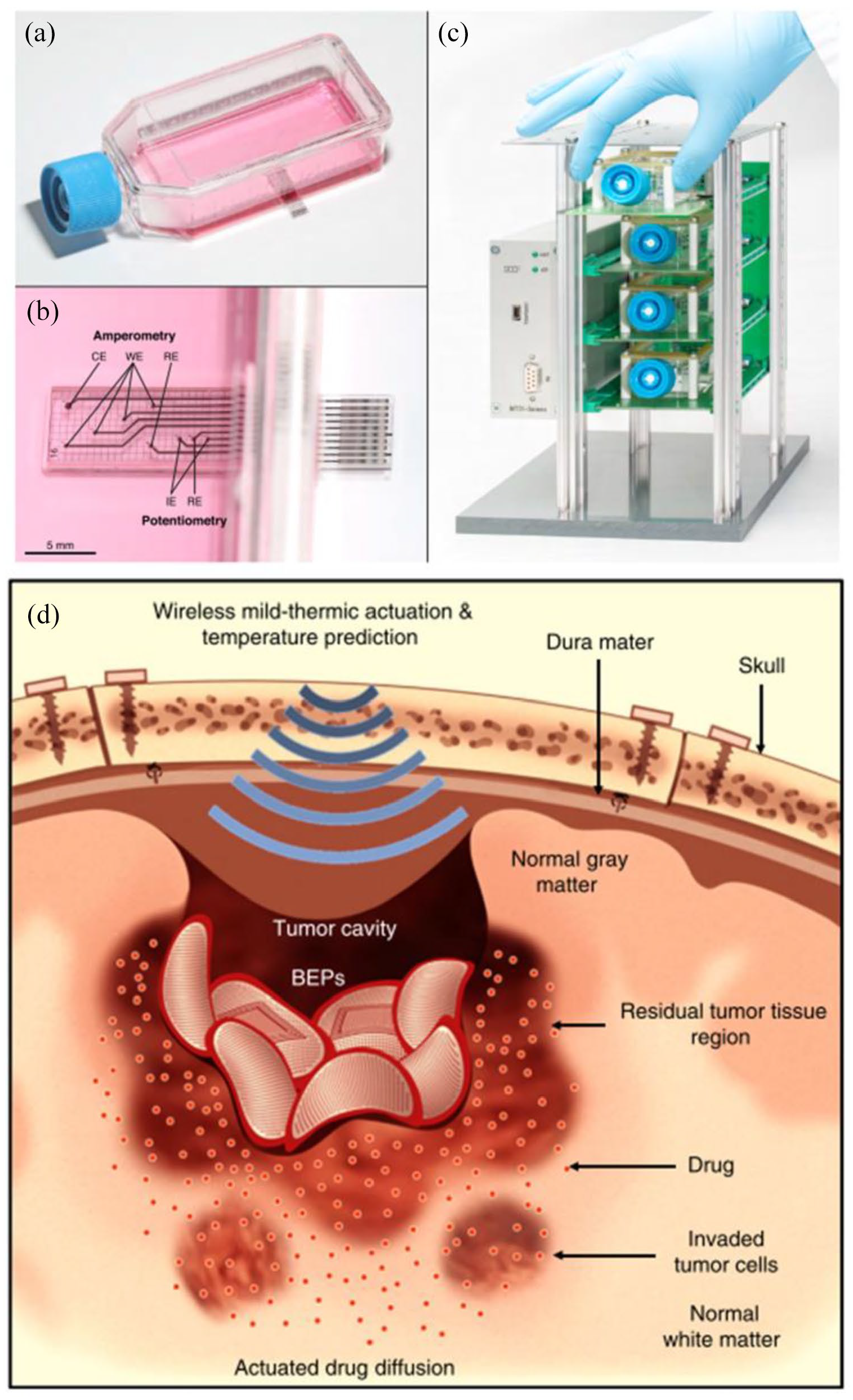
Engineered platforms for enhanced study of the tumor microenvironment: (a) sensing cell culture flask is designed around standard cell culture flasks to minimize protocol adjustment, (b) embedded electrodes allow for detection of a variety of biologically relevant chemicals; companion rack systems allow parallel real-time monitoring, and (d) the bioresorbable electronic patch uses wireless thermal actuation for enhanced drug delivery into glioblastomas. (A color version of this figure is available in the online journal.)
Manipulation of the TME
To improve the success rate of therapeutic translation, more accurate modeling of clinically relevant cancers and treatment outcomes are needed. Still cancer characteristics, including systemic spread, multiscale heterogeneity, and acquired resistance, present formidable complexity when recapitulating a comprehensive cancer model. Thus, the use of practical, application-specific cancer models is still warranted, particularly in basic research. However, the predictive efficacy of translational models needs work. To this end, greater experimental control in vivo and more representative in vitro cancer models would allow for effective modulation of the TME.
Pharmacotherapy targeting the TME
Pharmacotherapy in combination with radiation and surgery is the primary treatment strategy used in the treatment of cancer. It is comprised of cytotoxic chemotherapy and radiation therapy, which non-specifically interfere with cell division, immunotherapy, which promotes the immune system’s anticancer activity, and hormonal therapy and targeted therapy, which interfere with cancer growth signaling pathways. These drugs encompass the majority of clinical effectors used to treat cancer yet challenges such as drug resistance and nonspecific toxicity limit their clinical effectiveness.121,122 To alleviate these issues and improve the efficacy of pharmacotherapeutics, various drug delivery methodologies have been demonstrated.
The most prolific method of selective pharmacotherapy is via biomolecular recognition of target sites. Surface targeting ligands, as mentioned previously in the discussion on analyte recognition, allow for targeting of differentially expressed receptors on specific cell types and the capture of signaling molecules. This directed behavior is used to selectively eliminate cancer cells, promote non-cancerous cell behavior, and tune aspects of the TME for therapeutic benefit.123–125 For instance, radiopharmaceuticals, which serve as calcium analogs or chelators, are used for their preferential accumulation in bones to target bone metastases with localized radiation. 126 Oftentimes, however, a single anticancer therapeutic is not potent enough to eliminate cancer on its own. In these cases, combinations of anticancer drugs and adjuvant TME therapies can have synergistic effects. TME modification with antiangiogenic therapy can transiently improve nanotherapeutic delivery by reducing the interstitial fluid pressure of solid tumors to restore convective transport. 127 The result is higher drug bioavailability in the tumor, which impacts drug efficacy and therapeutic response.
Cell-mediated therapy is another targeting approach that has explored the loading of tumor-homing cells with therapeutics or receptors to circumvent typical barriers to nanoparticle delivery and immunosurveillance.128–130 Implantable cell encapsulation technology can be used to isolate populations of cells for genetically directed secretion of therapeutics (e.g. prodrug activators, cytotoxic agents, and immunostimulants) into the TME. 131 Immunotherapy uses checkpoint inhibitors and T-cells that are genetically modified with chimeric antigen receptors (CAR T-cells) or T-cell receptors (TCR T-cells) to overcome the immunosuppressive TME and tumor evasion mechanisms. 132 This therapeutic strategy is not universally applicable, however, as complications including T-cell production, specificity, and exhaustion, as well as side effects (e.g. cytokine release syndrome and neurotoxicity) limit patient compatibility. 133 Efforts to alleviate these shortcomings and improve target selectivity include combinatorial antigen recognition to reduce bystander cell recognition and exhaustion-resistant phenotypes.134–136
Alternatively, TME-specific release can also be achieved by sensitizing drug delivery systems (DDSs) to endogenous physiochemical conditions, such as hypoxia and pH.137–140 In these cases, localized therapeutic release can be realized without altering the drug itself. This targeting is possible due to the significant remodeling that occurs during tumor progression, producing abnormal vasculature, nutrient gradients, and metabolic states. 141 The accumulation and retention of many large drugs and nanomedicines in solid tumors due to leaky vasculature and non-functioning lymphatics, called the enhanced permeability and retention (EPR) effect, is one such method of tumor targeting that has been widely explored.142,143 Recent work has shown that this effect is not universal, however, and that intratumoral drug distributions are heterogenous. To overcome these limitations, Li et al.144,145 used size-switching nanoparticle superstructures to accumulate in tumor tissue via the EPR effect, and dissociate into small molecule particles to diffuse more readily through the TME. For extended delivery applications, controlled degradation of drug-loaded hydrogels has been used to minimize systemic exposure. 146 Here, tuning the composition of the hydrogel and the degradation rate of the individual components, allows for variable control over the release of multiple drugs. Exogenous triggers (e.g. light, radiofrequencies, and ultrasound) can also be used to control drug kinetics or perform photothermal therapy, but target accessibility can limit viable signaling modalities.147,148
Engineered tissue models for recapitulating the TME
Like biosensors, transducers are capable of operating at the cellular scale, improving in vitro modeling and affording direct control over the neighboring TME. These devices can bridge the gap between experimentally robust in vitro models and physiologically relevant in vivo models through the incorporation of pertinent biophysical and biochemical conditions. This approach allows the study of crucial aspects of tumor biology that are not easily observed otherwise, such as metastasis and angiogenesis. Methods to accomplish this feat include functional and structural improvements to existing cancer model technology. 149 One such example uses a magnetic actuating platform to mimic respiration-induced tissue stretching in vitro. 150 Results show actuation of breast cancer cells decreases metabolic activity and inhibits matrix degradation, indicating a potential role in dormancy and reactivation. 150 To improve the structural composition of tumor models, microscale organization of cells, and bioactive materials can be achieved using bioprinting and scaffold technologies. This strategy has enabled the management of local cell–cell interactions as well as cell confinement and ECM stiffness through variable cross-linking.151–153 Other engineering solutions seek to improve the analysis of 3D tissue models without the use of sectioning or isolation methods. TRACER (tissue roll for analysis of cellular environment and response) uses a stackable cell culture design to enable rapid disassembly and layer-by-layer analysis of 3D tissue constructs.154,155 The miniaturization of sensors and fluid handling technology to the cellular scale and beyond has empowered the development of microfluidic systems for novel cell culture platforms and drug delivery applications. These organ-on-a-chip (OOC) devices are capable of dynamically managing mechanical signals, biochemical gradients, and cellular interactions to improve cell differentiation and tissue organization over conventional 3D culture techniques. 156 OOCs have been used to model angiogenesis, tumor progression, drug exposure, and crosstalk between cells, as well as visualize spatial heterogeneity.156–159 In one implementation, metastatic and intravasation potential were evaluated by producing dynamic oxygen gradients across a collagen barrier to observe matrix breakdown. 160 This OOC thus provides functional control over physiologically relevant conditions that are not manageable in conventional cancer model formats. OOCs also provide an opportunity for the development of personalized cancer models using patient cells for treatment screening and predicting therapeutic response. 161 More advanced microphysiological systems are also being explored for modeling multiple cellular compartments (body-on-a-chip technology) to experimentally validate pharmacokinetic models as an intermediate to clinical testing.162,163
Microfabricated system for manipulation and study of the TME
For in vivo applications, improvements to therapy administration and control factors are of broad interest. Implantable drug reservoirs, used to extend therapeutic release profiles, circumvent many issues associated with repeated injection regimens and prevent the rapid clearance of therapeutics from the target site.146,164–167 Another strategy to limit systemic exposure incorporates collagen-binding moieties to limit drug transport out of injection sites by anchoring to generalizable target sites in tumors.168,169 For situations where drug distribution is insufficient, actuated drug delivery has been used to improve cellular uptake. 120 This approach allows dynamic control over therapeutic concentrations in the TME. The use of biodegradable materials can also be selected to obviate the need for retrieval surgery and complications associated with chronic implants (Figure 2(d)). 120 Improvements to tumor grafting methods using cell sheet transplants are a novel way of subcutaneously engrafting tumor cells in biologically intact structures with high efficiency compared to enzymatically treated cell cultures. 170 Presumably, this technology could be extended to graft tumoroids and bioprinted tissues for directed TME formation in vivo. The application of micro-control systems in vivo presents many benefits as localized manipulation can enable internal controls, reducing the number of animals need for a study, and improve data collection. 171 The nano-intravital device (NANIVID) is one implementation that has been used for an array of studies in vitro and in vivo including hydrogel-mediated release of the chemotaxis agents (epidermal growth factor), hypoxia mimetics (deferoxamine and cobalt chloride), and ROS inductors (H2O2), as well as for cell collection (Figure 3(a)–(h)).166,172–175 Further work in this direction has aimed at extending experiment duration through the integration of fluidic control with intravital imaging windows (ported mammary imaging window) (Figure 3(i)).63,176 Recently, the development of integrated micro-nozzles for enhanced control over localized delivery has provided additional impetus for high resolution study in vivo (Figure 3(j)). 177

Implantable devices for simultaneous imaging and drug delivery in vivo: (a) the induction nano-intravital device (NANIVID) next to a US dime, (b) the NANIVID is designed to penetrate solid tumor tissue for passive delivery, (c) insertion is facilitated by an applicator which aligns the device with the tumor surface, (d) a 3D render demonstrates the device orientation during insertion, (e) a cross-sectional view of an implanted NANIVID depicts the location of the outlet and generated diffusion gradient, (f) top–down view of the insertion site for imaging, (g) alternative NANIVID design for cell collection, scale bar = 500 μm, (h) magnified view of the device outlet where green fluorescing cells were collected, scale bar = 100 μm, (i) exploded view of the microfluidic imaging window for active reagent delivery, (j) demonstration of improved dye localization in hydrogel tissue mimics with a micro-nozzle outlet. (A color version of this figure is available in the online journal.)
Genome editing to manipulate the TME
For functional analysis of genetic alterations in cancer, CRISPR/Cas9 (clustered regularly interspaced short palindromic repeats) has enabled specific, efficient, and affordable genome editing. Building off of earlier genome editing techniques (e.g. RNA interference, transcription activator-like effector nucleases, and zinc finger nucleases), pooled screening of novel therapeutic targets with CRISPR/Cas9 can identify essential driver genes and genotype-specific vulnerabilities. 178 This technology has been used to identify potential tumor suppressor genes and a gene involved in tumor metastasis in colorectal cancer organoid models. 179 Genome editing with CRISPR/Cas9 systems has also expedited the creation of novel genetic cancer models.180,181 Somatic gene editing in live animals provides a scalable alternative for model generation but has limited targeting capabilities currently. 182 Rapid progress in this field, however, already promises additional genomic editing tools and applications, such as point mutations and epigenetic editing.183–186 Finally, targeted delivery of CRISPR/Cas9 systems in the TME has the potential to identify critical intercellular interactions by disrupting communication with cancer-associated cells.
Future considerations of TME manipulation
Improvements to TME model controls for increased physiological relevance are imperative for improving the translative success of preclinical data. Molecular and microphysiological manipulation allow probing of all aspects of cell signaling which feed into development and therapeutic response. Ongoing research in pharmacotherapeutics is centered on drug screens and formulation technology. Synthesis of novel drugs face issues in maintaining activity, stability, toxicity, and delivery while balancing regulatory and manufacturing hurdles. Larger macromolecular and nanoparticle formulations currently must contend with poor biodistribution profiles due to high drug clearance and poor interstitial diffusion. Still, novel biopharmaceuticals, including RNA and recombinant proteins, have tremendous potential for revolutionizing medicine due to their flexible design and sequence recognition properties. Efficient delivery of miRNAs, siRNAs, and antisense oligonucleotides also have gene silencing capabilities that would allow for targeting of previously undruggable non-coding RNAs. 187
Although engineered cancer models encompass a wide range of techniques, general design principles for comparing methodologies exist. As a model’s main objective is to faithfully convey the behavior of a target system, model–user interactions must be weighed against model accuracy. With increasing model complexity, a concomitant increase in specialized knowledge and loss in throughput are typical. These factors, as well as the use of intensive fabrication techniques and equipment, can limit utilization by the scientific community. 188 To avoid this limitation, integrated compatibility with standard data collection techniques, including those technologies mentioned previously, can mitigate specialized training and startup costs.45,158,189 Application-oriented design can also reduce peripheral systems that have little or no role in the process of interest. 190 Standardization of components and protocols is also necessary to perform meaningful comparisons across multiple modeling techniques and improve reproducibility.
Conclusions
Despite the availability of a diverse field of cancer models, systems with reliable indicators of clinical success are still lacking. The low predictive power of current preclinical cancer models stems from their ineffective recapitulation of the human TME. To resolve this situation, advancements in preclinical model relevance and accessibility through technological innovation are necessary. This review surveys conventional model systems and advancements in both monitoring and manipulation techniques for enhanced experimental control. Balancing biological complexity and model practicality are critical for optimal model identification and selection. Still, the largest obstacles to the adoption of new technologies are regulatory barriers and accessibility. For regulatory approval, the safety and stability of novel drugs and medical devices must be established, often without clear guidance for evaluation. Accessibility comes in both the scalability of the manufacturing process, companion equipment cost, and the ease of operation. Accessibility also plays a significant role in uptake by the scientific community, which is driven largely by performance comparisons to equivalent systems and standardization of protocols and analysis. Nevertheless, the number of cancer modeling technologies will undoubtably continue to grow, and the development of sophisticated cancer models in novel preclinical workflows will require collaboration across institutions and disciplines to pool resources and expertise.
Footnotes
Acknowledgements
The authors thank Yubing Xie for reviewing the manuscript.
Authors’ Contributions
The first draft of the manuscript was written by TH and all authors commented on previous versions of the manuscript. All authors read and approved the final manuscript.
Declaration of Conflicting Interests
The author(s) declared no potential conflicts of interest with respect to the research, authorship, and/or publication of this article.
Funding
The author(s) disclosed receipt of the following financial support for the research, authorship, and/or publication of this article: This work was supported by the RNA Institute at the University at Albany, SUNY (grant no. 5T32GM132066-03); and the National Institutes of Health/National Cancer Institute in collaboration with Albert Einstein College of Medicine (AECOM) (grant no. 5R01CA216248-06).
