
Abstract
The enzyme Na+/K+-ATPase (NKA) is important in the heart. Reductions in NKA activity and expression have often been observed in chronic kidney disease (CKD)-related heart injury. Previously, our group found that an antibody targeting the NKA1α1 subunit’s DR extracellular region (897DVEDSYGQQWTYEQR911) stimulated NKA activities and produced cardioprotective effects against ischemic injury and isoproterenol-induced cardiac remodeling. In here, we assessed whether DRm217, a specific DR antibody, exhibits cardioprotective effects in chronic renal failure models. In 5/6 nephrectomy (5/6 Nx) surgery to mimic CKD in Sprague Dawley rat, we observed that NKA activity and expression were depressed in the hearts of 5/6 Nx rats. DRm217, an NKA DR region antibody, alleviated heart hypertrophy and cardiac fibrosis under 5/6 Nx conditions. Further studies revealed that DRm217 inhibited Src activation and reduced reactive oxygen species (ROS) levels in hearts under 5/6 Nx conditions. Our findings imply that NKA could be a treatment target in CKD-related cardiac diseases. Prevention of CKD-induced myocardial injury by DRm217 provides an appealing therapeutic alternative.
Impact Statement
Na+/K+-ATPase (NKA) is an important dual functional enzyme that is involved in diseases of different organs, including kidney, heart, and brain. NKA DR region antibodies are NKA activators and play protective roles in ischemic heart disease, excitotoxicity and stroke, Parkinson’s disease, and hepatic insulin resistance. This study details novel research on DR antibodies in protecting against chronic kidney–related heart injuries. This research will be beneficial for future drug development.
Introduction
Na+/K+-ATPase (abbreviated as NKA) is a 10-times transmembrane protein that actively transports K+ into and Na+ out of the cell. 1 The heart exhibits high levels of NKA, which is charge of regulating the proton electrochemical potent gradient. 2 However, in many heart disorders, the levels of NKA are lowered. For example, according to RH Schwinger report, NKA protein levels and activity were lowered in human heart failure. 3 Decreased NKA activity and expression were also found in the hearts of mammals following myocardial infarction, obesity, insulin resistance, diabetes, and hypertension.4 –6 The regulation of NKA activity and expression could be important in the treatment of cardio-diseases.
Previously, we and others reported that antibodies targeting the NKAα1 subunit’s DR extracellular region (897DVEDSYGQQWTYEQR911) stimulate NKA activities as well as protect hearts against oxidative injury, ischemic injury or isoproterenol-induced injury by stabilizing NKA expression on cellular membrane and closing NKA/Src/reactive oxygen species (ROS) amplifiers.7 –10 In patients with chronic renal failure, heart enlargement and fibrosis are prevalent. 11 Reductions in NKA activity and protein levels were also observed in the rats’ hearts with chronic kidney failure. 12 Considering the importance of NKA in heart and the function of DR antibodies on NKA activities, we conducted a number of tests to determine if DRm217, a specific DR antibody, 8 has cardioprotective effects in chronic renal failure models and assess its potential mechanisms.
Materials and methods
Chemicals and antibodies
Sigma-Aldrich (St. Louis, MO, USA) provided all of the chemicals, including ouabain. Rabbit antibodies against p-Src (Tyr416), t-Src, β-actin, and goat antirabbit antibody with HRP (Horseradish Peroxidase) labeling were provided by CST. Inc (MA, USA). Primary antibody against collagen III was purchased from Abcam Company (Shanghai City, China). RIPA (Radio Immunoprecipitation Assay) buffer and bicinchoninic acid (BCA) assay kits were provided by Fude Biological Technology Company (Hangzhou, China). Bioss Company (Beijing, China) provided mouse immunoglobulin G (IgG). HiTrap Protein G HP columns (GE Healthcare, Shanghai, China) were used to purify DRm217 antibodies from mouse ascites. The chemical reaction kits were provided by Nanjing Jiancheng Bioengineering Institute (Nanjing, China).
Animal experimental design
Sprague Dawley rats (male, seven weeks old, 225–250 g) derived from Xi’an Jiaotong University’s Laboratory Animal Center were employed in this investigation. The rats were housed in a pathogen-free, temperature-curtained facility with a 12-h light/12-h dark cycle and unlimited to food and water. The Animal Care Committee of Xi’an Jiaotong University authorized all animal care and experimental techniques, which followed worldwide criteria on the ethical use of animals. At random, the rats were allocated into four groups: (1) Sham group; (2) 5/6 Nx + IgG group (2 mg/kg IgG on alternate days for four weeks starting on the second day after 5/6 Nx surgery, intraperitoneal); (3) 5/6 Nx + DRm217 group (2 mg/kg DRm217 on alternate days for four weeks starting on the second day after 5/6 Nx surgery, intraperitoneal); (4) 5/6 Nx + ouabain group (30 µg/kg ouabain on alternate days for four weeks starting on the second day after 5/6 Nx surgery, intraperitoneal). The animal surgery and parameter determination were finished in blind method.
Partial nephrectomy surgery
Partial nephrectomy surgery (5/6 Nx) was established using a two-step method. Step one, 3% sodium pentobarbital were intraperitoneal injected to rats at a dose of 30 mg per kilogram of body weight for anesthesia. Then, the upper and lower spheres of the left kidney were removed, and just 1/3 of the left kidney size remained. Step two: seven days after the surgery, the rats were anesthetized again with 3% sodium pentobarbital, and the right kidney was dissected. The two-step operations for sham surgery were carried out in the same way as partial nephrectomy surgery, without removal of the right kidney and 2/3 left kidney. On the second day after Step two of surgery, animals in different groups underwent different treatments.
Tissue preparation
Mice were weighed and sacrificed at the end of the treatment. The hearts were taken out, rinsed in cold phosphate-buffered saline (PBS) to remove any blood. Then, the hearts were blotted dry and weight. The tibia length of every rat was measured after mice sacrificed.
Detection of serum creatinine, blood urea nitrogen, aspartate aminotransferase, lactate dehydrogenase, and creatine kinase-MB
The serum was collected by centrifugation at 4000 r/min for 13 min after blood was taken via the abdominal aorta. BUN (blood urea nitrogen), serum creatinine, serum aspartate aminotransferase (AST), serum lactate dehydrogenase (LDH), and serum creatine kinase isoenzyme-MB (CK-MB) were examined using a Beckman Coulter AU5431 automatic biochemical analyzer (CA, USA).
Detection of the cross-sectional areas of cardiac myocytes
Cardiac myocytes’ cross-sectional areas were used to assess cardiac hypertrophy. Briefly, hematoxylin and eosin-stained cardiac tissue sections (5 mm thick) were examined under a light microscope (Nikon, 200×). For every rat, 50 cardiac myocytes with clear cell boundaries and central nuclei were selected, and the area of the cross-section was computed. The average value of the cross-sectional area was then calculated.
Masson staining
Cardiac fibrosis was evaluated by Masson’s staining. Briefly, heart slices that had been paraffin-embedded were deparaffinized and rehydrated. Then, 0.1% Masson’s staining solution was used to stain deposited collagen on the sections. ImageJ (Media Cybernetics, Rockville, MD, USA) was used to digitally quantify the sections’ Masson-stained regions (green, fibrosis) (Masson-positive area = green area/total area × 100%).
Immunohistochemistry
Deparaffinized and rehydrated paraffin-embedded sections were soaked in 0.01 M citric acid buffer (pH = 10.0). Then, they were treated for 10 min with 3% H2O2, blocked for 30 min with rabbit serum at a comfortable temperature. After that, different antibodies (p-Src [1:100], NKA [1:200], and collagen III [1:100]) were coated on the slides at 4 °C above 12 h. The slides were coated again with goat antirabbit antibody-HRP labeled for 1 h. The sections were washed and stained with DAB and counterstained with hematoxylin.
Western blotting analysis
RIPA buffer was used to lyse heart tissues. Sodium dodecyl sulfate-polyacrylamide gel electrophoresis (SDS-PAGE) was used to separate 50 μg of protein samples, which were then shifted to nitrocellulose (NC) membrane. After blocking in 10% milk in TBST buffer, the membrane was treated with various primary antibodies at 4 °C overnight. Membranes were rinsed three times in TBST before being incubated at room temperature for 2 h with a 1:8000 dilution of antirabbit IgG-HRP-conjugated. Then, the membranes were rinsed in TBST thrice. The blot images were presented using an enhanced chemiluminescence kit (Fude Biological Technology Company, Hangzhou, China) and chemiluminescence imaging system (Syngene Company, G:BOX Chemi XRQ). ImageJ software was used to calculate the density of the bands.
Detection of hydroxyproline, malondialdehyde, oxidized glutathione, and NKA activity
Hydroxyproline, malondialdehyde (MDA), oxidized glutathione levels, and NKA activity were determined separately using hydroxyproline content assay kits, MDA assay kits, oxidized glutathione assay kits, and NKA assay kits, respectively, in preference to manual handling.
Statistical analysis
The results were given as mean values with standard errors. SPSS software was used to analyze the results. A one-way variance study accompanied by Tukey’s test (post hoc) was used for co-operation of multigroups variables. Difference was considered as P < 0.05.
Results
DRm217 decreased serum creatinine, BUN, LDH, and CK-MB levels under 5/6 Nx conditions
BUN and serum creatinine are two common markers of renal function. In the 5/6 Nx + IgG conditions and 5/6 Nx + ouabain conditions, serum creatinine and BUN were enhanced. DRm217 alleviated the increase in creatinine and BUN under 5/6 Nx conditions (Figure 1(a) and (b)). AST, LDH, and CK-MB are three common cardiac enzymes and biomarkers of myocardial injury. Serum AST levels in rats from different groups had no significantly different (Figure 1(c)). Serum LDH and CK-MB levels were elevated in 5/6 Nx + IgG and 5/6 Nx + ouabain groups. However, DRm217 blunted the increase in serum LDH and CK-MB levels under 5/6 Nx conditions (Figure 1(d) and (e)).
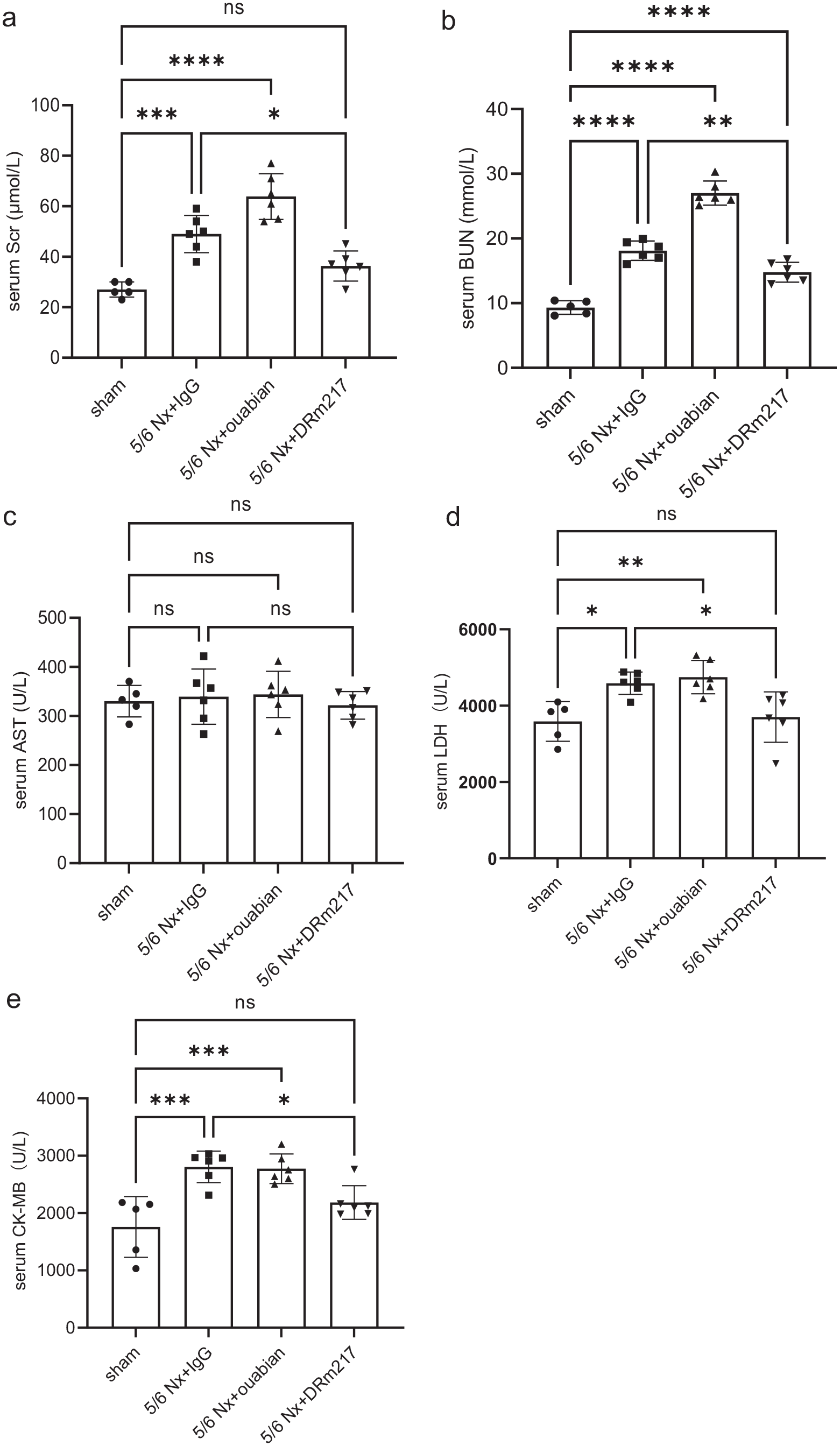
Myocardial enzymes content in every group. (a) Serum creatinine, (b) serum BUN, (c) serum AST, (d) serum LDH, and (e) serum CK-MB.
DRm217 alleviated 5/6 Nx-induced heart hypertrophy
To confirm whether DRm217 has an antihypertrophic effect, we first detected the rats’ body weight and the ratio of HW/BW (heart weight/body weight). There was no significant difference in the rats’ body weight in different groups (Figure 2(a)). In comparison to the sham group, 5/6 Nx dramatically raised the HW/BW ratio. DRm217 treatment alleviated HW/BW ratio under 5/6 Nx conditions (Figure 2(b)). To reduce bias due to diet changes with various treatments, we also detected the ratio of HW/tibia length. It showed the same trend with the amount of HW/BW (Figure 2(c)). The area in cross-section of cardiac myocytes was also dramatically raised in the 5/6 Nx + IgG group. DRm217 treatment inhibited the increase of cardiac myocyte cross-sectional areas (Figure 2(d) and (e)).
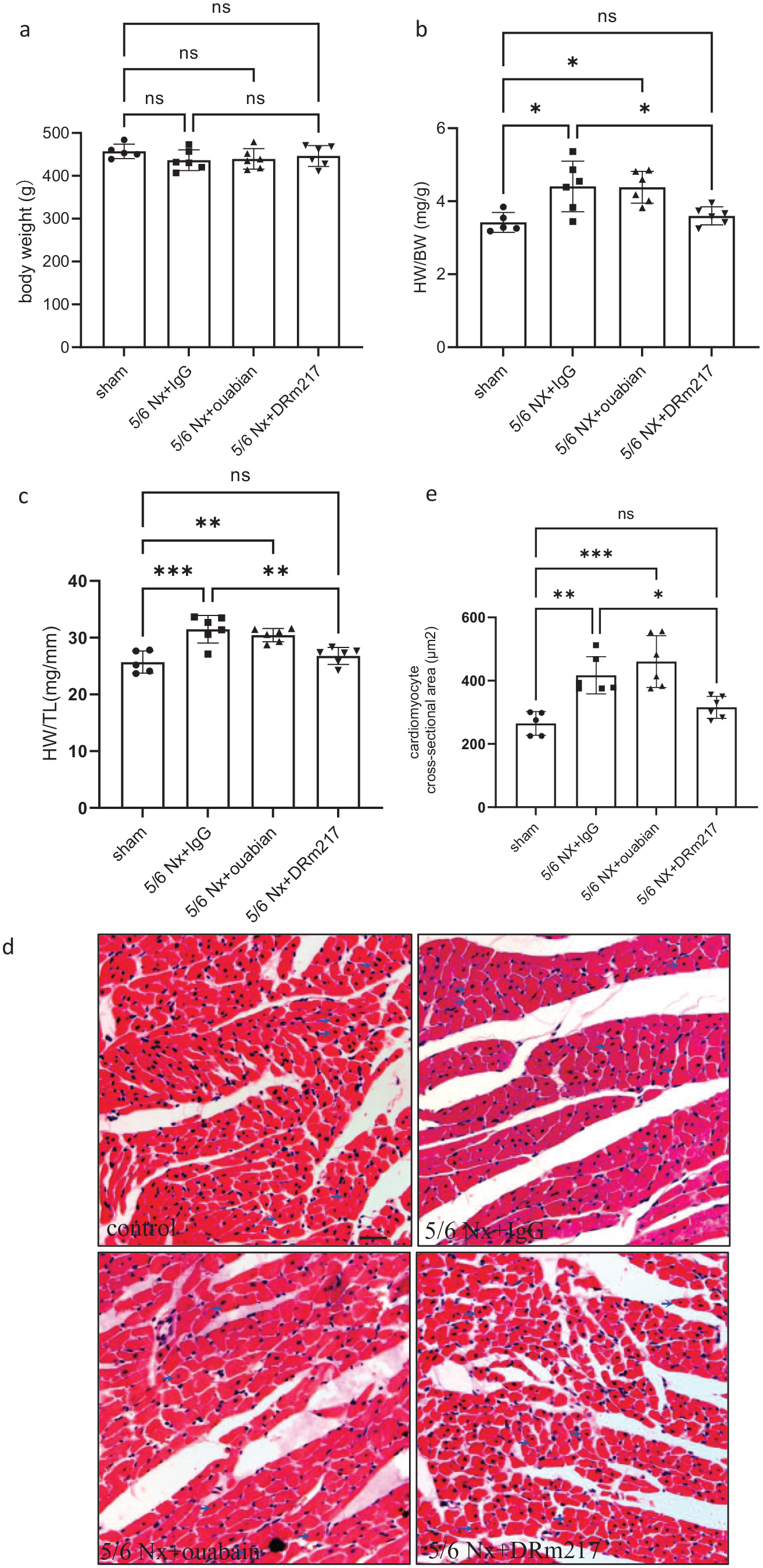
DRm217 alleviated 5/6 Nx-mediated heart hypertrophy. (a) Analysis results for the body weight, (b) statistical results for HW/BW, (c) statistical results for HW/TL, and (d) representative images of HE staining of hearts in each group; The scale bar is 100 μm (arrow represents cardiomyocytes). (e) Analysis results of the cross-sectional area of cardiomyocytes. (A color version of this figure is available in the online journal.)
DRm217 alleviated 5/6 Nx-induced cardiac fibrosis
Masson’s staining results revealed no obvious morphological changes in the sham group. In 5/6 Nx + IgG and 5/6 Nx + ouabain conditions, rats’ hearts had disordered arrangement of cardiomyocytes and obvious collagen accumulation in the heart, as evidenced by more green colors in the slides. The Masson-positive staining area was drastically depressed in hearts collected from 5/6 Nx + DRm217 rats (Figure 3(a) and (b)). Collagen III is also a common indicator for fibrosis. Immunohistochemical staining results showed that collagen III-positive staining was mainly observed in the perivascular region in the sham group. However, the level of collagen type III was greatly increased in the heart tissues collected from 5/6 Nx + IgG and 5/6 Nx + ouabain rats, as evidenced by the increased brown color present in the myocardial interstitium. In contrast, DRm217 treatment reduced collagen type III levels under 5/6 Nx conditions (Figure 3(c)). The hydroxyproline content was also not increased in 5/6 Nx + DRm217-treated rat hearts, but dramatically elevated in the 5/6 Nx + IgG-treated rat hearts and 5/6 Nx + ouabain-treated rat hearts (Figure 3(d)).
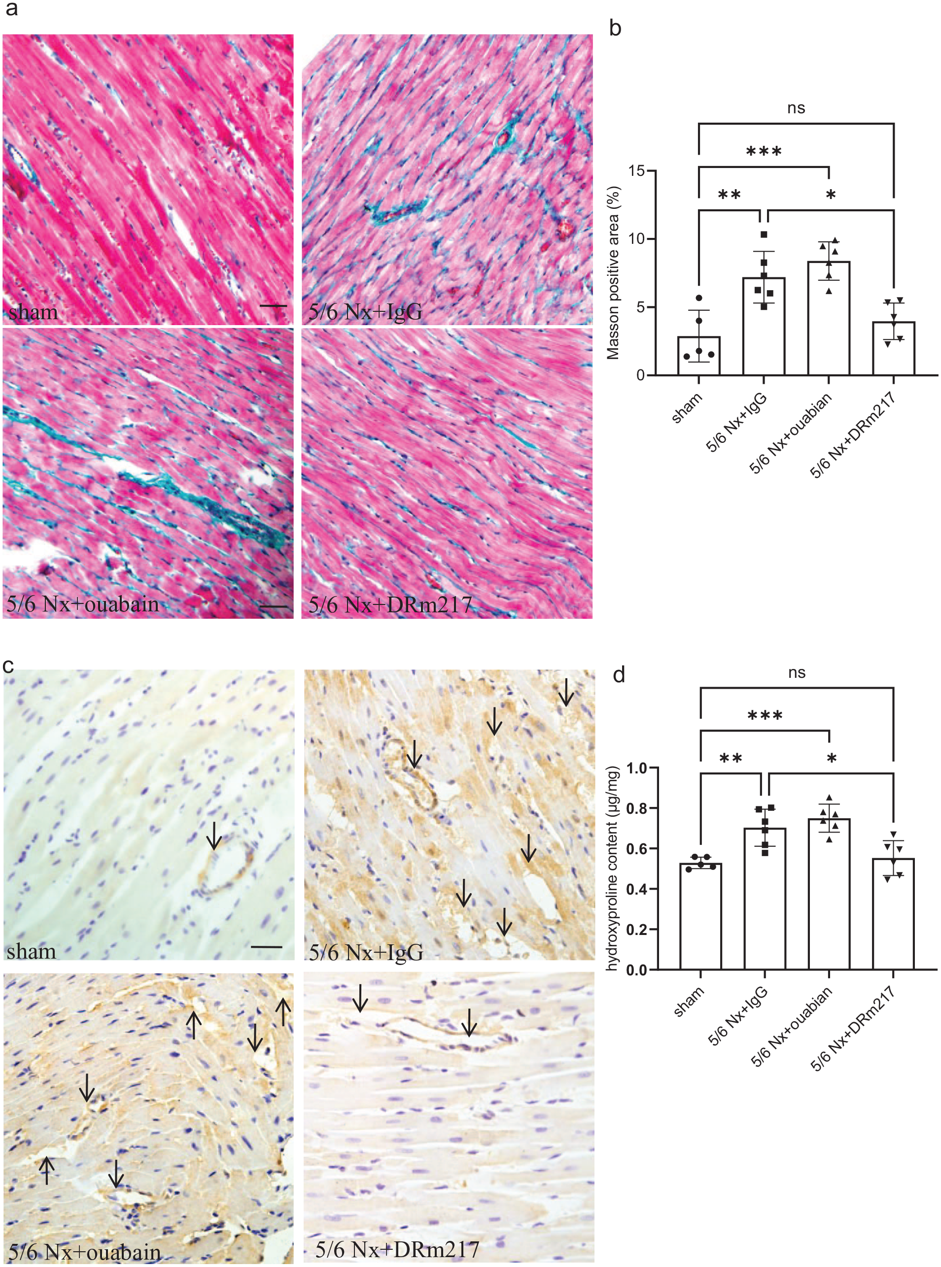
DRm217 reduces 5/6 Nx-mediated cardiac fibrosis. (a) Masson staining results in hearts from every group (scale bar is 100 μm). (b) Masson-positive stained area quantitative analysis. (c) Representative immunohistochemical staining of collagen III in heart tissues (scale bar is 100 μm). (d) Hydroxyproline contents in different heart tissues. (A color version of this figure is available in the online journal.)
DRm217 increased NKA activity and expression under 5/6 Nx conditions
NKA activity was depressed in the 5/6 Nx + IgG and 5/6 Nx + ouabain conditions. DRm217 increased NKA activity under 5/6 Nx conditions (Figure 4(a)). Immunohistochemistry staining showed that NKA expression was reduced in rat hearts from 5/6 Nx + IgG and 5/6 Nx + ouabain conditions, and DRm217 treatment alleviated the reduction in NKA expression under 5/6 Nx conditions (Figure 4(b) and (c)).
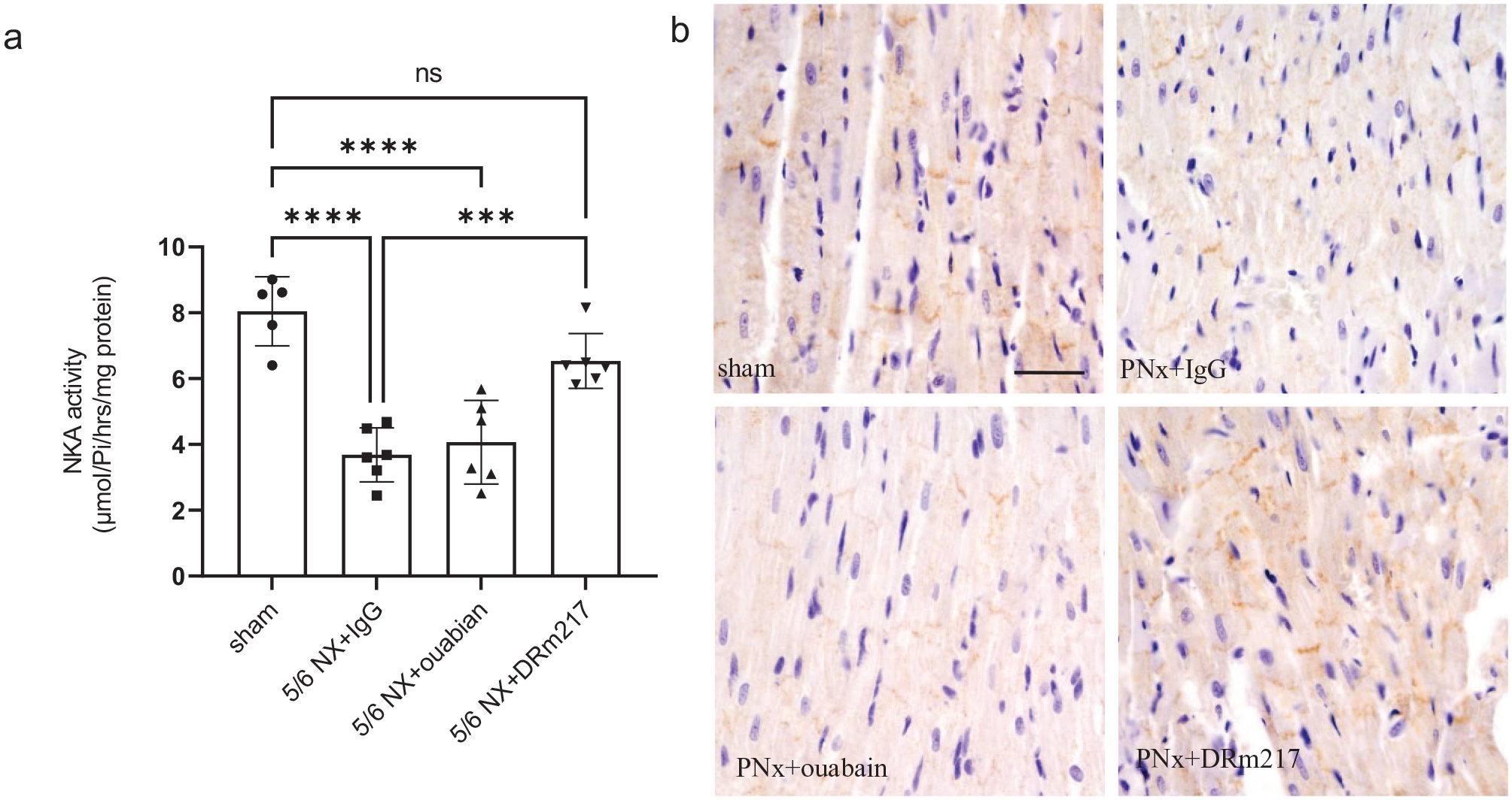
DRm217 increased NKA activity and expression. (a) NKA activity in hearts from every group. (b) Representative immunohistochemical staining of NKA expression in heart tissues (scale bar is 100 μm). (c) Integration optical density quantification (IOD) in immunohistochemical staining results. (A color version of this figure is available in the online journal.)
DRm217 inhibit p-Src expression in heart tissues
Western blotting showed that, in rat hearts collected from 5/6 Nx + IgG and 5/6 Nx + ouabain conditions, p-Src was elevated. DRm217 significantly reduced the expression of p-Src under 5/6 Nx conditions (Figure 5(a) and (b)). Immunohistochemical staining showed that the p-Src-positive staining area (yellow particles) increased significantly in the heart slides of the 5/6 Nx + IgG conditions and 5/6 Nx + ouabain conditions. In the 5/6 Nx + DRm217 conditions, the positive staining area of p-Src was less than in the 5/6 Nx + IgG conditions (Figure 5(c) and (d)).

DRm217 inhibits Src phosphorylation. (a) Representative western blotting bands of p-Src. (b) Quantitative analysis of the western blotting images of p-Src. (c) Immunohistochemical results of p-Src in heart tissue (scale bar is 100 μm). (d) Integration optical density quantification (IOD) in p-Src immunohistochemical staining results. (A color version of this figure is available in the online journal.)
DRm217 decreased oxidative stress in the heart tissue under 5/6 Nx conditions
MDA and oxidized glutathione levels in cardiac tissues reflected oxidative damage. The cardiac tissue collected from the 5/6 Nx + IgG and 5/6 Nx + ouabain conditions had dramatically higher levels of oxidized glutathione and MDA when compared to the sham animals. DRm217 treatment decreased considerably oxidized glutathione (Figure 6(a)) and MDA levels (Figure 6(b)).
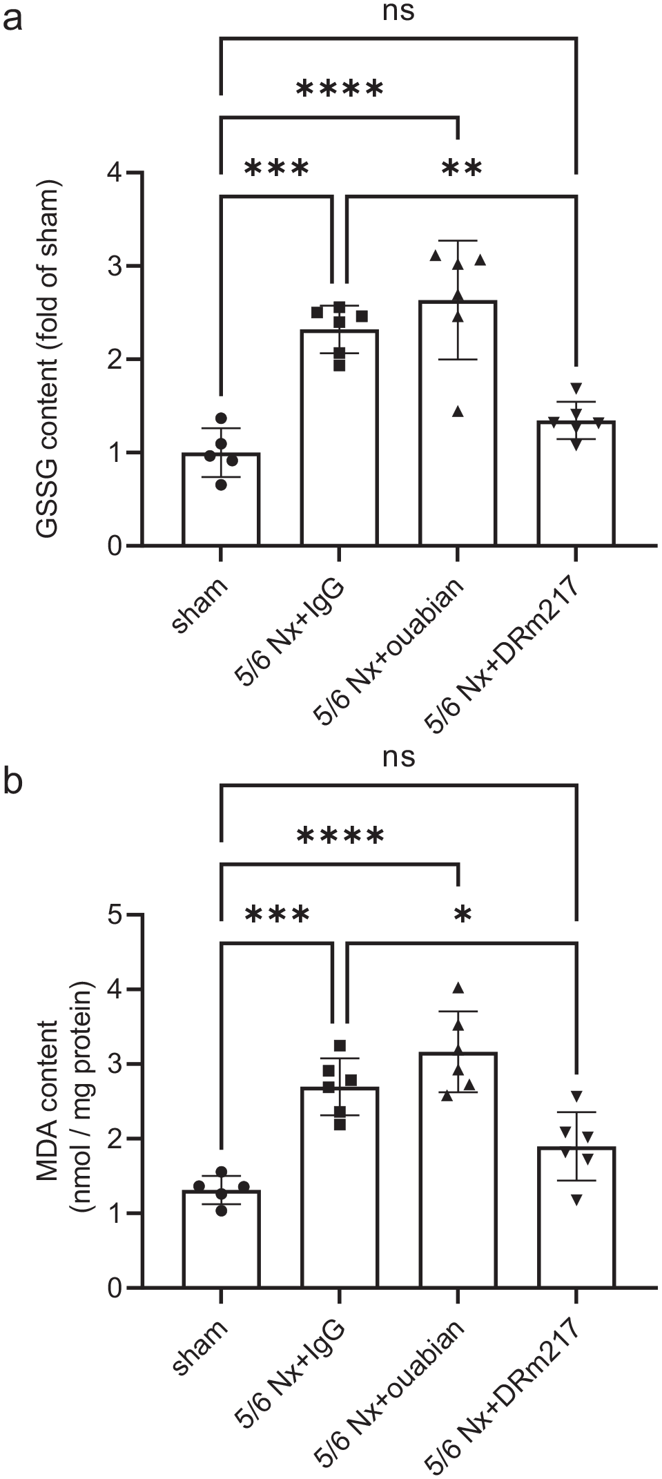
DRm217 inhibited the oxidative stress state. Oxidized glutathione (a) and malondialdehyde (b) levels in different heart tissues. DRm217 reduces the levels of malondialdehyde and GSSG in the hearts of 5/6 NX rats.
Discussion
In CKD patients, cardiovascular illness is the dominant cause of sickness and mortalities, accounting for over half of all deaths. 13 Myocardial hypertrophy and fibrosis are two main pathological features of CKD-related heart disease. As patients often have chronic kidney dysfunction, many drugs are not available for use in the treatment of CKD-related heart disease. Therefore, seeking effective strategies for CKD-related heart disease remains a hot research point.
NKA is an important bifunctional enzyme in the heart that participates in both ion transport and signal transduction. 14 Decreased NKA activity and expression occur for different reasons and have been noted in CKD-induced cardiac disease. 12 Management of NKA activity and expression is a possible therapeutic candidate for cardiomyopathy. The DR-region-specific antibody has been shown to activate the NKA. Previously, Xu and Zheng et al. reported that DR antibodies could stimulate heart contractility.7,15 We and other groups found that DR antibodies could protect cardiac cells against ischemic injury.8 –10 Recently, our group also demonstrated the effect of DR antibodies on isoproterenol- and angiotensin-induced cardiac remodeling.11,16 However, whether DR antibodies have a protective effect on CKD-related heart disease remain unknown. The partial (5/6) nephrectomy animal model is a typical chronic kidney disease (CKD) model. In the current investigation, we found that DRm217, a particular DR antibody, decreased serum LDH and CK-MB levels, inhibited HW/BW and myocyte size, and collagen deposition in the myocardial interstitium under 5/6 Nx conditions. Serum LDH and CK-MB are two biomarkers of myocardial injury. HW/BW and myocyte size are two indicators for cardiomyocyte hypertrophy. These findings indicated that DRm217 protects against heart damage, hypertrophy, and fibrosis. It has reported that, compared to sham rats, end systolic diameter of the left ventricle as well as end diastolic diameter of the left ventricle are significantly higher, and ejection fraction are depressed, in 5/6 Nx rats. 17 Left ventricular hypertrophy and fibrosis are the pathological reason for depressed cardiac function. So, we can speculate that DRm217 have beneficial effect on heart function. DRm217 has a potential treatment effect on CKD-related cardiac disease.
Cardiorenal syndrome (CRS) is a predominant cause of death in persons with CKD. Primary chronic renal disease causes ventricular hypertrophy, decreased heart function, and increased risk of cardiovascular events. Previously, we have demonstrated that administration of DRm217 protects against renal injury, preserves kidney function, and prevents renal interstitial fibrosis in a 5/6 nephrectomy rats model. 18 Here, we further proved that DRm217 also could prevent fibrosis and hypertrophy in cardiac tissue, displaying multiorgan beneficial effects in CRS. It is hard for us to pinpoint the exact mechanism participant in the cardiac benefits of DRm217 in 5/6 Nx rats are due to some of the kidney benefits or not. Inflammation, oxidative stress, and damage are all possible causes of CRS. In kidney, we have observed DRm217 can inhibit Src activation and ROS accumulation. 18 So, we further detected whether these mechanisms also present in DRm217 protective effect on CKD-related heart disease.
Cardiotonic steroids (CTSs) levels have been discovered to be higher in individuals with CKD and/or chronic cardiovascular illnesses by a number of laboratories. 19 Cardiac glycoside/NKA signaling and ROS amplification are connected with the progress of left ventricular hypertrophy and heart fibrosis in CKD-related cardiomyopathy. 20 Src is a key downstream signal in NKA signaling pathway. 21 Prolonged activation of Src was also revealed to cause heart hypertrophy and tissue fibrosis.22,23 The DR region includes the DSYG sequence, which seems to influence NKA enzyme activity and CTS binding with NKA.24,25 Therefore, we highly suspect that the DR antibody exerts its positive effect by controlling Src activation. Both western blotting and immunohistochemistry staining results showed that Src was activated under 5/6 Nx conditions and that DRm217 inhibited Src overactivation. Documents have demonstrated that NKA/Src/ROS signaling is implicated in the etiology of uremic cardiomyopathy.20,26 As we observed that DRm217 inhibited Src activation, we then detected ROS levels. GSSG is formed by GSH oxidation. MDA is a by-product during lipid peroxidation. In heart tissues, ROS levels were reflected by GSSG and MDA levels. Despite a considerable increase in MDA and GSSG in the tissues of rats’ hearts in the 5/6 Nx + IgG group, DRm217 greatly reduced the MDA and GSSG contents under 5/6 Nx conditions. This finding suggested that DRm217 inhibits ROS accumulation in the heart in CKDs. Jiang Liu et al. 27 has used pNaKtide, a specific peptide that inhibits NKA/Src binding and further Src activation, in 5/6 NX-induced mouse model. Their experiment results showed that administration of pNaKtide reversed 5/6 NX-induced cardiac dysfunction, fibrosis, and inhibit oxidative stress. 28 Our group have demonstrated that DRm217 have ability on stabilizing NKA cell surface expression, attenuated Src (Tyr(P)418) phosphorylation, and closing NKA/Src/ROS signaling. However, the mechanism of pNaKtide and DRm217 is different. pNaKtide blocks NKA and Src binding, but DRm217 binds to the extracellular segment of NKA. NKA endocytosis is a primary step for Src activation. 29 The expression of NKA on the cell membrane can be stabilized by DR antibodies.9,10 Therefore, stabilizing the membrane NKA and then suppressing NKA/Src/ROS signaling are two probable mechanisms for DRm217’s beneficial impact.
CTSs, such as ouabain, are natural ligands of NKA. CTS inhibits NKA pump function or activates NKA signal cascade. Patients with chronic renal insufficiency have higher levels of CTS. 30 Endogenous CTS binding to the α-subunit of NKA may active NKA signaling cascades and cause renal failure.16,19 Previously, we observed that ouabain worsened kidney injury and enhanced kidney fibrosis in 5/6 Nx models. 18 Ouabain has controversial effects in heart. Some documents have shown that ouabain causes cardiovascular damage.31,32 Other studies have shown that ouabain protects cardiac myocytes against ischemia/reperfusion injury. 33 Here, we observed that ouabain did not cause significant cardiac hypertrophy or fibrosis. The mechanism involved requires further study.
In summary, our findings showed that DRm217 protected cardiac cells from hypertrophy and fibrosis caused by the 5/6 Nx surgery. Inhibition of Src phosphorylation and ROS accumulation is involved in this process. NKA could be a therapeutic target for CKD-related cardiac disorders, and DRm217 has a potential treatment effect.
Footnotes
Authors’ Contributions
XY and XH conceived of the experiments and drafted the manuscript. JZ and PL finished most of the experiments and organized the data. XM and M-CK established the animal model. The final version of the manuscript was read and approved by all authors.
Data Availability
The data used and/or analyzed during this study are available from the corresponding author on reasonable request.
Declaration of Conflicting Interests
The author(s) declared no potential conflicts of interest with respect to the research, authorship, and/or publication of this article.
Funding
The author(s) disclosed receipt of the following financial support for the research, authorship, and/or publication of this article: This work was supported by the National Natural Science Foundation of China (nos 81970220 and 82170768), the Key Research and Development (R&D) Project of Shaanxi (no. 2019SF-164), and the Science and Technology Project of Shaanxi (no. 2019JM-169).