
Abstract
Cellulose fiber–reinforced composite scaffolds have recently become an interesting target for biomedical and tissue engineering (TE) applications. Cassava bagasse, a fibrous solid residue obtained after the extraction of cassava starch and soluble sugars, has been explored as a potential source of cellulose and has been successfully used to enhance the mechanical properties of gelatin scaffolds for TE purposes. This study assessed the cytocompatibility of the cassava microfiber–gelatin composite scaffold using human embryonic kidney cells (HEK 293) and a breast cancer cell line (MDA MB 231) under ISO 10993-5 standards. The viability of cells within the composite scaffold was analyzed through MTT assay. The growth of HEK 293, as well as the cell morphology, was not affected by the presence of cellulose within the composite, whereas the growth of breast cancer cells appeared to be inhibited with noticeable changes in cell morphology. These findings suggest that the presence of the cassava fiber in gelatin is not cytotoxic to HEK 293 cells. Thus, the composite is suitable for TE purposes when using normal cells. On the contrary, the presence of the fiber in gelatin elicited a cytotoxic effect in MDA MB 231 cells. Thus, the composite may not be considered for three-dimensional (3D) tumor cell studies requiring cancer cell growth. However, further studies are required to explore the use of the fiber from cassava bagasse for its anticancer cell properties, as observed in this study.
Impact statement
In this article, we assess the cytocompatibility of a cassava microfiber–gelatin composite scaffold using human embryonic kidney cells (HEK 293) and a breast cancer cell line (MDA MB 231) in accordance with ISO 10993-5 standards. The results show that HEK 293 cells exposed to the samples demonstrated a significant increase in cell viability with little or no changes in cell morphology, while there was a significant decline in cell viability and changes in cell morphology for MDA MB 231 cells. These findings suggest that cassava fiber in gelatin is not cytotoxic to HEK 293 cells; thus, the composite can be considered for tissue engineering purposes when using normal cells. On the contrary, the fiber in gelatin is cytotoxic to MDA MB 231 cells. Thus, the composite may not be considered for purposes such as three-dimensional (3D) tumor cell studies that require the growth of cancer cells.
Introduction
The biocompatibility of cellulose fiber has made it an attractive candidate for tissue engineering (TE) applications, such as forming composite scaffolds for cell growth.1–3 Researchers are constantly exploring various applications based on cellulose due to its biodegradability.4,5 Its ability to serve as a sustainable renewable natural resource is vital in this era of eco-friendliness and sustainability. 6 Cellulose has a densely packed glucan chain structure which improves its mechanical strength to support cellular networks and introduce various surface modifications.7,8 The diverse sources from which cellulose can be obtained include bacteria, tunicates, and plants. 9 The possibility of modifying the mechanical as well as surface chemical properties of cellulose paves the way for a wide variety of material properties, making it a suitable candidate for TE since different tissues require different scaffold properties to support growth. 10 As a result, a wide array of plant-based cellulose sources are being explored for biomedical engineering applications.11,12
In a study conducted by Xing et al., 13 cellulose obtained from hardwood fiber sheets was incorporated into gelatin for the first time. Ramphul et al. 14 also obtained cellulose from sugar cane bagasse and formed a composite scaffold with polylactide and polydioxanone for TE applications. The presence of cellulose in both cases enhanced the mechanical properties of the scaffold and increased the viability of human mesenchymal cells seeded onto it. Further exploration of cellulose sources that can be used as composite material for scaffold formation in TE applications would be a valuable addition to existing knowledge. In addition, it is well-known that the pathway used for extracting the cellulose fibers, that is, mechanical-, chemical- or bio-pulping, affects the physiochemical properties of the fibers, 15 which could affect the cell growth properties. For example, in the case of mechanical pulping, secondary metabolites may still be present depending on the temperature used for fiber extraction. These may either enhance or impede cell growth when these fibers are used in TE applications. Therefore, it is necessary to investigate the cytocompatibility and cell growth properties when a new source of cellulose is considered for TE purposes.
Cassava bagasse, a solid by-product of the cassava starch industry, has been explored as a source of natural reinforcement filler in plastic industries.16,17 The isolated nanocellulosic cassava fibers have been incorporated in tapioca films, 18 nanocomposite rubber films, 19 thermoplastic starch matrices, 20 and low-density polyethylene matrix. 21 These fibers have also generally resulted in enhanced mechanical properties. Diabor et al. 22 reported for the very first time the use of cassava bagasse as a potential cellulose reinforcement fiber material for gelatin in TE application. They analyzed the effect of different cassava fiber weight fractions on the mechanical properties and microstructure of the fabricated composite scaffold. 22 In a previous study by Larbie et al., 23 a preliminary assessment of the cytotoxicity of destarched cassava fiber granules was performed. This was done by examining changes in the composition of simulated body fluid (SBF) resulting from immersion of cassava fiber samples and via a lactate dehydrogenase test. The results indicated little or no significant toxicity levels. 23 These findings showed that the composite scaffold could potentially support cell growth. Beyond this, however, there is a paucity of information on its cytocompatibility. Such information on cytocompatibility is needed to complement earlier studies and confirm that cellulose fibers obtained from cassava can be used in cellulose composite scaffolds for TE applications.
This study fills that research gap as it investigates the cytocompatibility of cassava fiber–gelatin composite scaffold (GELCAS) for the first time. Human embryonic kidney (HEK) 293 and MDA MB 231 cancer cell lines were used to determine cytocompatibility in accordance with International Standard Organization (ISO) 10993-5 standard. Our findings demonstrate the potential of using cellulose fiber from cassava bagasse as a reinforcement biomaterial for TE purposes.
Materials and methods
Preparation of scaffold
Cassava fiber isolation was done using the water retting method, which is an established protocol. 22 Following isolation, three-dimensional (3D) cassava microfiber–gelatin scaffolds and pure gelatin scaffolds (GEL only) were fabricated by a freeze-drying technique. 22 The 3% (w/v) gelatin solution was prepared by dissolving 1.20 g of gelatin (type B powder from bovine skin, Sigma-Aldrich G9391, St. Louis, MO, United States) in 40 mL of distilled water in a beaker at 50°C, stirring it for 1 h. An amount of the 2.8 g cassava microfiber, calculated to yield a 7% (w/v) composite, was weighed and added gently to the gelatin solution with continuous stirring for 1 h to form the scaffold. An amount of 0.03 g of N-hydroxysuccinimide (NHS) and 0.05 g of N-(3-Dimethylaminopropyl)-N’-ethylcarbodiimide (EDC) crosslinkers were added to the 3% (w/v) gelatin solution and 7% (w/v) composite and stirred using a magnetic stirrer at room temperature for 15 min. The crosslinked mixture (20 mL of mixture) was then pipetted into either a 10-mm polystyrene petri dish or 150 µL of the mixture was pipetted into each well of a 96-well plate. The samples were covered and sealed with parafilm, kept at 4°C for 12 h, and then later transferred to 20°C for 12 h. Samples were then lyophilized in a Labconco Freezone (Kanas City, MO, USA) freeze dryer for a maximum of 36 h, as shown in Figure 1, and stored in a desiccator until needed. Sterilization was done by adding 50 µL and 1 mL of 70% ethanol to samples in the 96-well plate and the 10 mm polystyrene petri dish, respectively. The samples were left overnight in a laminar flow hood to dry and washed with phosphate-buffered saline (PBS) twice prior to cell seeding.
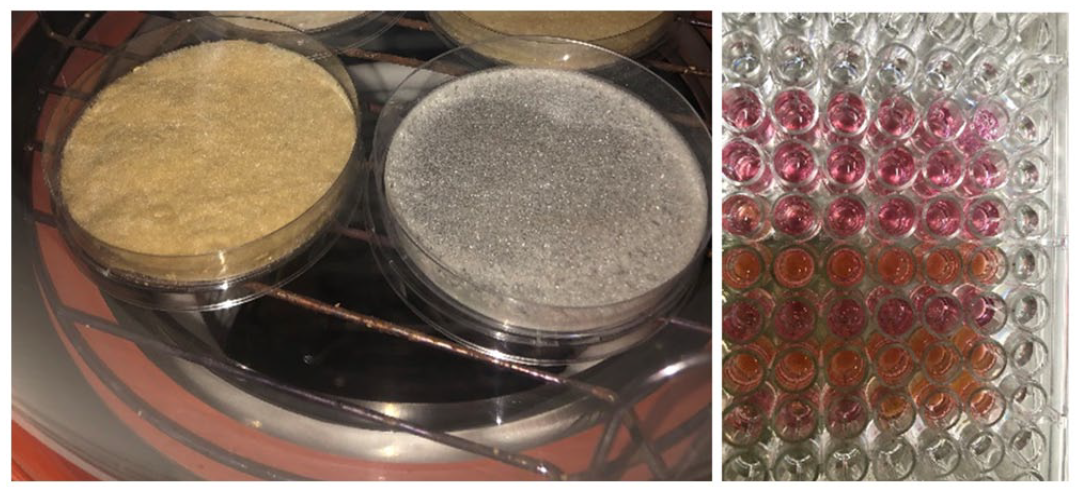
Photograph of the fabricated scaffold samples after freeze drying (left) and when placed in media (right).
Helium ion microscopy
A helium ion microscope (HIM) was used to obtain scanned ion microscopy images of fibers. Dried cassava fibers were dispersed onto a double-sided tape, attached to a sample holder and imaged under high vacuum pressure of about 1× Torr. The accelerating voltage used for imaging was 30 kV. The imaging aperture was 10 µm, and the beam current was between 0.7 and 1 pA. A flood gun was used for charge compensation on sample surfaces. Dwell time of 2 or 5 µs was used together with line averaging of 16 or 8 for image acquisition. ImageJ (version 1.53p) was used for fiber length analysis and surface roughness estimation. For surface roughness analysis, a background of 50 pixels was subtracted from each image, and a sampling length of 100 pixels was used. The surface roughness results were calculated from five different sample surfaces. HIM imaging was done at the University of Jyväskylä Nanoscience Center using the Zeiss Orion NanoFab (Jena, Germany) device in the cleanroom.
Cell culture
Cytotoxicity tests were conducted using HEK 293 and breast cancer cell line (MDA MB 231). Each cell type originally obtained from American Type Culture Collection (ATCC) was cultured in a T-75 flask with high glucose Dulbecco’s Modified Eagle’s Medium (D-MEM) with
Cytotoxicity assay
The cytotoxic effect was evaluated using two different methods: elution (extraction) and direct contact test according to ISO standards 10993-5. For both tests, cells cultured without scaffold were used as non-cytotoxic control. For the extraction test, cells treated with 70% ethanol were used as the positive/cytotoxic control. All experiments were performed in triplicate.
Elution test
This test evaluated the effect of the extract obtained from samples on the morphology and viability of the cells under sterile conditions. Using the recommended ISO surface area to extract volume ratio which is 125 mm2/mL, samples were incubated in the appropriate volume of media at 37°C for 24 h. Cells were also seeded in 24 well-plates at a density of 1 × 10⁵ cells/mL and grown to 80% confluency. After 24 h, the cell culture media was replaced with the extract media from samples. Cells with extract were further incubated for 24 or 48 h. At each time point, the extract was pipetted off and cells were washed with PBS to remove any remaining media. The cell morphology was then analyzed by viewing samples under an optical microscope. Cells were further incubated in calcein AM and propidium iodide (PI) staining solution for 30 min in the dark prior to imaging. Zeiss Axio Vert A1 Inverted Phase Fluorescence Microscope was used to obtain images of the cells.
Direct contact test
This test assessed the proliferation of cells seeded on the scaffold samples by quantitatively evaluating cell metabolic activity. MTT (3-(4,5-dimethylthiazol-2-yl)-2,5-diphenyltetrazolium Bromide) was used according to the manufacturer’s protocol. In this assay, the blank used was media only with no cells as well as the scaffold samples only with no cells, to account for any interaction between the scaffold and the dye. Cells were seeded at 1 × 10⁴ cells/mL on the sterilized samples placed in the 96-well plate and incubated for 3 h prior to the addition of 100 µL of media to each well. The cells seeded on the scaffold were incubated at 37°C with 5% CO2 for specific time points – day 1, 3, and 5. At each time point, 20 µL (2.5 mg/mL in PBS) of MTT solution was added to each well and incubated for 4 h. Afterwards, 100 µL of isopropanol solution was added and also incubated for 30 min to dissolve the purple formazan precipitate formed. The absorbance of each well in the plate was read at a wavelength of 590 nm. The true absorbance was then calculated by subtracting the blank absorbance from the sample absorbance. The mean absorbance ± SD which is directly proportional to cell viability was calculated and plotted for the various time points.
Statistical analysis
Statistical analysis of the results was carried out using one-way analysis of variance (ANOVA) (GraphPad Prism 8 Software) to determine statistical significance among the samples. A Shapiro–Wilk test was performed to test for normality of the data before using ANOVA, and all data obtained were normally distributed. Tukey’s post hoc test was used to further determine if the difference is significant at P value < 0.05. All tests were performed in triplicates (n = 3). The level of significance was represented by the number of “*” displayed on the graph; P > 0.05 (ns), P ⩽ 0.05 (*), P ⩽ 0.01 (**), P ⩽ 0.001 (***), and P ⩽ 0.0001 (****).
Results
This study was conducted to evaluate the effect of 7% (w/v) cassava microfiber in gelatin scaffold on cell viability and morphology. Prior to the fabrication of scaffolds, isolated cassava fibers were characterized using HIM. HIM images in Figure 2 show that fibers were randomly oriented, forming an intertwined mesh. Pores observed between the fibers were dependent on how densely the fibers were arranged on the imaging surface. The length of freshly isolated fibers was up to 10 mm; however, these fibers were filtered using a sieve with pore size of 180 µm to obtain uniform fiber lengths for scaffold fabrication as described by Diabor et al. 22 The average fiber diameter, measured from several sample points, was found to be 213 ± 121 µm. Average surface roughness obtained from defined sections that represented only the surface of fibers was found to be 27 ± 5 µm.
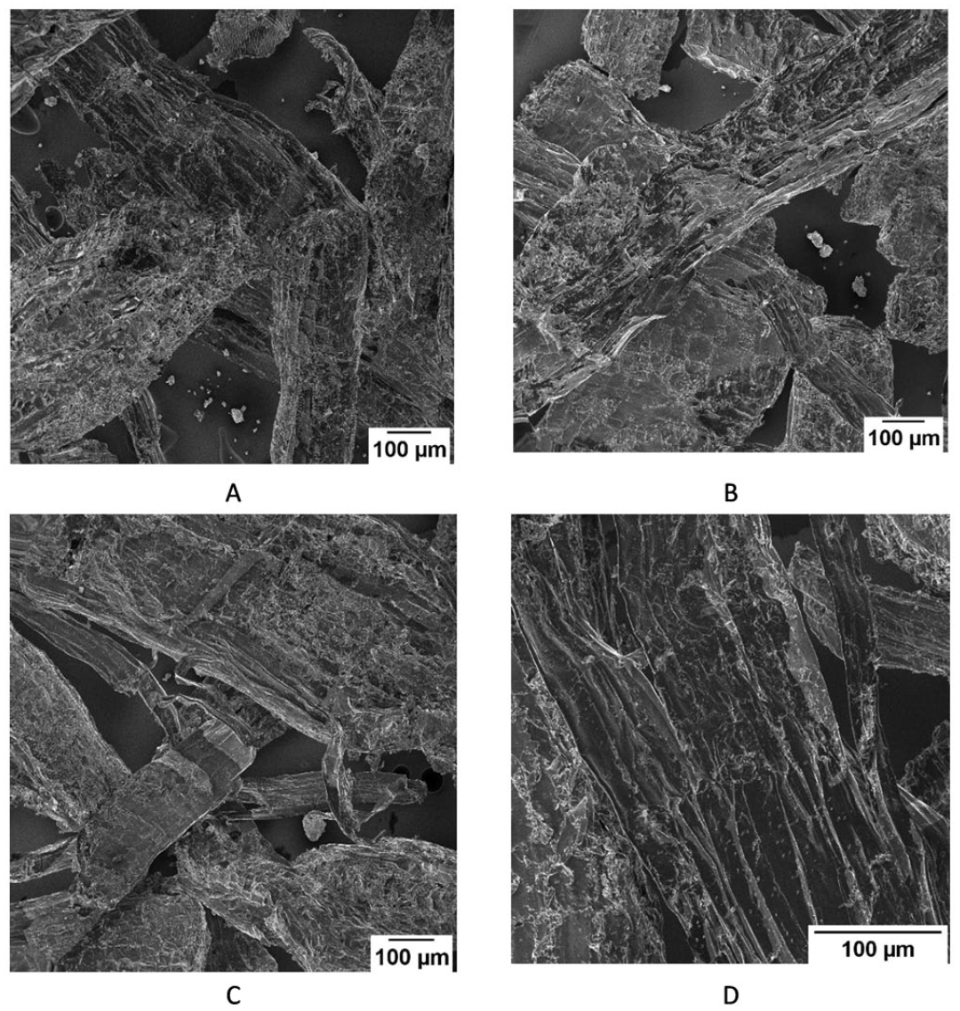
Helium ion microscopy images of different sample locations are shown in A, B, C and D. Images represent dried cellulose fibers extracted from cassava.
Evaluation of cell viability and morphology was done in accordance with ISO standards for in vitro cytotoxicity testing of medical devices. Results obtained from each of the cytotoxicity tests (elution test and direct contact) indicated that there was no significant change in terms of the cell morphology and viability of HEK 293 cells exposed to GELCAS and those exposed to GEL only which is known to be non-cytotoxic (Figures 3, 6, and 8). On the contrary, there was a significant change in the morphology and viability of the MDA MB 231 cells exposed to GELCAS compared to those exposed to GEL only (Figures 4, 7, and 9).
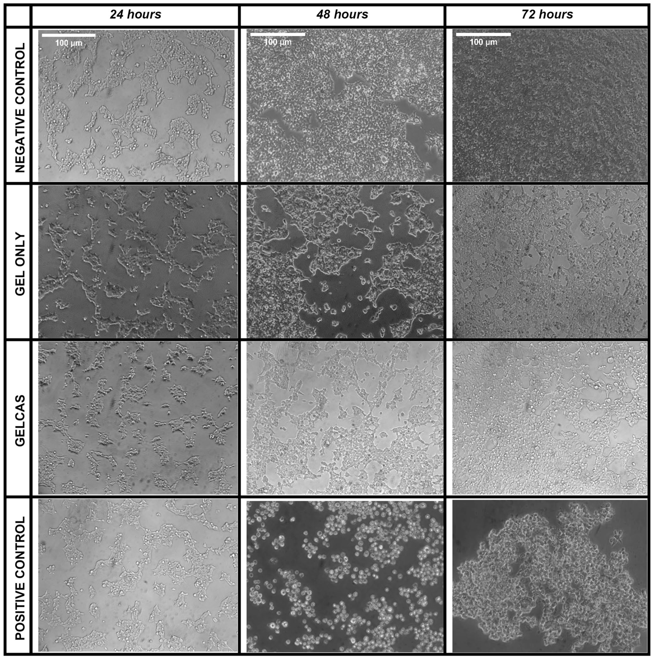
Morphological changes of HEK 293 observed under an inverted light microscope (100× magnification) after exposure to extract of samples from elution test, scale bar (100 µm).
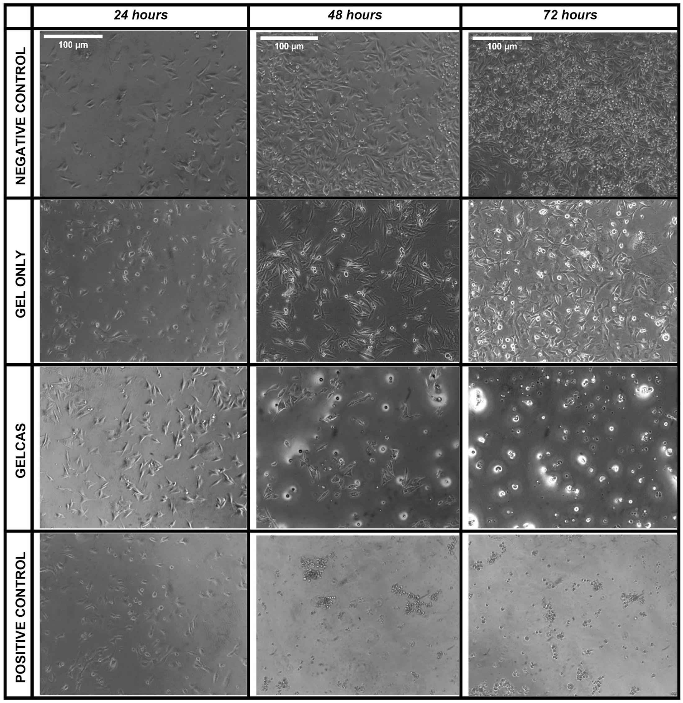
Morphological changes of MDA MB 231 cells observed under an inverted light microscope (100× magnification) after exposure to extract of samples from elution test, scale bar (100 µm).
Evaluation of cell morphology
Twenty-four hours prior to adding the extracts, the HEK 293 cells were observed to appear less circular in morphology, sparsely distributed and well attached to the well plate. After the extract from samples had been added to the HEK 293 cells, there was no significant difference in the cell morphology and attachment to well plate for cells exposed to GELCAS, GEL only extract, and the non-cytotoxic control (which is cells without extract) at all-time points (Figure 3). In addition, there was no significant difference in cell growth for GELCAS in comparison to GEL only. However, comparing the effect of GELCAS and GEL only to the non-cytotoxic control, there was a significant difference in cell growth. The cells had reached approximately 70–80% confluence for GELCAS and GEL only, while the non-cytotoxic control had reached approximately 90–100% confluence at time point of 72 h (Figure 3). For the MDA MB 231 cancer cells at time point 24 h, cells were observed to have the usual appearance of being spindle-shaped, spread, and well attached to the well plate. After the addition of extract to the cancer cells, GEL only and the non-cytotoxic control maintained the cell morphology while showing good attachment to the well plate with further increase in cell growth at all-time points. On the contrary, GELCAS extract resulted in a change in cell morphology from spindle shapes to spherical shapes, similar to that of the cytotoxic control (Figure 4). A further magnification (400×) of the images appeared to show cells with disrupted membranes for GELCAS and the cytotoxic control. After 72 h, majority of the cells showed apoptotic features such as cellular shrinkage and apoptotic bodies. Meanwhile, cells exposed to extracts of GEL only and the non-cytotoxic control exhibited cells undergoing mitosis (Figure 5).
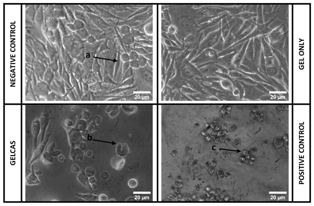
Optical microscopy images of cells after exposure to sample extract from elution test at 400× magnification, scale bar (20 µm); a denotes cell proliferation, b denotes cell disruption, and c denotes cellular shrinkage.
Evaluation of live-dead staining
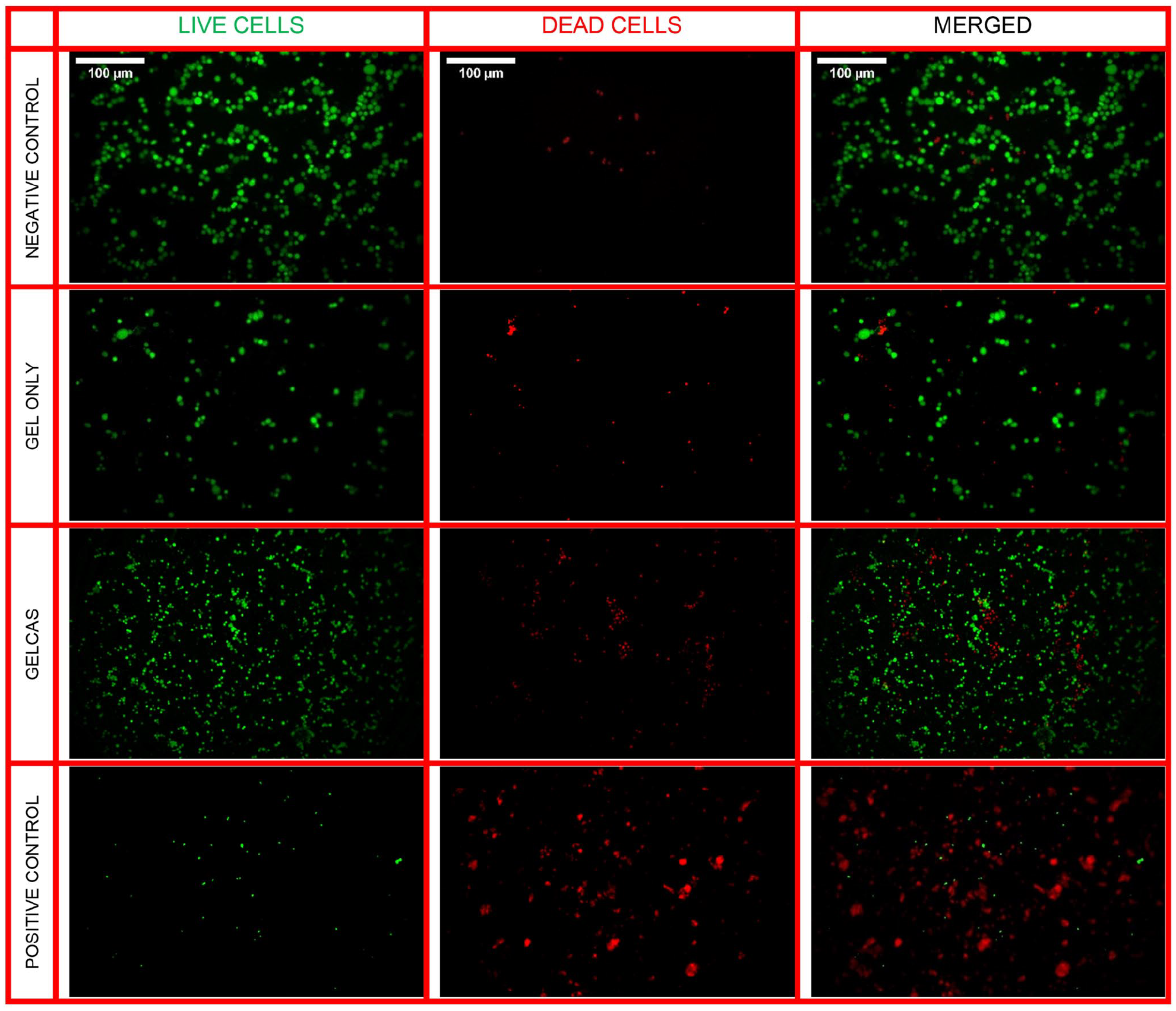
Fluorescence imaging was used to detect live and dead cells after the elution test. As seen in Figure 6 for HEK 293 cells, the non-cytotoxic control had the highest intensity of live cells. GELCAS and GEL only had similar live cell intensity but were slightly lower in comparison to the non-cytotoxic control, indicating a slight reduction in cell viability as observed in the optical microscopy images. Cells exposed to 70% ethanol appeared to have most cells stained red, indicating a significant decrease in cell viability. Similar results were observed in MDA MB 231 cells for GEL only and the non-cytotoxic control as seen in Figure 7. However, GELCAS and the cytotoxic control resulted in higher intensity of dead cells, indicating a significant decrease in cell viability for MDA MB 231 cancer cells.
Fluorescence microscopy images depicting live cells stained green with Calcein-AM solution and dead cells stained red with propidium iodide solution for HEK 293 cells after exposure to sample extracts from elution test, scale bar (100 µm).

Fluorescence microscopy images depicting live cells stained green with Calcein-AM solution and dead cells stained red with propidium iodide solution for MDA MB 231 cancer cells after exposure to sample extracts from elution test, scale bar (100 µm).
Evaluation of cell proliferation using MTT assay
The direct contact test measured cell metabolic activity as an indicator of cell viability on cells seeded directly on scaffold samples. The mean absorbance which is directly proportional to the measure of the cell viability of the samples and control was calculated and plotted at all the respective time points (day 1, 3, and 5). For HEK 293 cells, levels of metabolic activity were found be significantly higher in GELCAS than GEL only for all-time points (Figure 8). This further supports the lack of cytotoxic response recorded with GELCAS in the elution test for HEK 293 cells. Similar to the results obtained in the elution test for MDA MB 231 cancer cells, there was a statistically significant difference between the mean absorbance value recorded for GELCAS and GEL only. There was a gradual decline in the absorbance value from 0.132 ± 0.01 to 0.100 ± 0.01 for GELCAS across the time points. GEL only recorded the highest absorbance at 0.386 ± 0.03, although there was an unexpected reduction in absorbance to 0.310 ± 0.012 recorded on day 5 (Figure 9). Hence, it is interesting to note that while the presence of the fiber in gelatin enhanced cell growth in HEK 293 cells, it resulted in a decline in the growth of MDA MB 231 cancer cells.
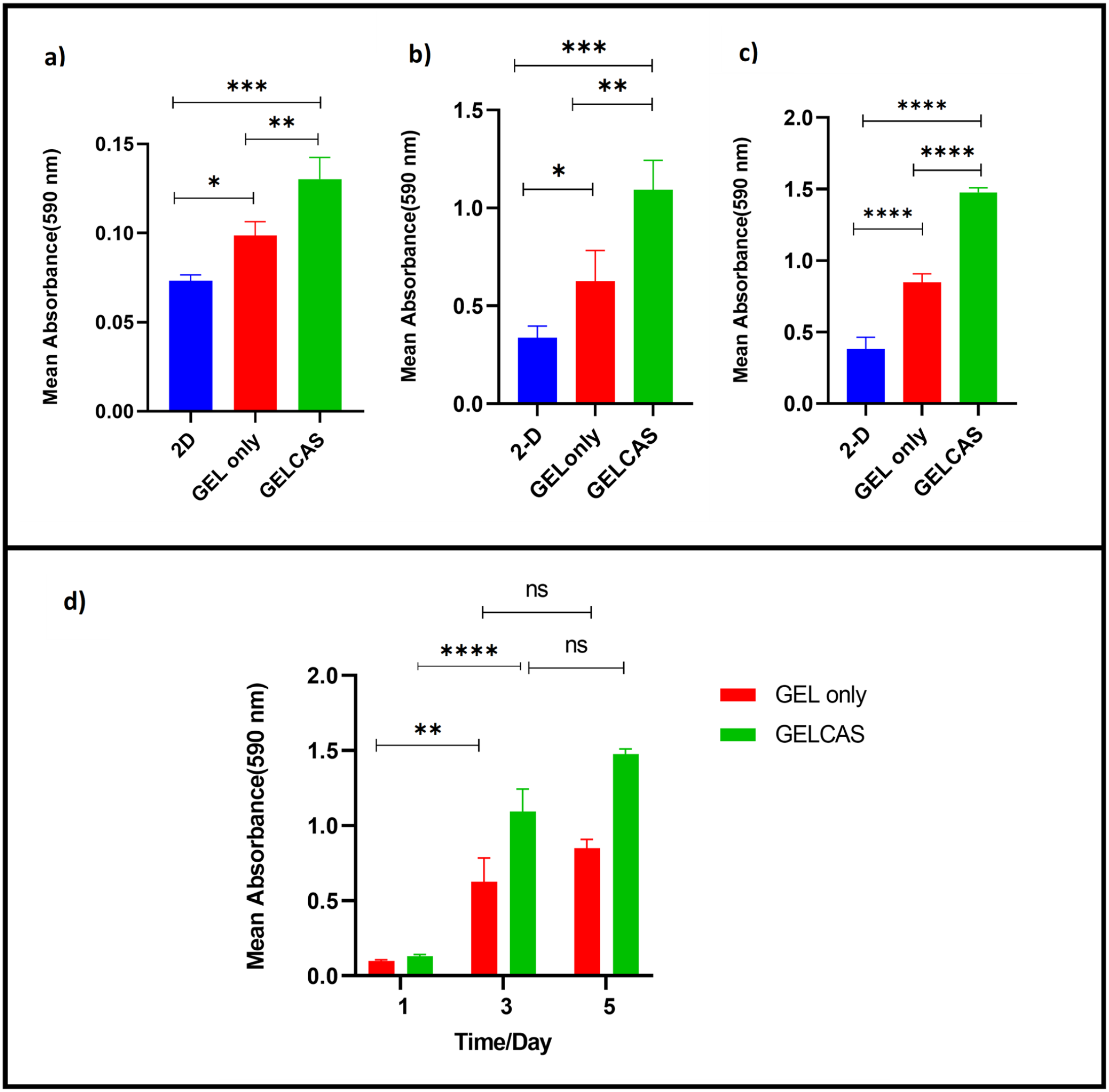
Cell viability determined by MTT assay. Formazan absorbance values at 590 nm expressed as a measure of HEK 293 cell viability when cultured on the scaffold samples for (a) day 1, (b) day 3, and (c) day 5 and (d) comparison between GELCAS and GEL only for all days.
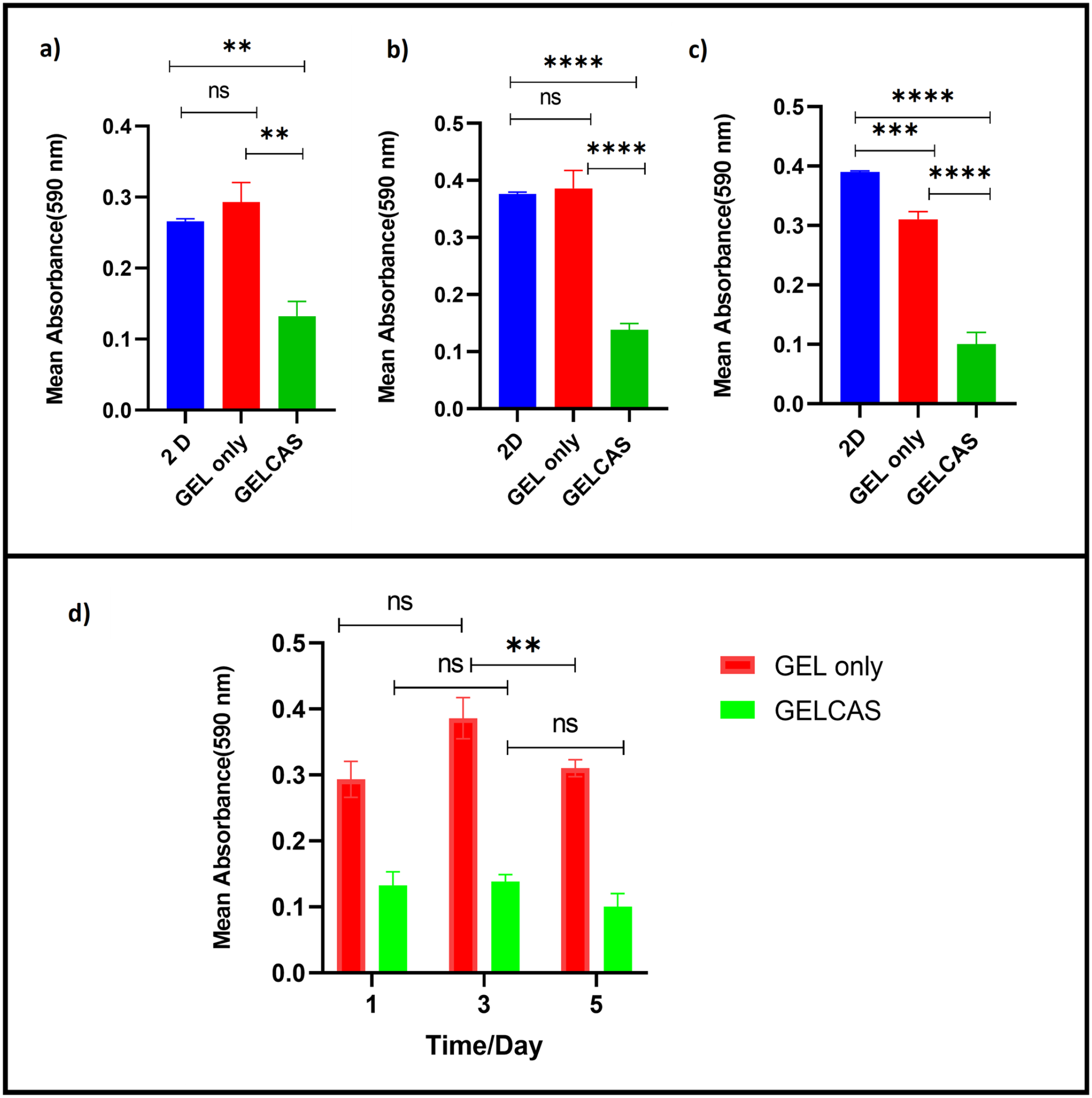
Cell viability determined by MTT assay. Formazan absorbance values at 590 nm expressed as a measure of MDA MB 231 cell viability when cultured on the scaffold samples for (a) day 1, (b) day 3, and (c) day 5 and (d) comparison between GELCAS and GEL only for all days.
Discussion
Medical devices that will end up implanted in the human body undergo preclinical testing through a variety of in vitro and in vivo examinations in order to be approved by regulatory bodies. 24 The main objective of such studies is to evaluate the biocompatibility of the various biomaterials that are used in fabricating the device.25–27 Considering that scaffolds interact directly with cells, it is essential to ensure the materials used do not have any toxic effect on cells or the human body in general if implanted.28,29 Thus, in vitro cytotoxicity tests are done according to ISO standard guidelines to ensure uniformity in results. In this study ISO 10993-5 30 standard was used as a guide. In choosing the cells for the study, cells commonly used in cytotoxicity tests, ease of culture, and availability were considered. 31 HEK 293 cells have been successfully used to test the cytotoxicity of bacterial cellulose/hydroxyapatite and poly-β-hydroxybutyrate (PHB) scaffold.32,33 In order to assess if the scaffold can be considered for biomimetic disease tissue models, MDA MB 231 cancer cells were also used.
Overall, the results of this study suggested that GELCAS had a non-cytotoxic effect similar to GEL only, which is a known non-cytotoxic material for HEK 293 cells. 34 However, GELCAS exhibited a cytotoxic effect against MDA MB 231 cancer cells contrary to what has been reported in other literature, considering that cellulose-based scaffolds have been used to support the growth of MDA MB 231 in other studies. 35 Prior evaluation of the GELCAS cytotoxicity was done using the elution test. This was to determine the effect of the presence of the cassava fiber extract on cell growth, morphology, and adherence. These factors were used to determine the cytotoxicity grade using ISO standards. Afterwards, a direct contact test was done to determine the viability of cells seeded directly on the scaffold, as this reflects better the use of the scaffold in a clinical setting.
The optical microscopy images (Figure 3) and fluorescence microscopy images (Figure 6) showed similar results for the cytotoxicity of GELCAS using HEK 293 cells. The slight decline in growth observed in the optical microscopy images was confirmed by the increase in the intensity of the red stain in comparison to the cells growing in the absence of the extract. Therefore, although the presence of the extract did not affect the morphology and interconnection of the cells, it caused a slight growth inhibition in the cells. Per the ISO 10993-5 30 standard cytotoxic grade, it can be inferred that GELCAS can be considered as Grade 1, with slight reactivity. However, since both test samples (GELCAS and GEL only) had similar reactivity, it can be concluded that the presence of the cassava fiber in the gelatin had little or no significant effect on HEK 293 cells. The metabolic activity of cells seeded on the scaffold further supported the elution test results. Besides, GELCAS exhibited a higher absorbance value than the GEL only at all the time points (Figure 8).
The enhanced cell growth in GELCAS compared to GEL only could be due to the differences in the structure of the scaffolds as reported in the previous study by Diabor et al. 22 The GELCAS scaffold had short discontinuous cassava fibers randomly dispersed in the gelatin matrix, and the surface morphology appeared much rougher compared to that of GEL only scaffold. From other studies, these properties are known to support cell adhesion to the surface and serve as contact guidance for directing and spacing cells to grow along the fibers.36–38 This is also expected to reduce the cluster growth of cells, preventing cells from competing for nutrients and ending up dying as may have been the case with GEL only.39,40 Furthermore, cell adhesion to mechanical structures within a growth matrix has been shown to facilitate multilayered cell formation, since there is a larger surface area for them to attach and grow. 41 The results obtained are also comparable to similar composite scaffolds found in other studies that recorded higher cell viability in cellulose-enhanced composite scaffolds. 13 In their study which is very similar to this work, Xing et al. 13 recorded higher cell viability in cellulose-enhanced gelatin than in gelatin only. All these suggest that the presence of cellulose fiber in composite scaffolds promotes the growth of cells. In addition, both 3D samples (GEL only and GELCAS) had higher cell viability than the negative control which had cells growing on a two-dimensional (2D) platform. This supports the fact that 3D platforms have multiple layers for cell attachment, and can therefore provide more room for cell attachment, unlike the negative control which allows cell growth in one layer.
Results obtained for MDA MB 231 cells widely contrasted with the results obtained for HEK 293 cells. There was a change in the morphology of the MDA MB 231 cells from their spindle shapes to spherical shapes (Figure 4) after exposure to GELCAS extract. The cell membrane looked disrupted and did not have similar cell density and morphology as seen in both non-cytotoxic control and GEL only cells (Figure 4). From the literature, this change indicated that the cells were undergoing apoptotic cell death. 42 After 72 h, the majority of the cells showed apoptotic features such as cellular shrinkage (Figure 5). 43 Similarly, fluorescence microscopy images indicated higher intensity of dead cells impacted by GELCAS than by GEL only. Results obtained from the metabolic activity of cells seeded on the scaffold were coherent with the elution test. GELCAS samples recorded the least cell viability at all-time points compared to the other samples (Figure 7). The difference between the absorbance value of GEL only samples and GELCAS samples was also statistically significant for all the time points, indicating that the presence of the fiber did not support the growth of the MDA MB 231 cancer cells unlike it did for HEK 293 cells.
These results for MDA MB 231 were unexpected because from the literature cellulose-based composites such as bacterial cellulose–gelatin composite scaffold supported and enhanced MDA MB 231 cells seeded on it.35,44 Since the cassava microfiber was not chemically treated to obtain pure cellulose, the difference in results may be attributed to the presence of other components such as hemicellulose and lignin.45,46 According to Wang et al., 47 some lignin–carbohydrate complexes formed as a result of chemical bonds between cellulose, hemicellulose, and lignin are known to exhibit antiproliferative properties at the cellular level. Their findings indicated that the extracts of two lignin–carbohydrate complexes from the chaga mushroom (Inonotus obliquus) had antitumoral activity on cancer cells. However, the mechanism by which cell apoptosis was induced was mostly by inhibiting nuclear factor kappa B (NF-κB), a protein transcription factor responsible for turning on the gene expression that prevents cancer cells from undergoing cell apoptosis. This particular protein transcription factor exists in an inactive form in normal cells but is active in cancer cells.47,48 Considering that the same material extracted from GELCAS for the elution tests resulted in the death of the MDA MB 231 cancer cells while supporting HEK 293 cell growth, it could be that the mechanism of cell death may be similar to what was reported by Gupta et al. 46 Plant extracts or compounds such as phenolics, alkaloids, polysaccharides and glycoproteins, lectins, tannins, and lignins have been reported to selectively induce apoptosis in neoplastic cells instead of normal cells.42,49 Ethanol cassava extracts have also been investigated for their cytotoxic effect against different cancer lines, and they all showed promising results as potential anticancer agents.50,51 Therefore, cassava may possess antiproliferative properties against cancer cells.
Ahead of the comparative study between HEK 293 and MDA MB 231 cells, cassava fibers obtained immediately after isolation from the root tuber were characterized and filtered to obtain uniform fiber length for the scaffold formation. Microscopy images from HIM aided in estimating the physical dimensions of the fibers used in scaffold fabrication. HIM imaging does not require sputtering so actual sample dimensions were obtained. Although fiber diameter was measured to be about 213 ± 121 µm, Figure 2(D) suggests that the measured diameter was still composed of multiple individual fibers that were bound together. Since the fiber isolation was done via a process of maceration, it is possible that the larger fiber bundles observed could be fibers that had not completely separated from each other during the maceration process. In addition, the fact that no chemical additives were used ensures that the fibers remained unadulterated, thereby maintaining their true mechanical properties as reported in the previous study – Young’s modulus ranging from 162.218 ± 37.788 MPa to 336.485 ± 130.803 MPa and strength of fiber ranging from 5.91 ± 3.43 MPa to 7.19 ± 4.26 MPa. 22
Surface roughness was measured from line profiles on the surface of different fibers. There are many reports of the influence of surface roughness on cell activity including one by Rahmat et al., 51 who estimated the effect of surface roughness on cell attachment. 52 The surface roughness results obtained in this study are not comparable to results obtained by Rahmat et al., 51 as their results reflected nanoscale roughness. Nevertheless, their results suggest a positive correlation between roughness and cell adhesion, which is also demonstrated by this study as incorporating the relatively rough cassava fibers into the gelatin matrix resulted in improved cell adhesion and viability of HEK 293 cells.
The outcome of this study suggests that the presence of the cassava fiber in gelatin is not cytotoxic to HEK 293 cells but to MDA MB 231 cancer cells. Further studies to examine the adherence of cells to the surface of the scaffold samples, to help verify and understand what caused the decline in cell viability for MDA MB 231 need to be done. These preliminary results form a strong basis for further in-depth studies that utilize cassava fiber for TE purposes and support further investigation of the potential of this bio-based sustainable material as an anticancer agent.
Conclusions
Cytotoxicity tests following both elution and direct contact method according to ISO standards were applied in this study. The effect of cassava microfiber on cell viability and morphology of HEK 293 cells and MDA MB 231 cells was analyzed. The results obtained indicated that 7% (w/v) cassava microfiber–gelatin composite scaffold (GELCAS) influenced the two cell types differently. The presence of the fiber supported the growth of HEK 293 cells while inducing the death of MDA MB 231 cancer cells. Possibly, the presence of other components such as lignin associated with the cassava fiber could be a contributing factor to the death of cancer cells. The incorporation of the fiber significantly enhanced HEK 293 cell growth. Since GELCAS is not cytotoxic to HEK 293 cells, it can be considered as a reinforcement material for gelatin for TE purposes. Therefore, these findings could help inform biomaterials engineers and materials researchers about the cytotoxicity of this material when considering it as a reinforcement material for TE purposes. The composite, however, may not be considered for purposes such as 3D tumor cell studies that require the growth of cancer cells. Rather researchers may consider exploring the material as a potential anticancer agent.
Footnotes
Acknowledgements
The authors wish to acknowledge Dr Godwin Amenorpe for providing cassava tubers for the work, Samuel Mensah Baffoe and Jessica Kugblenu for training first author in cell culture techniques, Temitayo Ademolue for assistance in fluorescence microscopy, Dr Isawumi Abiola for assistance with freeze drying of samples and Michelle Oti-Bronya and Miracle Ndego for assistance with cassava fiber extraction process.
Authors’ Contributions
The conceptualization of the research idea originated from EEK. Cellulose extraction, cytotoxicity measurement, statistical analysis, and imaging were done by PNAP, ARA, and KJB. All authors contributed to data acquisition, review of results, and writing and editing the manuscript. In addition, EEK worked on project administration, funding as well as supervision.
Declaration of Conflicting Interests
The author(s) declared no potential conflicts of interest with respect to the research, authorship, and/or publication of this article.
Funding
The author(s) disclosed receipt of the following financial support for the research, authorship, and/or publication of this article: This work was supported by a World Bank African Centres of Excellence Impact grant (WACCBIP-NCDs: Awandare).