
Abstract
Leiomyosarcoma of the inferior vena cava (IVC) is a rare tumor of mesenchymal origin, occurring most frequently in middle-aged women. Insidious complaints delay diagnosis, prognosis is poor, and the only curative modality remains an aggressive surgical resection yielding clear margins of disease. Commonly, radical tumor excision mandates caval repair or reconstruction, with significant related morbidity and mortality. We present a case of a 50-year-old woman with a leiomyosarcoma arising from the lower segment of the IVC, managed by surgical en-bloc resection of the tumor and IVC segment without further caval repair or reconstruction. During 14 months of follow-up the patient is well, had not had any complications, and is disease free.
Keywords
Introduction
Although rare, primary vascular tumors most commonly arise from veins, and the inferior vena cava (IVC) is the most commonly affected vein. 1 Leiomyosarcoma of the IVC (IVCL) is scarce with about only 400 cases reported in the literature so far. 2
Nonspecific mild abdominal pain is the most prevalent presenting symptom, often preceding diagnosis by several months to years, though clinical symptoms and manifestations may vary. A palpable abdominal mass may rarely be the presenting symptom. Peak incidence of diagnosis is at the sixth decade of life, with an obvious female predominance. Average tumor size at diagnosis is 10.8 cm. 1 This mass is usually detected using abdominal ultrasound (US), yet clarifying its origin requires further imaging and occasionally biopsy.
Various parameters such as tumor size, grade, mitotic rate, or other histological features that are known to be of prognostic value in the majority of mesenchymal neoplasms are unable to predict disease outcome in IVCL. 3 Nevertheless, cure may be achieved only after radical resection with negative surgical margins. 3 The 5- and 10-year survival rate for patients undergoing curative surgery is 49% and 30%, respectively. 1 Local disease recurrence is frequent—up to 53.7% and common metastases sites are the liver, lung, and bone. 1,3 The role of radiotherapy or chemotherapy is uncertain, and to date no large report provided evidence for a long-term benefit.
The proper planning of the surgical strategy is based on computerized tomography (CT) and cavography to determine tumor extent, vena cava lumen patency as well as evaluation of collateral venous circulation. Optimal surgical management is controversial, owing to frequent complications related to disruption or reconstruction of venous blood flow in the IVC.
Reported rates of surgical mortality varies between 2.5% and 8%, while minor and major morbidity ranges between 17.5% and 81% and 5.8% and 19%, respectively. 2,4
Case Report
A 50-year-old woman was referred for evaluation following several weeks of vague abdominal pain and ongoing exercise exertion. Past medical history was irrelevant, and physical examination and routine blood tests were unremarkable.
Abdominal US revealed a 6 × 3 cm hypoechogenic mass adjacent to the IVC beneath the right renal vein, yet a patent IVC was evident on a Doppler flow scan. The CT scan of the abdomen showed a 6 × 3 cm heterogenous retroperitoneal mass, with hypodensic core and a markedly narrowed IVC lumen. The mass extended 6 cm below the right renal vein and no metastases were seen on total body CT. Further evaluation by angiography exhibited a near complete obstruction of the IVC along with numerous collateral veins. The main collateral vein was an 11 mm wide left ovarian vein (Figure 1).
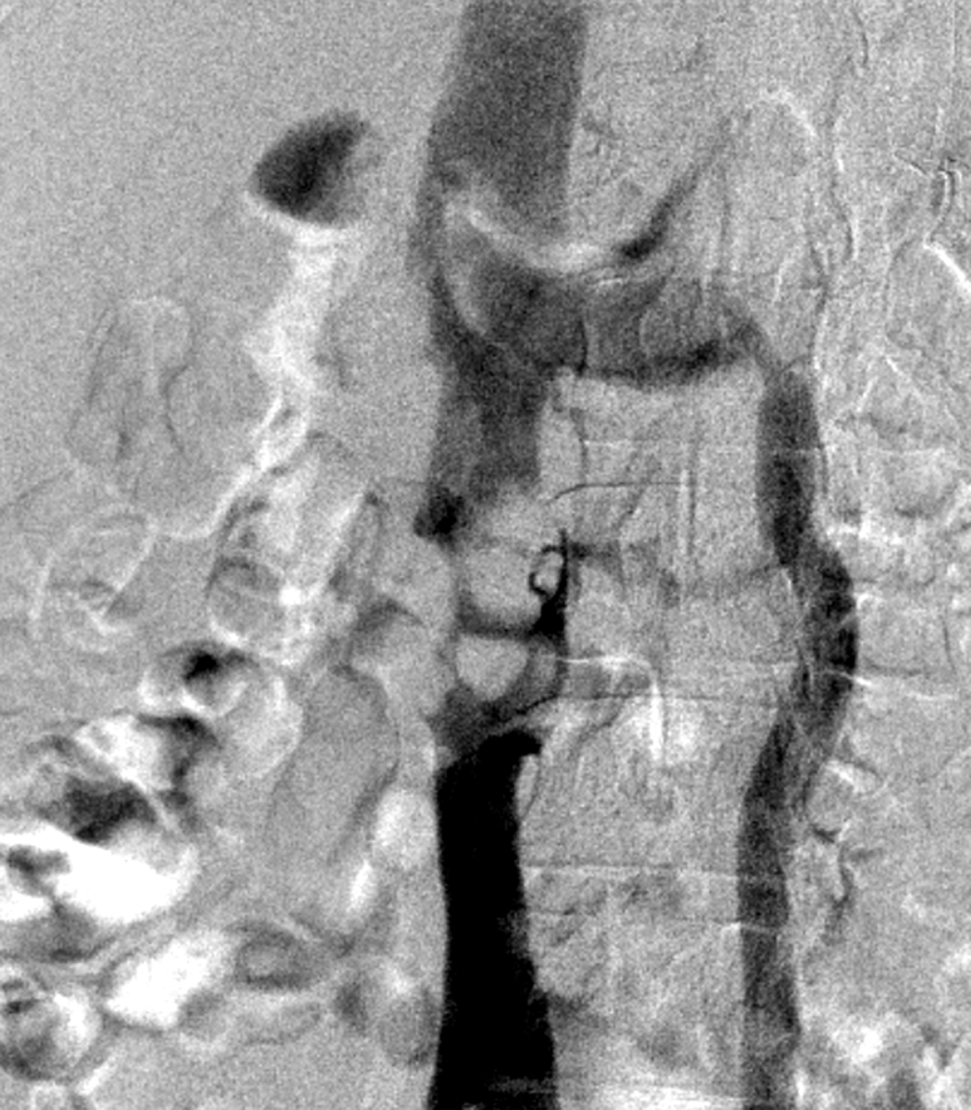
Inferior vena cavography demonstrating a major filling defect suggesting near complete obstruction of the infrarenal inferior vena cava (IVC). Filling of the suprarenal IVC is demonstrated via dilated left ovarian vein.
Differential diagnosis of the mass included an IVC leiomyosarcoma, neurogenic neoplasm, metastases as well as other retroperitoneal tumors. Based on the above findings, percutaneous biopsy was considered unsafe and a radical surgical approach was planned, including preparation for venous–venous bypass and IVC grafting.
Midline laparotomy was performed revealing a 6.5 × 3.8 cm irregularly shaped encapsulated tumor fixed to the lower segment of the IVC (Figure 2). Intraoperatively, hemodynamic monitoring was obtained by continuous venous and arterial pressure. Upon proximal clamping of the infrarenal vena cava neither hemodynamic changes nor distal IVC dilatation were noted. Therefore, en-bloc resection of the entire tumor along with a 6-cm segment of distal vena cava was carried out without the need for further reconstruction. The patient tolerated the procedure well.
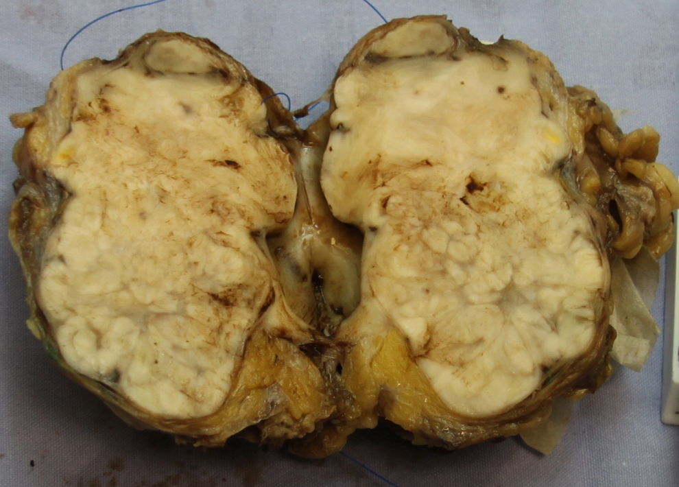
Inferior vena cava leiomyosarcoma—surgical finding.
Histopathologic examination confirmed the diagnosis of an IVC leiomyosarcoma of FNCLCC (French Federation Nationale des Centres de Lutte Contre le Cancer) 3 histologic grade 2: primarily composed of spindle cells, highly differentiated with several areas of severe differentiation loss, foci of necrosis complying <5% and low mitotic activity. Tumor was completely excised with free surgical margins.
Postoperative course was uneventful, the patient had neither lower extremity edema nor other venous-related complications. Twenty-two months postsurgery, the patient is well, with no evidence of local or distant disease recurrence.
Discussion
Leiomyosarcoma of the IVC are rare, slow-growing tumors arising from smooth muscle cells underlying blood vessel wall, and represent technical surgical challenge due to their size and location. Initial symptoms are often of minor nature unless generating veno-occlusive disease. Prognosis is usually poor, although an aggressive surgical approach combined with the absence of distant metastases can obtain long-term survival and even cure.
The IVCL grow either intra- or extraluminally rather than by infiltration. The IVCL are classified into 3 groups based on anatomic location: segment I—arising below the renal veins, segment II—arising in-between the renal veins and the retrohepatic portion of the IVC, and segment III—arising from the suprahepatic veins and above including lesions extending intracardially. Presenting symptoms, surgical technique, and prognosis highly depend on the involved segment. 1,5
An international registry of IVCL established in 1992 by Mingoli et al at La Sapienza University collected more than 400 cases published in several reports, the latter in 1997 summarizing more than 200 cases. This database accentuates the importance of complete surgical resection as the only therapeutic modality which can lead to long-term survival. 1,6
Laskin et al had recently published the second largest study consisting of 40 patients with IVCL, also concluding that complete resection of localized IVCL provides the best chance for long-term survival, reaching a 5-year survival of 50%. 3
Regarding surgical technique, small tumors can be excised with minimal caval defect suitable for a primary or patch repair. Large tumors however mandate circumferential caval resection, segment II and III tumors with patent vena cava usually require prosthetic graft reconstruction. Segment I tumors tend to slowly grow extraluminally and are more likely to incline caval lumen narrowing forming abundant secondary collateral venous supply, allowing segmental resection without further reconstruction. 1,4
There is limited experience in the literature regarding the proper technique following IVC resection. Refraining from caval reconstruction reduces operative time and avoids morbidity and mortality associated with a complex prosthethic graft repair. 5 However, reconstruction is mandatory in the absence of sufficient collateral venous flow or in cases of interruption of vital organ vasculature.
Hollenbeck et al 4 published a single center experience with 25 patients, mostly undergoing either ligation or primary repair. The rate of postoperative morbidity was low and consisted mainly of lower extremity edema. They preferred primary repair and cited ligation only as feasible in cases of preoperative caval thrombosis.
Daylami et al 5 profoundly addressed the necessity of reconstruction after resection. They cite the controversial literature and present their own experience of 6 cases, 4 of which had an occluded vena cava prior to surgery. Despite successful ligation without further reconstruction, they encountered high morbidity in their series, including acute renal failure and chylous leak.
In a recent letter to the editor by Mingoli et al, 2 he reanalyzed older data suggesting that reconstruction was associated with significant mortality and morbidity and proposed that the rich collateral venous supply may allow safe caval resection without reconstruction. However, no recommended evaluation method or decision making were indicated.
In our case, we present a lower segment IVCL safely managed by an aggressive en-bloc tumor excision and vena cava ligation without further reconstruction. This was possible due to the rich collateral supply the patient developed, mainly, a very wide left gonadal vein. The fact that there were no late consequences of the IVC segmental resection proves that the collateral venous system was sufficient.
As pointed out in the literature, and well demonstrated in our case, we believe that whenever possible, primary IVC resection without reconstruction in the management of lower IVC leiomyosarcoma should be preferred.
Footnotes
Declaration of Conflicting Interests
The author(s) declared no potential conflicts of interest with respect to the research, authorship, and/or publication of this article.
Funding
The author(s) received no financial support for the research, authorship, and/or publication of this article.