
Abstract
The epithelioid hemangioendothelioma (EHE) is a rare type of endothelial neoplasm found mainly in soft tissues and visceral organs and in extraordinary cases in large veins like the iliac veins. Currently, there is an active discussion in which EHE behavior, classification, new diagnostic tools, and treatment procedures are proposed. Here, we present 2 cases of EHE and discuss our experience in diagnosis and treatment of this neoplasm.
Introduction
Primary tumors of major blood vessels are uncommon and usually exhibit malignant behavior. The clinical presentation in many cases is vascular occlusion and the absence of palpable mass and shares clinical findings with other diseases and conditions, making its diagnosis difficult. 1 Generally, the neoplasm derived from the endothelial layer of blood vessels is classified as completely benign (like hemangiomas), malignant (like angiosarcomas), or intermediate grade (like hemangioendothelioma). The last category is the epithelioid hemangioendothelioma (EHE), a characteristically rare type of endothelial neoplasm, characterized by large “epithelioid” endothelial cells, with frequent localizations in soft tissues and visceral organs. 1–3
Enzinger and Weiss described EHE for the first time in 1981 as an intermediate (or borderline) malignancy neoplasm. 1 However, recently some authors proposed that EHE is considered as a fully malignant neoplasm; however, the current World Health Organization classification recognizes this neoplasm as intermediate grade. 2,3 The most common location is in soft tissues of the extremities. They have also been reported in skin, liver, lung, bone, testicle, larynx, thyroid, parotid, brain, heart, arteries, and veins. 1–4
Furthermore, the discussion about EHE behavior recently has been increasingly advanced in diagnostic tools that can be useful in the correct characterization and early diagnosis of this neoplasm. Here, we report and discuss 2 cases of EHE derived from the external iliac vein.
Case 1
A 23-year-old female student presented to the physician with progressive lower left limb edema, increasing with ambulation over 2 months. No relevant history was found. On physical examination, a left lower extremity edema with compromise from foot to groin was documented. The pulse and perfusion were conserved. A color duplex ultrasound was negative for deep venous thrombosis (DVT)and showed high-velocity blood flow in the external iliac vein, with a high-pitched Doppler signal suggesting stenosis. An ascending venography documented a 95% obstruction of the external iliac vein in cephalic proximity with the inguinal ligament (Figure 1a and b). Possible external compression by the inguinal ligament was considered, and open surgical exploration was performed via an extra peritoneal approach (Figure 1c). A nodular mass in the external iliac vein was found, and after longitudinal venotomy an endoluminal tumor (1.7 × 0.9 × 0.8 cm3) with luminal occlusion was observed (Figure 1d). The tumor that involved vein section was resected, and reconstruction was performed with a cylindrical saphenous vein section.
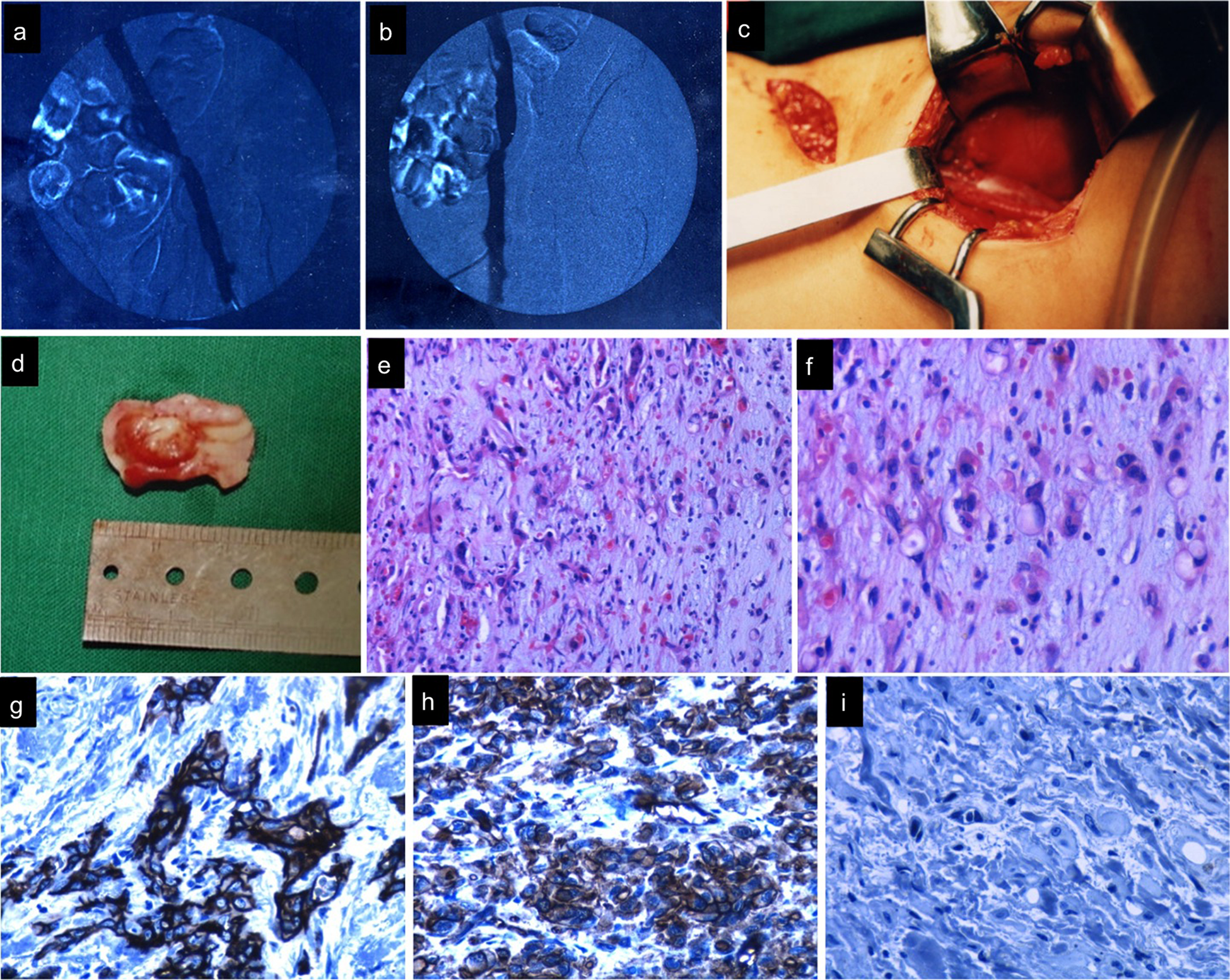
In case 1, an initial venogram documented a stenosis of the external iliac vein cephalic to the inguinal ligament (a and b). In the surgical exploration, a mass derived from the external iliac vein was identified (c and d). The histopathological examination shows a vascular neoplasm with prominent fibrohialine stroma (e, HE 10×), with round and spindled cells disposed in chords and nests (f, HE 40x), positives to CD31 (g, 40×), and CD34 (h, 40x). The proliferation index (Ki67) was very low (I, 40×). HE indicates hematoxylin and eosin.
In the histopathological analysis, an infiltrative vascular neoplasm was observed. The neoplastic cells had a round and spindled morphology with large vacuolated cytoplasm and eccentric epithelioid hyperchromatic nuclei and were distributed in nests, bands, cords, and sheets, forming rudimentary vascular structures, some with true lumen. The stroma was hialinizated with fibromixoid areas (Figure 1e). The mitotic index was low (<1 mitosis in 10 high power fields [HPF]). The neoplastic cells were positive for CD31 (Figure 1g), CD34 (Figure 1h), and Ulex-Europeus, and the proliferation index (Ki67) was 1% (Figure 1i). The surrounding soft tissues were free of neoplastic infiltration. A diagnosis of EHE was made.
The thoracic and abdominal computed tomography (CT) scans were normal. The patient received a 6-week course of warfarin anticoagulation. Currently, the patient is asymptomatic after 9 years of treatment, without recurrence of disease.
Case 2
A 44-year-old man presented with left leg edema pain and swelling. He had a history of DVT treated with warfarin 3 months prior to the actual symptoms being reported. A duplex ultrasound detected a high-pitched Doppler signal with severe velocity increase in the iliac vein without venous thrombosis, and a venogram documented stenosis of the external iliac vein just above the inguinal ligament (Figure 2a). A surgical exploration was performed. A left external iliac vein mass (2 × 1.5 × 0.8 cm3) with lymph node compromise was observed. The involved segment of vein and soft tissues was resected, and the iliac vein was reconstructed with a saphenous vein section.
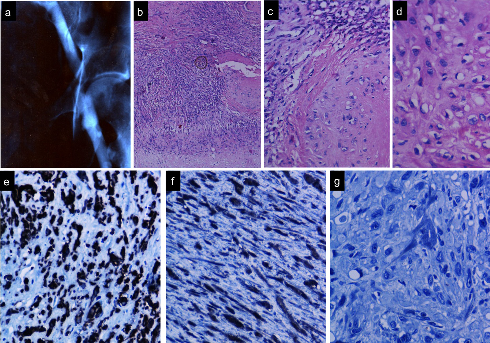
In case 2, a venogram showed stenosis of the external iliac vein (a). The microscopic analysis of the tumor identified was hypercellular composed by epithelioid and spindled cells (b, HE 5×), composing vascular structures with necrotic areas (c, HE 20×). The neoplastic cells exhibit atypia (d, HE 40×) and were positives to Ulex-Europeus (e, 40×) and CD34 (f, 40×). The proliferation index (Ki67) was low (g, 40x). HE indicates hematoxylin and eosin.
The microscopic study identified a vascular neoplasm formed by atypical epithelioid neoplastic cells (Figure 2a–d), positives for Ulex-Europeus (Figure 2e), CD34 (Figure 2f), and CD31, distributed in nests, bands, cords, and sheets, and forming rudimentary vascular structures in a hialinizated stroma. Necrosis and extensive areas of spindled diffuse pattern were observed (Figure 2c). A mitotic index of 2 mitosis figures of mitosis or mitotic figures (FM) in 10 HPFs, and a Ki67 of 2% was documented (Figure 2g). The lymph nodes demonstrated tumor invasion. A diagnosis of EHE was made.
The patient received anticoagulation with warfarin for 6 weeks. Postoperative metastatic evaluation was negative. Complementary local radiation therapy was given. Multiple bilateral lung and liver metastasis were found 31 months later, and the surgical resection was performed. The patient is currently alive and free of disease with a follow-up of 8 years.
Discussion
Increased discussion about the behavior of EHE is directed to a new classification of this neoplasm, as authors including Goh 2 are clamming an increase in the EHE grade, and others like Mentzel and Calonje 3 are suggesting that this neoplasm should be considered as fully malignant, due to its high metastatic potential (20%-30%; mainly to regional lymph nodes, lung, liver, and bone) and 10% to 20% mortality. In a recent publication, Deyrup et al 5 analyzed 49 cases and proposed that EHE can be stratified into 2 groups according to various pathologic criteria, including 5-year metastases and mortality. They concluded that tumors with >3 mitotic figures/50 HPFs and greater than 3 cm were associated with higher mortality and should be considered malignant. In the same line, Fletcher 6 believes that the term “border-line” is not appropriate, and that EHE must be considered totally malignant due to the number of metastasis and mortality rate reported. Fletcher also states that understanding the biological behavior of EHE is not possible using only its histological examination, based on the morphological appearance, because this examination does not necessarily accurately reflect the clinical course of this neoplasm (x).
In general, the clinical presentation is variable and depends on tumor localization and size. In large vessels, the principal symptoms are secondary to occlusion, embolism, or palpable mass as in our cases. The EHE has been mainly observed in adults, 40% under 40 years of age, of both sexes but predominantly in women. It has also been described in children. 7 Reported vascular primary locations that have been described EHE are heart, 7–9 thoracic aorta, 10 aorto-iliac segment, 11 radial artery, 12 vena cava, 13 azygos vein, 14 femoral vein, 15,16 and less frequently in iliac vein 4,17,18 (Table 1).
Reported Cases of Iliac or Femoral Vein Epithelioid Hemangioendothelioma.
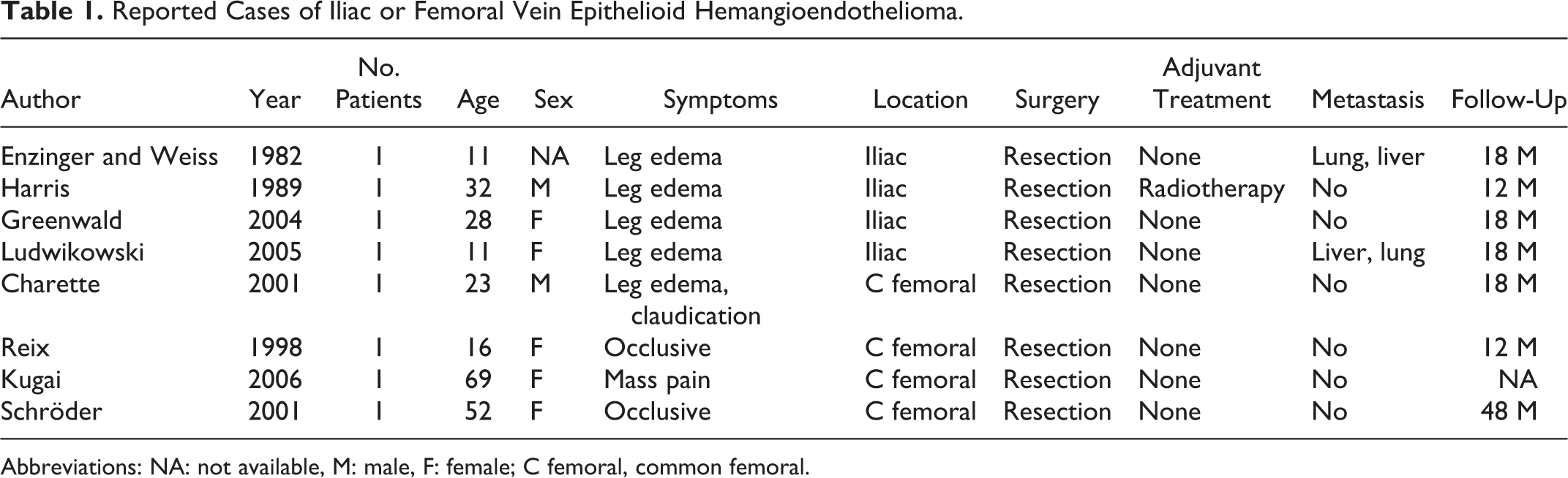
Abbreviations: NA: not available, M: male, F: female; C femoral, common femoral.
The clinical differential diagnosis needs to be done with edema of other etiologies, including venous claudication and DVT. 19,20 Furthermore, extrinsic comparisons with anatomical structures such as right common iliac artery, 20 retroperitoneal fibrosis, 21 Baker cysts, 22 other neoplasms, 23 and femoral hernia or arterial aneurysms need to be conducted.
In the initial evaluation of primary venous tumors, a duplex ultrasound should be done, since it allows for a morphological and a dynamic blood flow-based evaluation of the venous system. Highly suggestive findings include intraluminal defects, altered flow patterns (high velocity with a high pitch sound), Doppler waveform alterations, loss of plasticity, and velocity increase. Reix et al 16 described color duplex ultrasound criteria to diagnose the primary malignant venous tumors by observation of bulging veins (local increase in vein diameter), heterogeneous high-intensity endoluminal material, deviation of the vein, peritumoral vascularization, and localized increase in flow rate (patent vein). 24,25 Labropoulos et al reported the following as detection parameters: >50% stenosis and poststenosis to prestenosis peak vein velocity ratio of 2.5 as the best diagnostic criteria. 25
The CT scan and magnetic resonance imaging (MRI) are the preferred methods to study vascular tumors, as they can determine their venous origin and their relation to the surrounding structures, helping to propose a surgical procedure and provide an accurate diagnosis. 26–28 According to many authors, MRI provides better images and seems to be more effective for the diagnosis. The venography will help confirm a venous stenosis or occlusion and its hemodynamic compromise extension through measurement of the pressure gradient. However, it provides only vein lumen information and does not provide data about the endothelium, vein wall, or surrounding tissues.
Finally, the intravascular ultrasound (IVUS) has been described as a useful study in the diagnosis and management of iliac vein pathology, and we posit that it can help in the evaluation of possible tumors of the veil wall, from determining the tumoral etiology of the stenosis previous to the surgical procedure. 27,28 According to many authors, IVUS provides anatomic information and is correlated with other radiological studies.
The EHE treatment when located in iliac veins is surgically seeking for free margins. The reconstruction can be made by autogenous interposition or PTFE prosthesis. In our experiences, we favor reconstruction with a cylindrical vein panel as described recently by Kugai. 29 We used a similar technique in both of our cases, utilizing the greater saphenous vein with good results in our patients with oncologic disease and trauma. The difference in our technique is that we divide the harvested saphenous vein longitudinally and reconstruct it in situ, cutting the exact length and using fixed sutures, which are located proximally and distally. The advantage of in situ reconstruction is that it results in a better matching of the proximal and distal diameter of the reconstructed vein. Radiotherapy or chemotherapy is used to treat EHE, but the benefit is not clear. 30
Footnotes
Declaration of Conflicting Interests
The author(s) declared no potential conflicts of interest with respect to the research, authorship, and/or publication of this article.
Funding
The author(s) received no financial support for the research, authorship, and/or publication of this article.