
Abstract
The evolution of minimally invasive procedures to treat aortic aneurysms has expanded to include access interventions as well. Traditionally, groin exposures have been the standard approach for common femoral artery exposure with open cutdown; however, inherent and related complications to that approach have paved the road to the percutaneous approach. Current available evidence from the literature supports the feasibility and the safety of percutaneous endovascular aneurysm repair (PEVAR); however, predictors of success are not well defined. We should examine all available studies (both prospective and retrospective) in order to draw a conclusion and evidence-based outcome for selecting patients who would benefit the most from PEVAR.
Keywords
Introduction
In traditional open endovascular aneurysm repair (OEVAR), access is achieved through a surgical cutdown via a transverse groin incision placed one-third the distance from the pubic tubercle to the anterior superior iliac spine. Once the femoral fascia is reached, dissection proceeds longitudinally along the femoral sheath. The common femoral artery is then punctured anteriorly approximately1 cm above its bifurcation. Although this approach has worked well historically, recent advances in endovascular tool technology have evolved and permitted percutaneous access approaches as well.
Reports on percutaneous endovascular aneurysm repair (PEVAR) have been published for over a decade with mixed results as well as some uncertainty regarding durability and applicability. Technical success was reported to be between 46.2% and 100%. 1 There are only 2-randomized control studies available, and although data may be limited or biased secondary to the small patient numbers, the main ongoing message is related to safety and feasibility.
With the advent of suture-mediated arterial closure devices, along with ongoing small-sized profile sheaths and endografts, the trend toward the use of PEVAR has increased. Several studies have evaluated the percutaneous approach with different measures. 1–10 Although the safety, feasibility, and efficacy of PEVAR have been reported previously, controversy still exists regarding predictors of failure. 1,3–6,9,11–15 In this report, we sought to define predictors of failure and to examine the relevant literature with the goal being to clarify which patients are most appropriate for PEVAR as well as identify predictors of success and provide evidence-based recommendations for patient selection and technique.
Study Definitions
Obesity: body mass index >30 kg/m2.
Technical success: ability to complete the intervention using percutaneous suture-mediated closure devices.
Technical failure/conversion: when open cutdown is required to complete the intervention, factors that may contribute to all predictors of failure are included (see below).
Vessel calcification: the amount and distribution of calcifications within the vessel.
Smoking: current or past smoking.
Hypertension: defined as >140 mm Hg systolic blood pressure and >90 mm Hg diastolic blood pressure.
Estimated glomerular filtration rate (eGFR): defined as 186.3 × serum creatinine−1.154 × age−0.203 × 0.742 (if female) × 1.212 (if African American).
Proposed Technique
Using ultrasound guidance, each common femoral artery (CFA) is examined with regard to the severity and the location of calcification, takeoff of the profunda femoris artery, and vessel diameter.
The anterior portion of the CFA is punctured using the single-wall puncture technique with a 19-gauge needle, and a Supracore wire (Abbott, Illinois, USA) is passed under fluoroscopic guidance.
A small skin incision (about 4 mm in length) is made over the 19-gauge needle.
An 8F sheath is used to create a tunnel from the skin to the anterior arterial puncture site. In the authors’ opinion, this step is crucial to prevent any subcutaneous tissue interference during the final steps of the closure process.
For large sheaths (16-24F), 2 Perclose (Abbott, Illinois, USA) suture-mediated closure devices are deployed diagonally about 90° to 180° apart from each other. This 6F closure device is passed over a 0.035-in guide wire and is used to close 5 to 7F arteriotomies. Perclose devices utilize a 3/0 polypropylene suture and are deployed vertically via dual transarterial nitinol needle. Each device has 2 color-coded strands, the blue one is long and is used for tying, the other one is short and white and is used as a locking strand. Both strands are left extracorporeally and tagged until the EVAR/thoracic EVAR procedure is completed. Suture-mediated closure device (SMCD) deployment should be done while maintaining the guidewire in the vessel. Once hemostasis is achieved, the guidewire can be removed; however, in cases with significant back bleeding, additional SMCD can be used as needed to achieve adequate closure. On the other hand, for small sheaths (12-14F), 1 Perclose device should suffice.
After the guidewire is removed, the knot pusher is used again to tighten the slipknot more gsently and cautiously. Gentle forward pressure can be used to navigate the slipknot deeper into the wound in order to achieve hemostasis. Since a prolene-based stitch is used, the tying of the suture can be facilitated by keeping both strands moist with heparinized saline.
Pressure should be applied for 5 to 10 minutes, after which a small pressure dressing can be applied.
Protamine is not given routinely at the completion of the intervention.
Predictors of Failure
Vessel calcification: in a prospective review of 500 patients who underwent PEVAR using the Prostar 10F device, (Abbott, Illinois, USA), femoral artery calcification (odds ratio [OR] 74.5, 95% confidence interval [CI], P < .001), as well as the operator experience (OR 43.2, 95% CI, P < .001), was the most likely predictor of conversion. A reoperative groin was also predictive of conversion (OR 48.8, 95% CI, P < .001). Neither obesity nor sheath size was identified as independent risk factors for conversion to an open approach. 7
Vessel size: a second prospective randomized study 16 examined the size of the common femoral artery (4 mm) as a predictor of conversion (see Table 1).
Sheath size: in a meta-analysis of pooled data, technical success for PEVAR was superior to <18F sheaths when compared to procedures performed with sheaths ≥ 20F (OR 1.78, 95% CI 1.24-2.54, P = .002). Also, some benefit was observed when multiple, rather than single, preintervention SMCDs were deployed (OR 2.16 vs 1.64, respectively; P = .353). 4,5 Etezadi et al reported a PEVAR success rate of 89.7% for <16F sheaths and 80.4% for ≥16F sheaths (P = .15) 12 Also, Rachel et al, in a prospective nonrandomized study of 95 patients, reported that gender, previous femoral access, obesity, and iliac occlusive disease were not predictive of PEVAR failure. In that study, sheath size (>16F) was found to be associated with an increased chance of device failure. 22
Of note, other published studies have not found these factors to be predictive of the need for conversion. In a study of 445 patients who underwent EVAR over a 7-year period, 100 patients had PEVAR and 557 had OEVAR. The technical success rate for the PEVAR group was 85%. A body mass index (BMI) >30 kg/m2, vessel calcification, and sheath size were not predictive of technical failure. 12 Similar findings were reported by Smith et al. In addition, the authors demonstrated postoperative anatomic changes associated with PEVAR versus OEVAR using 3-dimensional computed tomographic (CT) reconstruction. In the patient population, there were no differences between the 2 groups with respect to femoral artery calcification (Agatston scores 667 vs 945, P = .37). Obesity rates (BMI > 30) were also similar (P = nonsignificant). However, the most impressive findings were the pre- and postoperative CT-derived anatomic data that showed significant decreases in the minimal vessel area in OEVAR versus PEVAR (P = .02). 6
Ultrasound-guided intervention: many reports have concluded that ultrasound guidance is helpful in achieving technical success. This technique not only aids in puncture precision but also provides a significant understanding of the arterial anatomy, the degree of calcification, vessel size, and that the puncture is above the common femoral artery bifurcation. A recent retrospective study reported that ultrasound increases PEVAR accessibility to almost all patients while decreasing access-related complications. This study was conducted over a 5-year period on 296 arteries in 186 patients, during which time there was an exponential increase in the number of PEVARs performed—increasing from 0% to 92% over the 5-year study period. This study concluded that a vessel size of 5 mm was the threshold for device-related mechanical failure. 17
Operator experience: intuitively, there is a direct relationship between operator experience and technical success. A study of 100 patients demonstrated a significant trend toward a decrease in the numbers of failures with increasing operator experience (P < .007). Also, the probability of closure failure decreased from 45% per patient at the initiation of the study to 5% per patient at the end of the study period. This report concluded that technical failure was inversely proportional to the experience of the surgeon with SMDC. 1
Device failure/malfunction: in a retrospective, single-center review of 101 patients, 2 the authors reported a 10% conversion rate with mechanical device failure as the only predictor of conversion. Other factors, such as obesity (defined as BMI >30 kg/m2), vessel calcification, smoking, hypertension, and age, were not predictive of the need for conversion. This study highlighted the role of ultrasound in the increasing technical success of PEVAR. Oguzkurt and colleagues also reported high technical success with ultrasound guidance. 24
Obesity: Starnes et al, in his retrospective review of 79 PEVAR large-bore-sheath access sites, reported technical success in 94% of the patients. Failure was attributed to sheaths larger than 20F and morbid obesity (>35% BMI; P = .02 and P = .01, respectively). 20
Literature Review If All Recent Studies That Outline Outcomes of PEVAR.
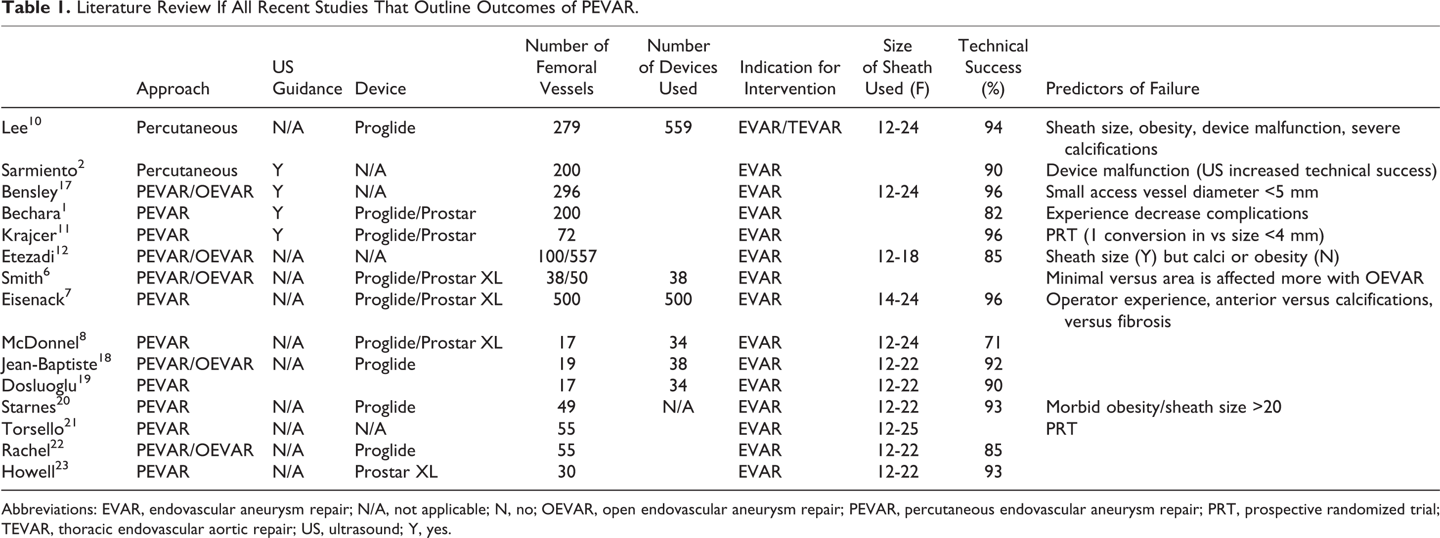
Abbreviations: EVAR, endovascular aneurysm repair; N/A, not applicable; N, no; OEVAR, open endovascular aneurysm repair; PEVAR, percutaneous endovascular aneurysm repair; PRT, prospective randomized trial; TEVAR, thoracic endovascular aortic repair; US, ultrasound; Y, yes.
Perceived Benefits of PEVAR
In an analysis of the American College of Surgeons National Surgical Quality Improvement Program database, the major advantage of PEVAR was a decrease in the operative time (159 ± 63 minutes vs 150 ± 68 minutes, P < .05); however, they indicated that there was no difference between the 1-month mortality and hospital length of stay between PEVAR and OEVAR. 25
Also, a shorter operative time (153.3 vs 201.5 minutes, P < .001), fewer wound complications, and larger minimal access vessel diameters (6.7 mm vs 6.1 mm, P < .01) were reported by Bensley and colleagues. 17 Wound-related complication rates were lower in the PEVAR group. Specifically, seroma formation was estimated to be lower in the PEVAR group than in the OEVAR group (11.4% vs 1.2%, P = .001). 12
Malkawi et al, in his systematic review of 22 published studies which included 1-randomized control trial, 10 prospective nonrandomized, and 11 retrospective studies, reported that PEVAR was performed in 1751 femoral vessels of 1087 patients with a technical success rate of 92%. The investigators concluded that PEVAR was associated with fewer access-related complications in comparison to OEVAR (RR 0.47, 95% CI 0.28-0.78, P = .004). 14
In another report, a decrease in wound complications was highlighted among 17 patients who underwent PEVAR, and there was a significant decrease in access-related complications (6% vs 10% OEVAR, P < .05, Mann-Whitney U test). 8 In addition, PEVAR decreases the invasiveness of EVAR and reduces not only the operative time but also the time to ambulation. Torsello et al also indicated that the mean surgery time is reduced with PEVAR (86.7 vs 107.8 minutes; P < .05) as well as the time to ambulation (20.1 vs 33.1 hours, P < .001). 21
There are 2 respected retrospective studies, which indicate that the possible benefits of a shorter length of stay associated with PEVAR are negated by the cost of the device. Jean-Baptiste et al reported a mean hospital stay of 5.8 days for PEVAR and 7.8 days for OEVAR (P = .05). According to the value of the Euro in 2008, the difference in the hospital stay was associated with a reduced cost for the PEVAR group (5579.60€ vs 7503.60€, P = .04), which subsequently offsets the cost of SMCD. 18 Similar results were reported 1 year earlier by Lee et al who demonstrated the safety and feasibility of 559 Proglide devices used during PEVAR. Despite the reduction in the procedure time, the cost effectiveness was negated by the cost of the Proglide devices ($295 per device in 2007). 10
Randomized Control Studies
PEVAR trial: a total of 38 consecutive patients with abdominal aortic aneurysm (AAA) in 19 centers were enrolled in a roll-in phase over a 12-month period. The PEVAR was conducted using the Proglide (Abbott, Illinois, USA) or Prostar XL (Abbott, Illinois, USA) closure devices. Technical success of the preclose procedure was 97% (37 of 38 patients). In 1 patient, Proglide devices failed and required surgical conversion. All endovascular aneurysm repairs were successful. No major adverse events occurred during the study time period. This study concluded that PEVAR is safe and feasible. 11
Second trial: a single institution, prospective, controlled evaluation was conducted in 57 consecutive patients with AAA who underwent PEVAR over a 2-year period. All interventions were performed with the adjunctive “preclose” use of the Prostar XL closure device in a hybrid endovascular suite and resulted in a technical success rate of 98%. One conversion, secondary to a small access vessel (4 mm) and major access-related complications, occurred in 8.8% of the patients within 30 days. 16
Tips for Success
Obtain appropriate preoperative computerized tomographic angiography evaluation of the groin vessels, as well as the iliofemoral vessels, specifically assessing the degree of calcification, stenosis, and vessel tortuosity. It is crucial to maintain a high index of suspicion in obese patients and in those with reoperative groin anatomy. When difficulty is encountered during the procedure, there should be a low threshold for conversion in these cases. 26
Use ultrasound guidance for access.
A hybrid room should be utilized, if available, in case conversion is necessary. 27
Keep the access wire in place until adequate hemostasis can be achieved.
Additional SMCD devices (>2) may be used if necessary, particularly in cases in which it is necessary to use a larger sheath.
Conclusion
The current published literature supports the feasibility, safety, and efficacy of PEVAR. The main predictors of technical failure and conversion to OEVAR include vessel calcification, vessel size, and sheath size. The advent of small-sized EVAR devices will allow for more applications of PEVAR and will likely become standard in the near future.
Footnotes
Declaration of Conflicting Interests
The author(s) declared no potential conflicts of interest with respect to the research, authorship, and/or publication of this article.
Funding
The author(s) received no financial support for the research, authorship, and/or publication of this article.