
Abstract
Iatrogenic cervical internal carotid artery pseudoaneurysm is a rare and potentially lethal complication following tonsillectomy. It can be complicated by thromboembolism, mass effect and eventually may rupture leading to death. Various endovascular treatment options are available for the management of these pseudoaneurysms, including coil embolization, detachable balloon occlusion, or stent graft placement. Parent artery occlusion using detachable balloons can be a therapeutic option in a subset of patients. However, evaluation of cross circulation with preprocedure balloon test occlusion is imperative in such cases.
Keywords
Introduction
Extracranial internal carotid artery (ICA) pseudoaneurysms are rare. These pseudoaneurysms may be complicated by thromboembolism, ischemic events, growth, and mass effect and eventually rupture. 1,2 Endovascular management is a well-established therapeutic option in most of these lesions. In particular, parent artery occlusion can be a simple solution in a subset of patients with adequate collateral circulation. We report the management of a post–tonsillectomy ICA pseudoaneurysm by using detachable balloons following successful balloon test occlusion (BTO).
Case Report
A 36-year-old male presented with left tonsillar fossa mass on first postoperative day. Clinical examination showed pulsatile, firm to hard swelling in left tonsillar fossa. Computed tomography angiography (Figure 1A) confirmed the presence of partially thrombosed cervical ICA pseudoaneurysm. Digital subtraction angiography showed a large pseudoaneurysm from left distal cervical ICA with very sluggish forward flow (Figure 1B). Balloon test occlusion of left ICA (Figure 1C) was done by clinical monitoring at 5-, 10-, 15-, 20-, and 30-minute intervals. Right ICA (Figure 1D) and left vertebral artery (Figure 1E) angiograms showed good cross circulation with patent anterior and posterior communicating arteries, respectively. Patient tolerated the BTO well. So parent artery occlusion of the left ICA proximal to the pseudoaneurysm sac was done using the same detachable balloon which was reinforced with another detachable balloon proximally (Figure 2A and B). Check angiograms showed occluded left ICA with no filling of the pseudoaneurysm sac (Figure 2C and D). Postprocedure the patient was neurologically intact and 1-year follow-up of patient was unremarkable. Although the patient was not put on anticoagulants in the follow-up period, no ischemic symptoms were reported by the patient.
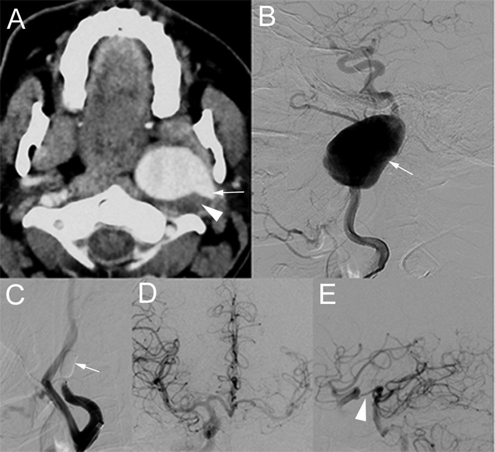
Axial computed tomography image (A) showing left internal carotid artery (ICA) pseudoaneurysm (arrow) with eccentric thrombus (arrowhead). Selective left ICA angiogram (B) showing large pseudoaneurysm (arrow) of distal cervical ICA with sluggish forward flow. Inflated detachable balloon (arrow) is seen in left ICA during balloon test occlusion (BTO; C). Good cross circulation with filling of the left anterior and middle cerebral artery via patent anterior and posterior communicating arteries (arrowhead) is seen on right ICA (D) and vertebral artery (E) angiograms.
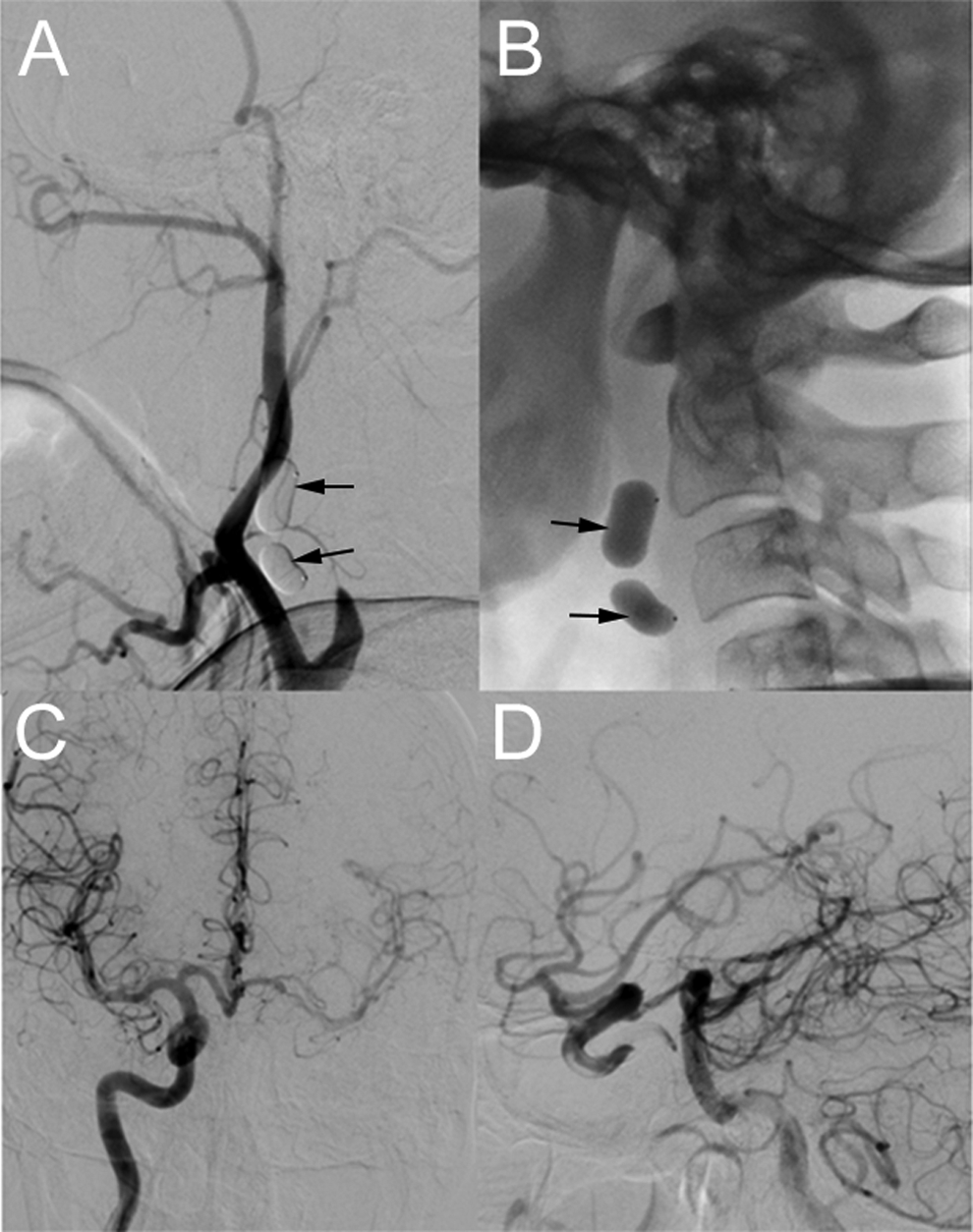
Selective left internal carotid artery (ICA) angiogram (A) after the deployment of 2 tandem balloons (arrows) in ICA showing no filling of distal ICA or the pseudoaneurysm sac. Fluoroscopic image (B) shows the tandem balloons (arrows) in situ. The right internal carotid (C) and the vertebral artery (D) angiograms show good cross circulation.
Discussion
Internal carotid artery pseudoaneurysm formation following tonsillectomy is a rare but potentially lethal occurrence. It represents a complete rupture of the vessel wall with a communication between the vessel lumen and the cavity limited by organized hematoma without true vascular wall. Common causes include penetrating or blunt trauma, postsurgical infection, vasculitides, and infiltrating neoplasms. 3 -5 They are associated with increased risk of continued expansion with mass effect, rupture, and distal embolism, necessitating early management.
Both surgical and endovascular management options are available and may involve parent artery occlusion or repair/pseudoaneurysm exclusion with vessel preservation. A detailed evaluation of the pseudoaneurysm and the brain circulation should be performed before establishing the best therapeutic strategy. If the brain circulation can be compromised during the ICA occlusion, then a superficial temporal artery–middle cerebral artery bypass followed by the occlusion of the ICA can be lifesaving. Traditional surgical repair involves high mortality and morbidity. 6
Temporary BTO is imperative to evaluate ischemic risks before attempting parent artery occlusion. The introduction of clinical BTO of the ICA is associated with a significant reduction in postocclusion morbidity. A review of the literature 7 comprising 516 patients demonstrated that the use of BTO of the ICA reduced the morbidity of permanent ICA occlusion from 26% to 13%. However, complications due to BTO have also been reported. The incidence of neurological deficits during BTO may range from 3.2% to 3.7%. 8,9 Our patient tolerated the BTO well and no focal neurological deficit was seen during or following the test.
Endovascular therapeutic options include coil embolization, detachable balloon occlusion, or endovascular stent graft insertion. A stent graft allows immediate occlusion of the lesion with maintenance of patency of the normal vessel. 10 Detachable coil embolization of the pseudoaneurysm sac can also be an alternative treatment. However, there may be a recurrence with delayed massive bleeding as it may be impossible to obliterate the sac completely. This may be due to the different clinical behavior of the pseudoaneurysm as it lacks complete limiting wall. Detachable balloons play a vital role in the management of these lesions. 11 Moreover, the same balloon used for BTO can be detached adding ease to the procedure. In our case, surgical management was limited by the location of the lesion and the technical difficulties of controlling the distal ICA. As there was good cross circulation and patient tolerated BTO well, occlusion of ICA was performed using detachable balloons. Moreover, in this case, further expansion of the pseudoaneurysm sac due to collateral circulation was unlikely, as there are no branches from cervical ICA and was no evidence of associated arteriovenous fistulae, causing a possible sump effect. In conclusion, endovascular detachable balloon occlusion is a good therapeutic option in the management of ICA pseudoaneurysm and can be performed safely in cases requiring ICA sacrifice. However, evaluation of cross circulation with preprocedure BTO is imperative in such cases.
Footnotes
Declaration of Conflicting Interests
The author(s) declared no potential conflicts of interest with respect to the research, authorship, and/or publication of this article.
Funding
The author(s) received no financial support for the research, authorship, and/or publication of this article.