






Abstract
Background:
Prosthetic vascular graft infection (PVGI) remains a severe and challenging complication in vascular surgery with high morbidity and mortality rates. Incidence has been reported between 1% and 6%. The aim of this study was to report our experience in terms of general and surgical management as well as outcome, over 15 years.
Methods:
A retrospective consecutive study was conducted of all patients treated in our department for PVGI between January 2000 and December 2015. We analyzed all data relative to primary operation, duration interval between initial surgery and infections signs, infection site, type of microorganism involved, and surgical treatment modality, as well as evaluation of short- and long-term results.
Results:
Sixty-two patients were admitted for PVGI. Primary revascularization procedures consisted of a peripheral bypass in 42 (68%) patients and an aortic bypass in the remaining 20 (32%) patients. Median interval between primary procedure and reintervention was 3 months (interquartile range 17 [IQR 17]) in the peripheral group and 48 months (IQR 70.5) in the aortic group. Complete excision of the prosthetic graft was carried out in 85% of the cases. Thirty-day mortality was 0% and 9.5% in the aortic and peripheral group, respectively. The overall survival rate was 62.3% at 2-years, 46.4% in the aortic group, and 69.7% in the peripheral group.
Conclusions:
Prosthetic vascular graft infection needs a multidisciplinary management with appropriate antibiotherapy, radical removal of the infected graft, and in situ reconstruction. This strategy gives satisfactory results in terms of mortality, morbidity, patency rates, and infection control.
Introduction
Prosthetic vascular graft infection (PVGI) is a rare complication of vascular surgery and occurs in 1% to 6% of cases after vascular graft implantation procedures. 1 Predisposing factors for PVGI have been identified, including exposure of femoral vessels in the groin, redo surgery, diabetes, obesity, hemodialysis, and Rutherford stage VI occlusive arterial disease. 1,2 Vascular graft infection is classified by its development in time (early infections, ≤4 months after graft implantation; late infections after >4 months), its relationship to the operative site (aortic or peripheral) and its severity. Prosthetic vascular graft infection is associated with high morbidity and mortality and accounts for major amputation rates from 12% to 70%. 3,4 Optimal treatment consists of radical removal of the infected graft. However, the surgeon is confronted with the problem of revascularization in a contaminated field. Extra anatomical bypass avoids working in a contaminated field but has the disadvantage of lower patency rates as compared to an anatomical bypass configuration. In order to perform an anatomical bypass in an infected site, various conduits have been used such as cryopreserved arterial allografts, autologous venous grafts, silver grafts, or antibiotics bounded synthetic grafts.
The purpose of this study was to analyze the indications, risk factors, and different surgical modalities along with short- and long-term outcomes after treatment of PVGI.
Materials and Methods
Population
We retrospectively analyzed, from our prospective maintained database, all patients treated for PVGI between January 2000 and December 2015. Table 1 lists the main cardiovascular risk factors of this population. Among the 62 patients admitted and treated for PVGI in our institution, 30 (48%) were transferred from peripheral centers. All patients were divided into 2 groups: those presenting with an aortic bypass graft infection (20) and those with a peripheral bypass graft infection (42).
Cardiovascular Risk Factors of the Population.

Abbreviations: BMI, body mass index; SD, standard deviation; IQR, interquartile range.
Preoperative Work-Up
Diagnosis was initially based on clinical findings, such as systemic (fever, tachycardia, hypotension, etc) and/or local symptoms (wound dehiscence, pus drainage, abdominal pain, etc). Various medical imaging investigations (Duplex, angio CT, leucocyte scintigraphy, and/or fluoro-d-glucose positron emission tomography [FDG PET-CT]) were performed to confirm the diagnosis of PVGI and are shown in Table 2. Angio CT was the most commonly used imaging technique (57.8%) upon which diagnosis of PVGI was based on presence of perigraft fluid, perigraft soft-tissue attenuation, ectopic gas (Figure 1, arrow a), and/or pseudoaneurysm development (Figure 1, arrow b). In uncertain cases, a complementary leucocyte scintigraphy or FDG PET-CT was performed. In more recent cases, the leucocyte scintigraphy was progressively abandoned in favor of FDG PET-CT (Figure 2).
Preoperative Differences Between the Peripheral and Aortic Group.

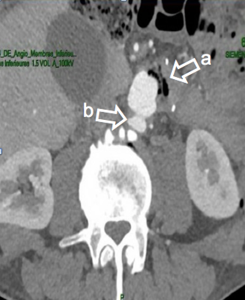
Computed tomography angiography (CTA) of an aorto-bi-iliac graft infection: showing ectopic air (arrow a) and a pseudoaneurysm (arrow b) at the proximal suture level.
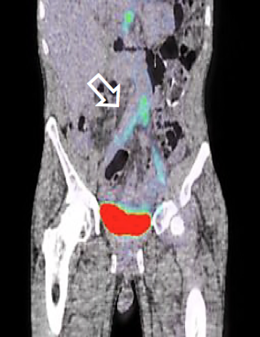
FDG PET-CT of an infected aorto-bi-iliac graft infection with high uptake of FDG on the aortic bifurcation and iliacs (arrow).
Treatment Strategy
Our treatment strategy consisted of complete removal of the infected graft, followed by extensive debridement of surrounding tissues and irrigation of the site with an antiseptic solution. Partial removal of the infected prosthetic graft was occasionally performed in selected patients who didn’t present general signs of infection and in which the infections’ process was well localized. Tissue samples were sent for microbiological evaluation. Subsequently, we performed in most cases (81%) an in situ reconstruction using cryopreserved arterial allografts or autologous veins.
Cryopreserved Arterial Allograft
The European Allograft Bank provided all cryopreserved arterial allografts (Figure 3), which were obtained either during multiple organ harvesting or from fresh cadavers. The interval between harvesting and preparation of the allograft never exceeded 18 hours and the warm ischemia period for cadavers was less than 6 hours. Donor age limits comprised between 18 and 45 years and the following exclusion criteria were strictly applied: transmissible illness, ongoing infection, cancer, immunodeficiency, corticosteroid therapy, connective tissue disease, and death of undetermined causes. The modalities of allograft preparation have been described in detail by Goffin et al 5 and mainly consist of rinsing in culture medium followed by decontamination with various antibiotics (cefoxitin, lincomycin, polymyxin, and vancomycin). Allografts then were frozen and stored at −150°C in liquid nitrogen.
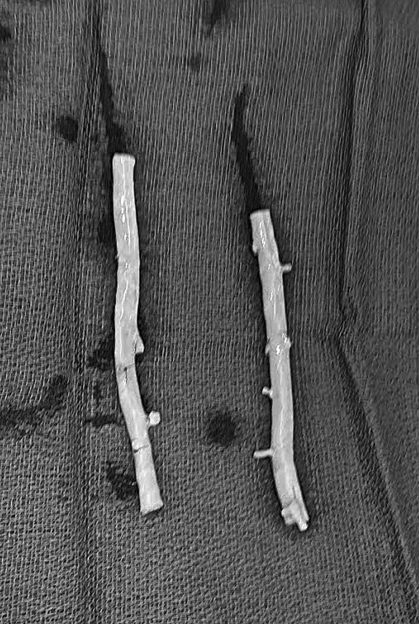
Two cryopreserved superficial femoral arteries homografts used to reconstruct an infected femoropopliteal bypass above the knee.
Postoperative Course and Follow-Up
All patients with unidentified microorganism prior to surgery received empirical broad-spectrum antibiotherapy. Thereafter, antibiotics were adapted according to bacteriology data and interdisciplinary debated with microbiologists.
Follow-up at 2 months included clinical examination and angio CT in the aortic group and duplex scanning in the peripheral. It was then followed by duplex scanning yearly or angio CT in case of adverse outcome.
Results
Over a period of 15 years, 62 patients with a median age of 75.5 (interquartile range 14.5 [IQR 14.5]) years were treated for PVGI. The primary reconstruction site involved the aorta in 20 (32%) cases and the peripheral arteries in 42 (68%) cases. Table 3 presents the major preoperative differences between the peripheral and the aortic group. The majority of patients transferred from peripheral centers to our department were infected at the level of the aorta. All primary aortic bypasses were performed with a Dacron graft. Only 4 (6%) patients had no inguinal incision during primary surgery. The median interval between the primary procedure and the PVGI treatment was 48 months (IQR 70.5) in the aortic group and 3 months (IQR 17) in the peripheral group.
Medical Imaging Used to Confirm the Diagnosis of PVGI.

Abbreviation: PVGI, prosthetic vascular graft infection; PTFE, Polytetrafluoroethylene.
Eleven (17.7%) patients were septic at the moment of diagnosis. Sixteen (80%) patients were hemodynamically stable in the aortic group and 39 (92.8%) in the peripheral group. Groin infection was found in 38 (90%) patients in the peripheral group as compared to 14 (70%) in the aortic group. Five (25%) patients with an aortic PVGI had an underlying aortoenteric fistula.
Procedural Characteristics
All procedures were performed under general anesthesia with a median operative time of 273 minutes (IQR 213 minutes). Complete prosthetic graft removal was performed in 19 (95%) and 34 (81%) patients in the aortic group and the peripheral group, respectively. Table 4 shows the percentage of graft types used for the various reconstructions, graft removal extension, and revascularization technique in both groups.
Differences in Graft Removal and Revascularization Between the Peripheral and Aortic Group.

In the aortic group, in situ revascularization was obtained in 16 (80%) and synthetic extra-anatomical revascularization in the remaining 4 (20%) cases. In situ aortic reconstruction was performed with cryopreserved arterial allograft in 13 (65%) cases and with autologous vein in 3 (15%) cases. In the event of prosthetic-enteric fistula, abdominal surgeons performed a duodenoraphy with additional omentoplasty in all cases.
In the peripheral group, revascularization was performed using a cryopreserved allograft in 24 (57.1%) cases and an autologous vein in 10 (23.8%) cases; 8 cases (19%) had no revascularization procedure after removal of the infected graft.
Microorganisms
Peroperative tissue culture revealed polymicrobial infection in 23 (37%) cases. About 30% of the microbiological culture remained negative. The most commonly found bacteria in the aortic group were Escherichia coli and Enterobacter in 19 (30%) and 16 (25%) cases, respectively, and Staphylococcus in 35 (57%) peripheral cases.
One-Month Results
We had no perioperative death. The median hospital stay was 22 days (IQR 15.75 days) in the aortic group and 23 days (IQR 15.5 days) in the peripheral group. The 30-day mortality amounted to 6.4%. In the aortic and peripheral group, the mortality was 0% and 9.5% (4 patients), respectively. The causes of death in the peripheral group were unknown in 2 cases, sepsis and encephalopathy in the other 2 cases. Complete infection remission was achieved in 39 (92.8%) cases in the peripheral group and in 18 cases (90%) in the aortic group. In the allograft subgroup, we reached 100% decontamination in both groups within the first 30 days. Furthermore, no graft dilatation, no graft occlusion, and no major amputation were observed during the first 30 days after surgery.
Long-Term Results
The median follow-up period of the 58 patients who survived the first postoperative month was 8 months (IQR 17 months, confidence interval 95%). Eight patients were lost to follow-up. In the remaining 50 patients, 19 patients belonged to the aortic group and 31 to the peripheral group. Only 1 allograft dilatation was observed in the peripheral group, after 57 months. The primary patency rate was 86% and assisted primary patency rate was 90%, with 7 occluded bypasses during follow-up, of which 2 were successfully treated by thromboembolectomy.
In 3 cases, a major amputation was required. Locale wound complication was a major problem with wound dehiscence and/or abscess drainage in 17 patients spread across both groups. All different general and local complication after graft removal are summarized in Table 6. The cause of death was a recurrent de novo aortoenteric fistula in 2 patients in the aortic group but unknown or unrelated to the infection or redo surgery in the other death patients. The 12- and 24-month survival rates (Figure 4) were higher in the peripheral group (80.2% and 69.7%, respectively) compared to the aortic group (61.9% and 46.4%, respectively). The overall survival rates at 12 and 24 months were 74.1% and 62.3%, respectively.
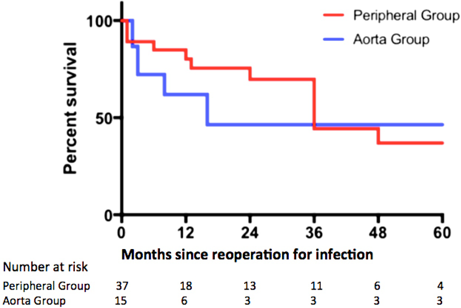
Kaplan-Meier survival curve at 12 and 24 months for the peripheral group (red) and aorta group (blue).
Discussion
Prosthetic vascular graft infection, although infrequent (1%-6%), remains one of the most adverse complications of open vascular surgery with an important mortality and amputation rate of up to 70%. 4 The pathogenesis of vascular graft infection is multifactorial with 4 main etiologic causes. Firstly, technical pitfalls during the operative procedure can lead to perioperative contamination of the graft as well as early postoperative wound dehiscence. In these cases, the infection is frequently located in the groin and related to obesity, diabetes, or redo surgery. As demonstrated in our series, up to 69% of the patients in the aortic group and as many as 90% of the patients in the peripheral group presenting with groin infection carried at least 1 of these risk factors. For these reasons, in high-risk patients, we advocate avoiding groin access wherever technically feasible and using autologous vein or arterial allograft when available.
Less frequently, graft bacterial seeding originates from hematogenous contamination following any procedures distant from the graft. In this setting, infection of the vascular graft is delayed and manifests with general clinical signs of bacteremia. These procedures should therefore take place under antibiotic prophylaxis in any patient with a vascular prosthetic graft.
Thirdly, endovascular procedures requiring direct puncture of the graft can induce its contamination via inoculation. Finally, the most deleterious graft infection can be due to aortoenteric fistula, which develops from chronic mutual erosion of the synthetic graft and the bowel. Extreme care during the primary procedure should be taken in order to avoid any contact between the graft and the intestine by performing an omental interposition and an end-to-end rather than a side-to-end aorto-graft anastomosis.
As far as between primary surgery and the onset of the infection process was concerned, we found a significant difference between both groups: 3 months for the peripheral group versus 48 months for the aortic group. This supports a different infection mechanism, with on the one hand, peripheral vascular grafts are contaminated perioperative and clinical manifestation will occur rather early after primary surgery. On the other hand, contamination of an aortic prosthetic graft is a slow progressive erosion between the prosthetic graft and the gastrointestinal tract and unravels over a longer period of time. These aortoenteric fistulas are rare (0.3%-1.6%), delayed (mean 75.5 months), and difficult to diagnose. 6 Gold standard for radical treatment is open surgery although Endovascular aneurysm repair (EVAR) can be used as bridging to surgery in hemodynamically unstable patients. 7 Surgery consisting of graft resection, resection of the intestinal fistula, and meticulous closure of the bowel, in situ vascular reconstruction and omentoplasty, is associated with higher morbidity but favorable long-term outcomes. 7
While PVGI remains a dreaded and challenging complication after vascular reconstructive procedures, it meets with poor medical literature guidelines, mainly because current practice recommendations are based on small case series and expert opinions. As a matter of fact, there is no validated criteria that properly defines PVGI. The Szilagyi classification established in 1972 seems to be progressively abandoned to the advantage of the Samson classification (Table 5), 8,9 although the use of a classification system for PVGI is not widely spread among practitioners. 10
Szilagyi and Samson Classification for PVGI.

Abbreviation: PVGI, prosthetic vascular graft infection.
Postoperative Complications After Specific Types of Bypass Graft Excisions and Implantation.

Whenever a vascular graft infection is suspected upon clinical criteria, a multidisciplinary approach and management should prevail and involve radiologists, nuclear medicine physicians, microbiologists, and vascular surgeons. Discussions between the various physicians will allow appropriate investigations, precise imaging, diagnosis, and treatment both in terms of accurate antibiotherapy initiation and surgical timing.
After graft removal, revascularization through a contaminated region is the major concern. The outcomes of extra-anatomic bypass surgery seem to have improved in recent series as compared to historical results but the disadvantages, especially in terms of patency, remain. 11,12 Furthermore, blowup of the aortic stump has been described in several series. 10 Our policy is to perform an anatomical bypass through the infected area using an infection resistant conduit whenever feasible. The choice of conduit depends on the patient’s characteristics, surgeon’s expertise, availability of arterial allograft, and the anatomy of the patient.
Autologous veins have shown better results in terms of patency, resistance to infection, and enlargement but are associated with longer operative duration and higher postoperative morbidity. Furthermore, the use of femoral veins to replace an infected graft is not recommended in patients with previous deep vein thrombosis. 4 If the patient’s general status is satisfactory, our first choice is reconstruction with autologous vein when available.
Arterial allografts are a good alternative to autologous vein and have shown excellent short and midterm results in terms of patency and resistance to infection, but in the long term, they carry the disadvantage of degeneration and dilatation. Some authors advocate their use as a temporary solution. 13,14 Furthermore, allografts are not always suitable for all vascular reconstructions, especially at the aortoiliac level. However, suturing several allografts to create a bifurcated conduit is perfectly feasible. There are 2 types of allograft preservation techniques; either the fresh allograft which is preserved at 4° as described by Bahnini et al 15 or the cryopreserved allograft which is frozen and stored at −150°C in liquid nitrogen and supplied by an organ bank. The main advantage of cryopreservation over preservation at 4°C on a histological and practical standpoint is to allow viral testing an unlimited storage of graft material. 16 Because of unsatisfactory results with high reinfection rates, we do not suggest the use of Rifampicin-bounded prosthesis. 4
In localized and early-stage graft infections in the groin, the use of muscular flaps can be useful, with promising results. Some authors consider prophylactic use of vascular flaps during initial grafting procedures. 17,18 The use of negative pressure wound therapy has shown good results in the groin, with high rates of graft salvage. However, it should be used in selected cases because of longer in hospital stay and risks of severe complications such as bleeding. 19
Antibiotherapy in PVGI has been well studied, regarding empirical and therapeutical treatment. 20 All efforts should be made to isolate the responsible germs at the very early stages of the infections process. Initial antibiotherapy should start with empirical coverage of Gram-positive cocci, including methicillin-resistant staphylococcus aureus (MRSA), Gram-negative bacilli, and anaerobes. As soon as microbiological results are available, the antibiotherapy should be gradually reduced and adapted to antibiogram results. We recommend the use of antimicrobial agents such as rifampicin, quinolones, or daptomycin (especially for Gram-positive cocci infection), combined with optimal surgical treatment and followed by long-term antibiotherapy.
Finally, preventive measures are of crucial importance to prevent PVGI. Different characteristics such as obesity, advanced age, smoking, and diabetes have proved to be risk factors for surgical site infection (SSI) therefore increasing the risk of graft infection. 21,22 Perioperative blood transfusion is associated with higher risk of infection. 23,24 Operative duration of 260 minutes increases the risk of SSI by 50% as compared to 150 minutes in femoral popliteal bypasses surgery with autologous vein. 25,26 The importance of postoperative wound care cannot be overemphasized. Silver-coated wound dressings should be applied to groin incisions in the operating room and not changed for 24 to 48 hours unless wound drainage occurs.
Limitations and Strengths
This is a retrospective study with all associated limitations. The number of patients is low and the long-term follow-up included a limited number of patients at risk. However, due to the infrequent condition of PVGI, sample size will always be a concern.
Conclusions
The management of PVGI should be multidisciplinary with an accurate diagnosis and treatment, consisting of complete removal of the infected graft, debridement of surrounding tissues, in situ reconstruction with autologous vein or arterial cryopreserved allograft, and appropriate perioperative antibiotherapy. This approach gave us satisfactory results in terms of mortality, morbidity, and patency rates.
Footnotes
Declaration of Conflicting Interests
The author(s) declared no potential conflicts of interest with respect to the research, authorship, and/or publication of this article.
Funding
The author(s) received no financial support for the research, authorship, and/or publication of this article.