






Abstract
Aims:
Stenting of the popliteal artery (PA) is generally considered inappropriate due to the high mechanical stress and bending of the artery during knee flexion. Nevertheless, vessel recoil remains problematic following angioplasty procedure for chronic total occlusions (CTOs) and adjunctive stenting may be required. The purpose of this study is to compare balloon angioplasty alone versus bailout stenting for isolated CTO of the PA.
Materials and Methods:
Between March 2012 and October 2016, 43 patients were treated with percutaneous transluminal angioplasty with balloon alone (PTA, n = 16) or bailout stenting percutaneous transluminal angioplasty and stenting (PTAS, n = 27) for de novo CTO of PA. There was no statistically significant difference between both groups with regard to patient demographics and lesions characteristics (calcification severity and lesion length). The median lesion lengths were 67 mm (39.5-78.5) in the PTA group and 94 mm (50-114) in the PTAS group (p = 0.14). The primary outcome measure was primary patency; secondary outcomes were technical success, primary assisted patency, major amputation, and increased Rutherford classification.
Results:
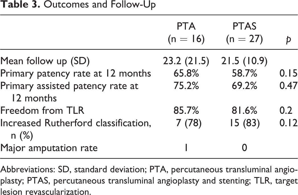
Technical success rate was 37% and 96.3% in the PTA and PTAS groups, respectively. There was no statistical difference in 12-month primary patency rate (65.8% versus 58.7%; p = 0.15) and primary assisted patency at 12 months (75.2 versus 69.2; p = 0.47) between the 2 groups. Freedom from target lesion revascularization at 12 months was not significantly different, with 85.7% and 81.6% (p = 0.2) in the PTA and PTAS groups, respectively. One amputation occurred in the PTA group.
Conclusion:
This small cohort suggests that stenting as a bailout procedure in CTO of the PA provides similar results to successful balloon angioplasty. Stenting should only be performed after suboptimal balloon angioplasty with vessel recoil. Due to the large lost to follow-up, strong evidence of a therapy over the other cannot be formulated. Larger studies with longer and stronger follow-up are needed to confirm those results.
Introduction
The advantages of endovascular therapy are well known, and its use as first-line therapy in obstructive femoropopliteal arterial lesions in patients with symptomatic peripheral arterial disease (PAD) is well established. 1 The popliteal artery (PA) is subject to high mechanical stress, significant bending, and foreshortening during knee movement. It remains a challenging area to treat endovascularly and generally considered a no-stenting zone. 2,3 However, in complex cases with suboptimal results such as significant recoil, flow-limiting dissection, or significant residual stenosis after balloon angioplasty alone, bailout stenting has to be considered. 4 Stenting of isolated popliteal arterial lesions can result in accelerated in-stent restenosis (ISR), stent fracture, and occlusion, limiting the endovascular approach when treating this arterial segment, 5 and patency rates are even worse in chronic total occlusion (CTO). 6 Despite the robust literature supporting the endovascular approach for treatment of femoropopliteal lesions, little data are available on endovascular treatment of isolated CTO in PA.
The purpose of this study was to compare angioplasty alone with satisfactory result versus bailout stenting after unsatisfactory balloon angioplasty of isolated CTO in popliteal lesions in symptomatic patients with PAD.
Materials and Methods
The present study complies with the principles outlined in the Declaration of Helsinki. The local ethics committee (B 403) of our institution approved this study.
Study Design and Patient Population
The studied participants came from a prospectively maintained database of our institution and were retrospectively analyzed. Between March 2012 and October 2016, 43 patients with symptomatic PAD with de novo isolated CTO of the PA were identified. Their clinical status affected their quality of life, despite exercise and optimal medical treatment. The exclusion criteria included arterial stenosis (≤99%), bypass occlusion, in-stent re-occlusion, absence of outflow vessels, popliteal entrapment syndrome, popliteal occluded aneurysms, and trauma injury. A diseased superficial femoral artery (SFA) was not an exclusion criteria as long as the popliteal lesion was not a continuous femoropopliteal lesion, with a lesion free zone >5 cm. All patients had at least 1 runoff vessel below the knee. The severity, location, and revascularization strategy of the popliteal lesions were assessed by computed tomography angiography (CTA) or magnetic resonance angiography in addition to duplex ultrasound (DUS).
Two groups of patients were defined. One group included all lesions treated successfully with percutaneous balloon angioplasty alone (PTA group; Figure 1). A second group included the lesions treated with bailout stenting (PTAS group) due to unsatisfactory angiographic result after balloon angioplasty alone (Figure 2).

(A) Angiogram illustration of a long (186 mm) popliteal (segment 1, 2, and 3) total occlusion and (B) the patent popliteal artery after percutaneous transluminal balloon angioplasty.

(A) Angiogram illustration of popliteal (segment 1) total occlusion, (B) suboptimal result after balloon angioplasty alone of the popliteal total occlusion, and (C) the patent popliteal artery after additional stenting of the popliteal artery.
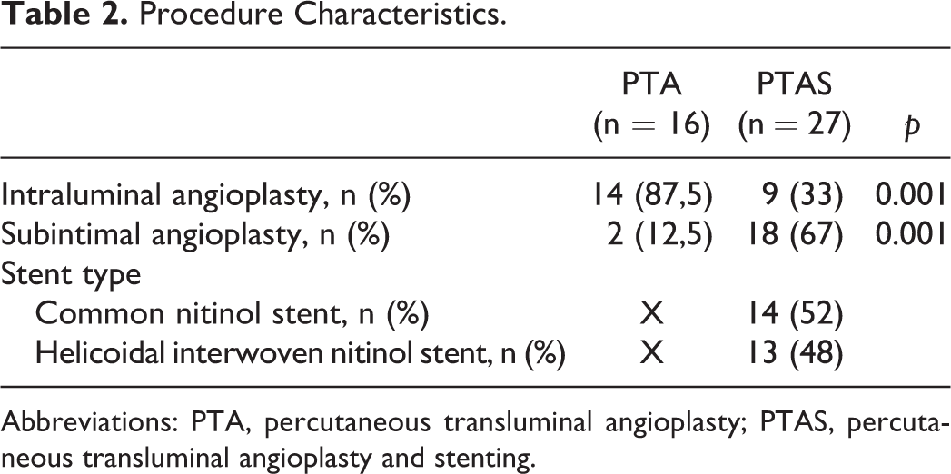
Patient demographics and lesions characteristics are summarized in Tables 1 and 2, respectively. All lesions were de novo PA lesions. There were no significant differences between groups regarding the degree of calcification (p = 0.09), lesion length (p = 0.14) and lesion localization (p = 0.16). Twenty-nine (67%) lesions were severely calcified and the mean lesion length was 81±44 mm. Twenty-eight (65%) patients were severe claudicants (Rutherford category 3), 8 (19%) patients presented ischemic rest pain (category 4), and 7 (16%) trophic lesions (category 5 and 6).
Patient and Lesion Characteristics.

Abbreviations: IQR, interquartile range; PTA, percutaneous transluminal angioplasty; PTAS, percutaneous transluminal angioplasty and stenting; SD, standard deviation.
Procedure Characteristics.
Abbreviations: PTA, percutaneous transluminal angioplasty; PTAS, percutaneous transluminal angioplasty and stenting.
Study Treatment
In de novo lesions, we opt for an endovascular approach at first intention. All patients received acetylsalicylic acid (ASA 80 mg/d) prior to the intervention. A digital subtraction angiography of the target lesion area was performed at the time of intervention to confirm the lesion (Figure 1A and 2A). Intraluminal recanalization was attempted primarily with conversion to the subintimal approach if this failed; no distal retrograde approaches were needed. No adjunctive debulking techniques such as mechanical or laser atherectomy were used. Dilatation with a standard uncoated balloon was performed with 4- to 7-mm balloons, the same size depending on the reference artery diameter. Balloon inflation times varied from 60 to 180 seconds at a pressure from 8 to 12 atm. The balloon selection was left to the discretion of the surgeon; balloon catheter used included the Passeo (Biotronik AG, Buelach, Switzerland), Sterling (Boston Scientific, Natick, Massachusetts), and Advance (Cook Medical, Bloomington, Indiana). Repeated prolonged dilatation for at least 120 seconds or stenting of the lesion was performed if flow-limiting dissection or vessel recoil responsible for a residual stenosis >50% occurred. The used stents were also left to the discretion of the surgeon and included common nitinol stents in 14 (52%) and interwoven stents in 13 (48%) cases.
Dual antiplatelet therapy with ASA 80 mg/d and clopidogrel 75 mg/d was prescribed for 2 months, followed by ASA monotherapy lifelong. Patients previously on oral anticoagulant or new oral anticoagulants (NOACs) were maintained with additional clopidogrel for 1 month, followed by a switch to ASA lifelong. Triple therapy (ASA, clopidogrel, and oral anticoagulant or NOAC) was not favored.
Follow-Up Protocol
Follow-up consisted of a clinical evaluation at baseline and day 1 after the endovascular procedure and both clinical and duplex evaluation to assess the patency at 2 months and annually thereafter or in case of clinical worsening.
Endpoints and Definitions
The primary endpoint of this study was to evaluate the primary patency in both groups, defined as freedom from significant restenosis (>50%) or occlusion based on DUS evaluation in the absence of target lesion revascularization (TLR), bypass of the target lesion, or major amputation of the target limb. Secondary endpoints were technical success, clinical success, primary assisted patency, and freedom of TLR.
Chronic total occlusion was defined and diagnosed when no intraluminal antegrade flow was observed in the angiogram (Figure 1A). Technical success was defined as a residual stenosis <30% in the absence of flow-limiting dissections and arterial perforation of the treated segment (Figure 1B and 2C). Clinical success was defined as improvement of Rutherford classification of 1 class or more and a subjective improvement of the quality of life reported by the patient. Significant restenosis was indicated by a peak systolic velocity ratio ≥2.5 on DUS. Primary assisted patency was defined as freedom from significant restenosis after endovascular redilation in the treated segment after restenosis (>50%) or occlusion.
The degree of calcification was graded on the basis of arterial wall calcium deposits on preoperative CTA or preoperative angiography. Mild calcification referred to calcifications <1 cm long on ≤50% of the arterial wall. Severe calcification referred to calcifications longer than 1 cm or >50% of the arterial wall. 7
The PA was divided into 3 anatomical segments: P1, from the intercondylar fossa to the proximal edge of patella; P2, between the proximal part of the patella and the center of the knee joint space; and P3, between the knee joint space and the origin of the anterior tibial artery.
Statistical Analysis
Continuous variables are presented as means (standard deviation, SD) for normally distributed variables or median with interquartile range for nonnormal variables, while categorical data are given as the counts (percentages). Continuous variables were compared with the unpaired Student t-test, while categorical variables by means of the χ2 test. Cumulative primary and primary assisted patency rates, as well as freedom from TLR, were estimated using the Kaplan-Meier method. Differences in time to event were compared with the log-rank test. The threshold of statistical significance was p < 0.05 2-tailed. Analyses were performed using STATA 11.2 (StataCorp, College Station, Texas).
Results
Between March 2012 and October 2016, 43 (26 males) patients with isolated CTO of PA underwent endovascular recanalization and balloon angioplasty alone (n = 16) or bailout stenting (n = 27). Patient and lesion characteristics are listed in Table 1. There was no statistically significant difference between both groups concerning the patient demographics (age, gender, and comorbidities) and lesion characteristics (calcification severity, lesion length, and lesion localization). The majority of patients in both groups presented with lifestyle-limiting claudication, 60% versus 70%, respectively. The median lesion lengths were 67 mm (39.5-78.5 mm) and 94 mm (50-114 mm) in the angioplasty group and in the stent group, respectively (p = 0.14).
Initial Outcomes
Technical success rate was 37% (16 lesions) in the angioplasty group and bailout stenting was needed in the 27 remaining lesions. In the stent group, technical success was 96.3%, with 1 (3.7%) residual stenosis of approximately 40% despite aggressive postdilatation up to 14 atm for 3 minutes. There never was a second stent deployment to cover the same lesion. Distal embolization or vessel perforation was not observed in any cases. Fifteen common nitinol and 13 helicoidal interwoven nitinol stent were deployed in the stent group. Vessel intraluminal recanalization was statistically significantly different between both groups, with 14 (87.5%) intraluminal recanalization in the angioplasty group versus 9 (33%) in the stent group (p = 0.001).
Outcomes and Follow-Up
The mean follow-ups were similar in both groups, with 23.2 (21.5) months and 21.5 (10.9) months in the angioplasty group and stent group, respectively (Table 3). Seven (43.8%) patients were lost to echographical follow up in the angioplasty group and 8 (29.6%) in the stent group. The 12-month primary patency rate was not significantly different (p = 0.15) between the angioplasty group (65.8%, 95% confidence interval [CI]: 23.8-86.6) and the stent group (58.7%, 95% CI: 32.5-67.4), as well as primary assisted patency at 12 months (75.2% vs 69.2%; p = 0.47). Freedom from TLR at 12 months was not significantly different (p = 0.2), with 85.7% (95% CI: 33.4-97.9) and 81.6% (95% CI: 58.0-92.7) in the angioplasty and stent groups, respectively. No increase in the Rutherford classification in any patient was noticed during follow-up; even in case of loss of patency, the Rutherford classification remained at the initial level.
Outcomes and Follow-Up
Abbreviations: SD, standard deviation; PTA, percutaneous transluminal angioplasty; PTAS, percutaneous transluminal angioplasty and stenting; TLR, target lesion revascularization.
In the angioplasty alone group, 2 TLRs were performed at 8 months; the restenosis were treated with a nitinol interwoven stent (Supera; Abbott Vascular, Santa Clara, California). One additional clinically driven TLR was performed at 5 months, due to target vessel occlusion. The patient suffered from acute limb ischemia finally leading to major amputation. The overall mortality in the angioplasty alone group was 19% (3 patients); none of them was associated with the endovascular procedure and happened after 30 days.
In the bailout stent group, 6 stent occlusions and 3 ISR occurred during follow-up, resulting in rest pain in all cases. Treatment could be achieved by 6 endovascular revascularizations (4 occlusions and 2 ISR), with 5 drug coated balloons and 1 stent in stent placement. In 2 occluded cases, bypass surgery was needed. One patient with an ISR received best medical treatment with good outcome. Limb salvage could be obtained in all cases. The overall mortality in the stent group was 11% (3 patients) and no deaths were associated with the endovascular procedure and happened after 30 days.
A subanalysis revealed no statistical differences (p = 0.9) in 12 months primary patency rate between interwoven and common nitinol stents. Kaplan-Meier curve estimates for primary and primary assisted patency rates for both groups (PTA and PTAS) during follow-up are depicted in Figure 3A and B.

Primary patency rate (A) and primary assisted patency rate (B) at 12 and 24 months for the percutaneous transluminal angioplasty group (PTA) versus the percutaneous transluminal angioplasty and stenting (PTAS) group (P = .6 and P = .47).
Discussion
Focal total occlusive atherosclerotic lesions of the PA are less common manifestation of PAD. Both PA and SFA lesions are often studied together as femoropopliteal disease. 8,9 Randomized trials have shown that nitinol stent placement is superior to plain balloon angioplasty alone in terms of TLR and clinical improvement in patients with occlusive disease in the femoropopliteal segment. 10,11 Nevertheless, the axial shortening, elongation, and bending to which both vessels are exposed vary according to their specific anatomical location. 2 Hence, it makes sense to study the PA distinctly from the SFA. Only a few studies assessed endovascular treatment results exclusively for atherosclerotic occlusive disease in the PA. 12,13 The endovascular treatment of isolated chronic total occluded PA is a paradox and challenging problem; on the one hand, total occluded lesions are more prone to be stented after balloon angioplasty due to higher risk of vessel recoil and residual restenosis. But, on the other hand, repetitive flexion and extension of the knee create high mechanical stress and stent compression in the PA, inducing higher degree of stent fracture and ISR in this segment. 3,14
Stenting of the PA has recently been reconsidered with the introduction of the Supera interwoven nitinol Stent System (Abbott Vascular). The interwoven stent design confers superior radial strength, multidimensional flexibility, durability, and vessel conformability compared to traditional nitinol tube stents, making this stent ideal for treatment of complex atherosclerotic PA disease. 15 Scheinert et al investigated 101 patients with atherosclerotic popliteal lesions and 48 (47.5%) lesions were total occlusions, treated with Supera stent. No stent fracture was observed and primary patency rate at 12 months was 87.7% compared to 58.7% in the present study. 15,16 Shorter lesions, lower calcification degree, and the Supera stent design could explain these differences in results. Despite this innovative stent design, Cambiaghi et al reported in 2017 a single case of an interwoven nitinol stent fracture with lost in patency following treatment of a popliteal arterial stenosis. 17 This last report suggests that even with new stent technologies, the isolated PA may still be problematic.
In our cohort, both groups were statistically comparable in terms of patient and lesion characteristics (p ≥ 0.05). Treating popliteal lesions for claudication could be questioned, but was performed only in cases where the lesion did not exceed P2, preserving the possibility for future bypass surgery if the endovascular therapy failed or symptoms worsened. The 12-month primary patency rate, primary assisted patency, and freedom from TLR were satisfactory in view of the complexity of the lesions and, when comparing the 2 groups, did not reach statistical significance. To our knowledge, no other study assessed endovascular treatment of isolated atherosclerotic CTOs of the PA. The ETAP trial, published in 2013 and 2015, comparing balloon angioplasty versus primary stenting in isolated popliteal lesions, included 81 total occluded lesions (32.9%), but no separate analysis of this subgroup was performed. 18,13 At 12 months, the ETAP trial showed that primary stenting achieves inferior technical success and lower primary patency compared to balloon angioplasty with bailout stenting in isolated popliteal lesions if bailout stenting during the index procedure was not considered as TLR and loss of patency. 18 In comparison with the ETAP trial, our study showed similar primary patency rates at 12 months in the stent and angioplasty groups.
Our data suggest that angioplasty alone in isolated CTO of the PA has comparable results to bailout stenting only if the angiographic control image after dilatation is satisfactory. In the stent group, those results were slightly lower, however, Kaplan-Meier analysis did not find a statistically significant difference between the 2 treatment strategies.
Limitation
First, the number of included patients in this study is relatively small, which prevents us from making broader conclusions. Second, this is a retrospective study and lacks randomization, but makes it possible to study PTA alone as treatment of the lesions without bailout stenting. Finally, this analysis is limited to current technology, which is rapidly changing. Specifically, we believe that the comparison of PTA alone versus stenting with BMS in CTO cases will soon permute to drug-coated balloon versus drug eluting stent with the new era of drug technologies implemented on balloons and stents.
Conclusion
Endovascular treatment of total occluded popliteal lesions is a challenging problem but seems to be safe and relatively effective with satisfactory revascularization, technical, and clinical results. Stenting as a bailout procedure in isolated CTO of the PA may provide acceptable results, similar to those of a successful balloon angioplasty. Stenting should only be performed in this no stent zone when the per-procedural results of balloon angioplasty are suboptimal. A greater cohort with stronger follow-up could significantly favor a group to another.
Footnotes
Declaration of Conflicting Interests
The author(s) declared no potential conflicts of interest with respect to the research, authorship, and/or publication of this article.
Funding
The author(s) received no financial support for the research, authorship, and/or publication of this article.
