
Abstract
Purpose:
Ultrasound-facilitated, catheter-directed, low-dose fibrinolysis (USAT) appears to provide promising results for the management of acute submassive pulmonary embolisms (ASMPEs) at tertiary care centers. This study assessed outcome measures at a community-based hospital systems and compared results to known studies.
Materials and Methods:
This is a single-center, retrospective study assessing clinical outcomes of the EkoSonic Endovascular System intervention for ASMPEs performed by three surgical 3 subspecialties (interventional radiology, interventional cardiology, and vascular surgery) part of a pulmonary embolism response team (PERT). We reviewed 146 PERT activations from June 2013 to December 2017. Eighty-three patients with ASMPEs underwent USAT.
Results:
Our study showed greater differences (P = .01) between baseline and follow-up pulmonary artery systolic pressures (20.9 ± 9.8 mm Hg [n = 14]) compared to the ULTIMA study (12.3 ± 10 mm Hg [n = 30]). Our length-of-stay measures were shorter (6.1 ± 5.1 [n = 83]; P = .0001) compared to the SEATTLE II study (8.8 ± 5.0 [n = 150]). Preprocedure transthoracic echocardiograms (TTEs) were performed for 54 (65%) of 83 patients. Postprocedure TTEs at 48 hours was performed for 52 (62%) of 83 patients. Use of TTEs before and after intervention did not change outcomes. Intracranial hemorrhage was not observed in our patient population. There was no difference in outcomes between the three subspecialties in our study.
Conclusions:
Use of USAT in a community-based hospital PERT has similar outcomes to tertiary care centers. Furthermore, similar outcomes were observed between the three subspecialties suggesting development of a comprehensive care team for management of ASMPEs.
Introduction
With over 275 000 hospitalizations a year, pulmonary embolisms (PEs) are the third major cause of cardiovascular morbidity and mortality, behind myocardial infarction and stroke. 1 -6 PE management is based on classification into three categories: low risk (nonmassive), intermediate (submassive), and high risk (massive). Consensus exists regarding the management of low- and high-risk PEs in the medical community, heparin and systemic fibrinolysis, respectively. There is no accepted method of management for submassive PEs. The parameters for a submassive PE include hemodynamic stability with evidence of right ventricular (RV) dysfunction shown by elevated cardiac biomarkers and/or RV strain (RV:left ventricle (LV) strain > 0.9). 3,7,8 Substantial literature has demonstrated that while systemic fibrinolysis is effective in clot resolution for submassive PEs, there is an undesirable risk of an intracranial hemorrhage. 1,4,8,9,10
The use of catheter-directed therapies (CDTs) for the management of submassive PEs has gained widespread popularity due to its ability to resolve clots with local application of fibrinolytics. 10,11,12 CDTs have demonstrated improved RV dysfunction, decreased pulmonary artery pressures, and reduced risks of major bleeds. 1,4,13,14,15,16 The recent development of ultrasound-assisted, low-dose fibrinolysis therapy (USAT), a variation of CDT, has provided another treatment modality for the management of acute submassive PEs (ASMPEs). 17 USAT incorporates ultrasound-generated acoustic energy to facilitate disruption of the PE allowing for improved fibrinolytic penetration. 16 Two landmark studies, SEATTLE II and ULTIMA, demonstrated the efficacy and safety of USAT for ASMPEs while maintaining low rates of bleeding complications. 8,9 Another study, OPTALYSE PE, determined the lowest optimal tissue plasminogen activator (tPA) dose and delivery duration using USAT for the treatment of ASMPEs. It showed improved RV function and reduced clot burden compared to baseline. 18 Another treatment intervention for ASMPEs includes catheter-directed thrombectomy. A recent prospective study evaluated the safety and efficacy of this intervention using the FlowTriever System (Inari Medical, Irvine, California) and observed significant improvement in RV:LV strain and few major bleeding events. Of note, this intervention did not use fibrinolytics. 19
USAT therapy appears to provide promising results for the management of ASMPEs; however, the generalizability of the SEATTLE II and ULTIMA studies remains uncertain. The clearance of USAT by the Food and Drug Administration (FDA) was based on these two landmark studies that consisted of a total of 209 patients, conducted at tertiary care centers, and had limitations of lacking a comparator group and selection bias. Furthermore, there is a paucity of data regarding USAT performance in community-based hospitals. The purpose of this study is two-fold. The first objective is to assess the performance of USAT at a large academic community hospital and compare findings to the SEATTLE II and ULTIMA studies. Second, we performed an analysis of the three surgical subspecialties (interventional cardiology, interventional radiology [IR], and vascular surgery) using USAT and compared outcome measures.
Methods and Materials
Study Design
This is a retrospective study done at Ascension Providence Health System (APHS), a community hospital, in the Detroit Metropolitan area. From June 2013 to December 2017, we reviewed the electronic medical records of patients who presented to the emergency department with a PE and required activation of Ascenion Providence's Pulmonary Embolism Response Team (PERT). Only data for massive and submassive PEs were collected. At APHS, management of ASMPEs with USAT (EkoSonic Endovascular System [EKOS], Bothell, Washington) is performed by vascular surgery, IR, and interventional cardiology. Data were collected for all three subspecialty groups. The study received institutional review board approval on May 1, 2017, from APHS.
Study Population
Patient eligibility for the study consisted of the presence of a submassive or massive PE on computed tomography (CT) showing greater than 50% of main pulmonary artery involvement or lobar thrombus burden with presence of RV dysfunction as identified on CT or TTE. Symptoms of PE needed to be within 14 days of onset. Criteria for acute submassive PE patients included: RV dysfunction (RV:LV >0.9 on CT or TTE) and absence of hemodynamic instability. Criteria for massive PE included one or more of the following: systemic arterial hypotension, cardiogenic shock, cardiopulmonary resuscitation, pulseless electrical activity, or requirement of vasopressor support. The on-call specialist deetermined the need to perform USAT. Not all patients who presented with massive or submassive PEs received USAT catheter placement.
Anticoagulation
Anticoagulation with unfractionated heparin (UFH) was initiated upon identification of PE on CT or high clinical suspicion on ventilation–perfusion (V/Q) scans. Target activated partial thromboplastin time (aPTT) of 60 to 80 seconds was attempted prior to initiation of USAT. A reduced aPTT of 40 to 60 seconds was targeted during USAT. Prior to removal of access site sheaths, systemic anticoagulation was held for 2 hours and an aPTT level was obtained. Following completion of USAT, all patients were restarted on therapeutic anticoagulation 2 hours after removal of catheter sheaths. Most patients were transitioned to oral anticoagulation within 48 hours of USAT completion. Instances when patients were not transitioned to oral anticoagulation included contraindications due to bleeding complications or in the setting of malignancy. The decision of oral anticoagulant agent was left to the discretion of the treating physician and patient.
Ultrasound-Assisted, Low-Dose Fibrinolysis Therapy Intervention (EKOS)
The EKOS ultrasound-accelerated infusion catheters were placed in all patients through the femoral vein in the cardiac catheterization laboratory, operating room, or IR suites; the location depended on the performing specialty. Bilateral or unilateral EKOS catheters were placed at the discretion of the specialist in the treatment area as indicated on CT angiography (CTA). In most cases, the catheters were placed in the main, lateral, and basal segmental arteries. The fixed-dose regimen of tPA was administered with a total treatment dose of 24 mg. If patients had bilateral catheters, the infusion was given at 1=mg/h/catheter for a total of 12 hours or at a rate of 1 mg/h in unilateral catheters for a duration of 24 hours. When bilateral catheters were placed, most patients had insertion of two 6F microaccess catheter sheaths in the femoral vein. Prior to placement of EKOS, preintervention pulmonary artery pressures were attempted to be measured and recorded. During alteplase infusion, patients received intravenous UFH for a goal aPTT of 40 to 60 seconds. After the treatment period, pulmonary artery pressures were again transduced through the infusion catheters before removal (like preintervention measurements, not all postintervention pressures were measured). After removal of the EKOS catheters, all patients received intravenous UFH for a goal aPTT of 60 to 80 seconds followed by transition to a long-term anticoagulation therapy as agreed upon by the patient and treating physicians. Bleeding complications were assessed in all patients until time of hospital discharge.
Data Collection
Data was obtained for all patients presenting with massive and submassive PEs for all three subspecialties. Data was compiled through database searches of the hospital electronic medical record system. Data was deidentified, stored, and password protected on computers at the hospital. Only patients receiving USAT intervention for the management of ASMPEs are reported in this study. Variables collected were pulmonary artery (PA) pressure prior to USAT removal, on-call proceduralist, performing subspecialty (interventional cardiology, vascular surgery, and IR), PERT activator, presence of sheath used with heparin, timing following USAT removal to initiation of anticoagulation (hours), presence of inferior vena cava (IVC) filter placement, oxygen discharge status, and anticoagulation discharge medication (dabigatran, apixaban, rivaroxaban, enoxaparin, warfarin, and no anticoagulation). Comorbidities and past medical history of interest consisted of venous thromboembolism/deep vein thrombosis (VTE/DVT), PE, no history of DVT/PE, obstructive sleep apnea, hypertension, myocardial infarction, congestive heart failure, peripheral vascular disease, chronic lung disease, connective tissue disease, diabetes mellitus, chronic kidney disease, and malignancy.
Outcomes
The primary outcomes were all-cause mortality and length of hospital stay (LOS) in patients who had USAT intervention. Secondary outcomes included all-cause complications, need for oxygen on discharge, change in PA pressure following USAT, bleeding events, change in RV systolic pressure on TTE, and rate of IVC filter placement.
Procedural and safety outcomes
The procedural measures were duration between CT scan and intervention (hours), fluoroscopy time (in minutes), and duration between intervention completion and anticoagulation (hours). Safety measures included average LOS, in-hospital deaths, nonlethal complications (bleeding), and number of IVC filters placed (Table 1).
USAT Outcomes Stratified by Specialty at APHS.
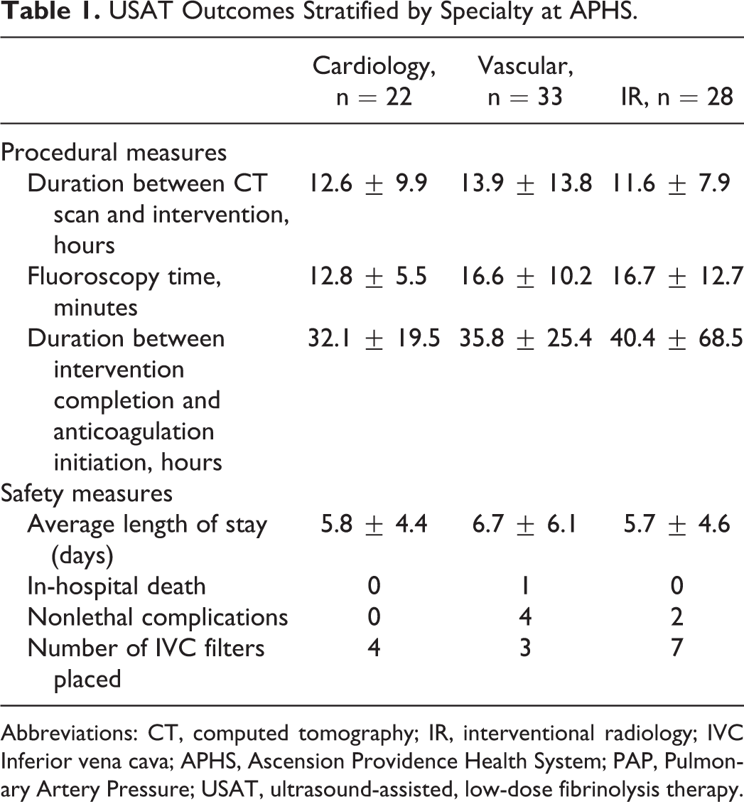
Abbreviations: CT, computed tomography; IR, interventional radiology; IVC Inferior vena cava; APHS, Ascension Providence Health System; PAP, Pulmonary Artery Pressure; USAT, ultrasound-assisted, low-dose fibrinolysis therapy.
Statistical Analysis
Analysis consisted of only ASMPEs in the study population. For comparisons of continuous data, Student t tests were performed. For categorical and proportional data, Fisher exact tests were completed.
Results
Patient Demographics
A total of 83 patients with ASMPEs received USAT intervention during the study period. The average age of the patient was 59.9 ± 13.5 years, and 42 patients were female. The average Pulmonary Embolism Severity Index score for the cohort was 3.0 ± 1.3 with a mean baseline RV:LV strain of 1.5. Baseline comorbidities ranged in prevalence with the two most common being hypertension (57.8%) and diabetes mellitus (31.3%). With the exception of peripheral vascular disease (P = .02), no significant differences in baseline demographics or comorbidities were observed between our study population and identical variables in the ULTIMA or SEATTLE II treatment groups (Table 2).
Baseline Demographics and Comorbidities of Study Population.
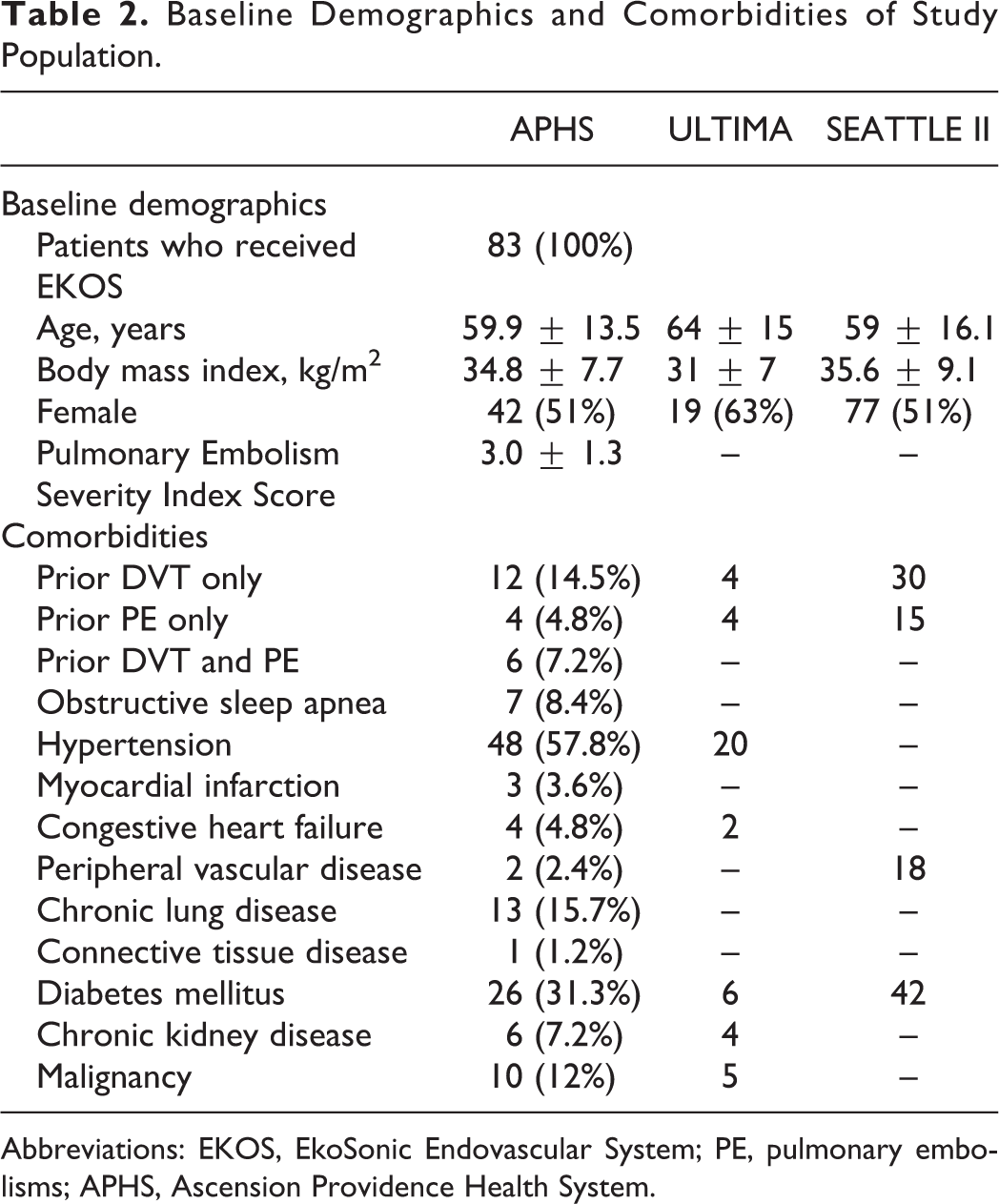
Abbreviations: EKOS, EkoSonic Endovascular System; PE, pulmonary embolisms; APHS, Ascension Providence Health System.
Primary Outcomes
At APHS, the average LOS for patients with ASMPE treated with EKOS (6.1 ± 5.1 days [n = 83]) was shorter compared to SEATTLE II (8.8 ± 5 days [n = 150], confidence interval [CI]: 1.343-4.057, P = .0001). No statistical differences were observed between in-hospital deaths and nonlethal adverse events. Fewer IVC filters were placed at APHS compared to SEATTLE II; however, this was not statistically significant (Table 3). At APHS, we observed a change in PA pressure of 20.9 ± 9.8 mm Hg (n = 14) after intervention which was greater compared to the ULTIMA (12.3 ± 10.0 mm Hg [n = 25], CI: 1.88-15.32, P = 001) and SEATTLE II (14.4 ± 15.4 [n = 147], no significant difference) studies. We did not measure the change in RV:LV strain after intervention. Of note, seven patients treated for ASMPEs with EKOS were discharged with oxygen, compared to 11 ASMPE patients discharged with oxygen who were not treated with EKOS. 31 patients presented with evidence of pulmonary infarction on CTA. Of those 31 patients, nine underwent EKOS with no complications.
EKOS Outcomes at Ascension, SEATTLE II and ULTIMA.

Abbreviations: IVC, inferior vena cava; LV, left ventricle; PA, pulmonary artery; RV, right ventricular; RVSP, right ventricular systolic pressure; APHS, Ascension Providence Health System.
a ULTIMA measured PA pressures within 18 hours of initiation of therapy, SEATTLE II measured pressures at 48 hours, and APHS measured pressures at 48 hours.
b Follow-up hemodynamic data were obtained at 48 hours.
c Follow-up hemodynamic data were obtained at 18 ± 3 hours after initiation of treatment.
d Significant at the .05 probability level when compared to APHS.
e APHS—Bleeding events in hospital by Global Utilization of Streptokinase and Tissue Plasminogen Activator for Occluded Coronary Arteries (GUSTO) criteria: minor (4), major (1). SEATTLE II—Bleeding events within 30 days by GUSTO criteria: severe (1), major (15). ULTIMA—Bleeding events within 90 days by study specific criteria: minor (3).
Analysis of Subspecialties and EKOS Performance
For all variables of interest, no statistically significant differences were observed between the subspecialties (Table 3). Both IR and vascular used more fluoroscopy time compared to cardiologists. The greatest duration between the completion of USAT and initiation of anticoagulation was seen with IR (40.4 ± 1.1 hours). The longest LOS was seen among vascular specialists at 6.7 ± 6.1 days. The most IVC filters were placed by IR, while the most complications were seen with vascular surgeons (Table 1). All of the subspecialties using USAT at APHS did not consistently measure pressures before or after intervention to assess objective improvement in patients (Table 4). No patient in our study underwent follow-up contrast-enhanced chest CT to assess PE clot resolution. TTEs were performed at 48 hours in 66% (n = 54) of patients. Of those performed, 44% (n = 24) failed to report data of right heart hemodynamics on TTE.
Completion of RVSP and PAP Measurements Before and After EKOS in Patients With Acute Submassive PEs (n = 80)a at APHS.
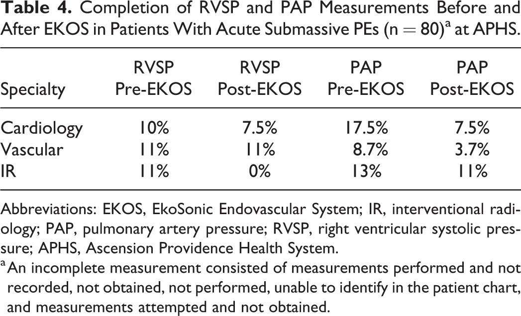
Abbreviations: EKOS, EkoSonic Endovascular System; IR, interventional radiology; PAP, pulmonary artery pressure; RVSP, right ventricular systolic pressure; APHS, Ascension Providence Health System.
a An incomplete measurement consisted of measurements performed and not recorded, not obtained, not performed, unable to identify in the patient chart, and measurements attempted and not obtained.
Complications Associated With EKOS
Five bleeding complications and one death were observed in our study. Four of the bleeding events were minor (Global Utilization of Streptokinase and Tissue Plasminogen Activator for Occluded Coronary Arteries criteria), while one was major. The one major bleeding event was a spinal hematoma which we attributed to a recent laminectomy surgery coupled with anticoagulation for PE resolution. The one death observed was due to a patent foramen ovale (PFO) that was not known to the patient or the performing specialist. The patient had significant clot burden in which a potential thromboembolism passed through the unknown PFO and resulted in a cerebrovascular accident.
Discussion
We conducted a retrospective review of 83 patients treated with EKOS for ASMPEs at an academic community-based hospital. We compared similar outcomes to studies conducted at larger tertiary care centers. We observed drops in PA pressures, hospital LOS, and an overall low complication rate. Furthermore, performance of EKOS did not vary significantly between various subspecialties. Taken together, our study suggests that EKOS can be adequately performed at a community-based hospital.
The USAT has serious implications for patient outcomes. Improved cardiac metrics following USAT can facilitate overall well-being and quality of life while avoiding the potentially devastating complications of systemic fibrinolysis. When combined with a PERT, EKOS can enable a fast and effective intervention for ASMPEs. While not the focus of our study, a key finding is that even in the absence of following the FDA-certified rules outlined in the ULTIMA and SEATTLE II studies, improvements were still observed in patients with ASMPEs. Part of implementing EKOS is performing pre- and post-TTEs; however, this is not routinely done by the subspecialties included in this study. To date, this appears to not have any detriment to the patient.
The management of ASMPEs is still controversial. The evidence behind EKOS is limited but growing. Many published studies have small sample sizes or limited follow-up times. Nonetheless, outcomes tend to be consistent in showing that EKOS has a positive impact for the patient in reducing pulmonary pressures and improving the underlying pathophysiology. With respect to physicians, this new technology might be an ideal balance between physical disruption of the clot and use of fibrinolytics. With further use of USAT, more data will be available for better assessment of the intervention.
This study is not without limitations. Being a retrospective study, not all data were complete. Furthermore, there was no control group, blinding, or randomization. Subanalysis based on subspecialties resulted in small sample sizes and these findings should be interpreted with caution. Additionally, the SEATTLE II study included submassive and massive patients, while our study and the ULTIMA study only consisted of submassive patients.
In conclusion, USAT intervention for the management of ASMPEs can successfully be performed in large academic community-based hospital centers. The development of a standardized USAT protocol still needs to be optimized, as this has implications for hospital LOS, health-care costs, and overall clinical outcomes. 20,21 Relying on ≥2 specialties did not sacrifice performance outcomes for USAT and can allow for a comprehensive management team that shares the responsibility of care. Lastly, additional prospective studies are needed for USAT in ASMPEs, especially in the setting of community-based hospital centers.
Footnotes
Acknowledgment
Thanks to Prahbhat Sinha, DO, Ascension Providence-Providence Park Hospital.
Declaration of Conflicting Interests
The author(s) declared no potential conflicts of interest with respect to the research, authorship, and/or publication of this article.
Funding
The author(s) received no financial support for the research, authorship, and/or publication of this article.