
Abstract
Retroperitoneal haemorrhage is a rare but potentially life-threatening event. It may occur either spontaneously or secondary to percutaneous vascular access procedures, trauma, or ruptured aortic, iliac, or mesenteric aneurysms. As a result, the clinical presentation is variable. Computed tomography and/or angiography are vital for diagnosis. Management may range from conservative treatment for stable patients to emergency laparotomy or embolization for catastrophic haemorrhage. Direct percutaneous puncture of a deep intra-abdominal pseudoaneurysm is an accepted but infrequently performed technique due to a number of diagnostic and technical challenges. We describe the successful percutaneous transabdominal angioembolization of a superior mesenteric artery rupture in a 77-year-old woman with a large retroperitoneal haematoma. This was performed after a conventional femoral transarterial approach was unsuccessful.
Case Report
A 77-year-old woman was admitted to a regional base hospital with a 2-day history of abdominal pain and vomiting. Her past history included partial paraplegia, congestive cardiac failure, and hypertension. There was no precipitating event or traumatic cause of her abdominal pain to the authors’ knowledge.
Laboratory tests revealed a severe metabolic alkalosis requiring aggressive electrolyte correction in the intensive care unit (ICU). An abdominal computed tomography (CT) scan showed a large retroperitoneal haematoma causing gastric outlet obstruction. A CT mesenteric angiography was then arranged. This showed active contrast extravasation from a branch of the superior mesenteric artery (SMA) and thus transferred to our facility for emergency radiological intervention (Figure 1A).
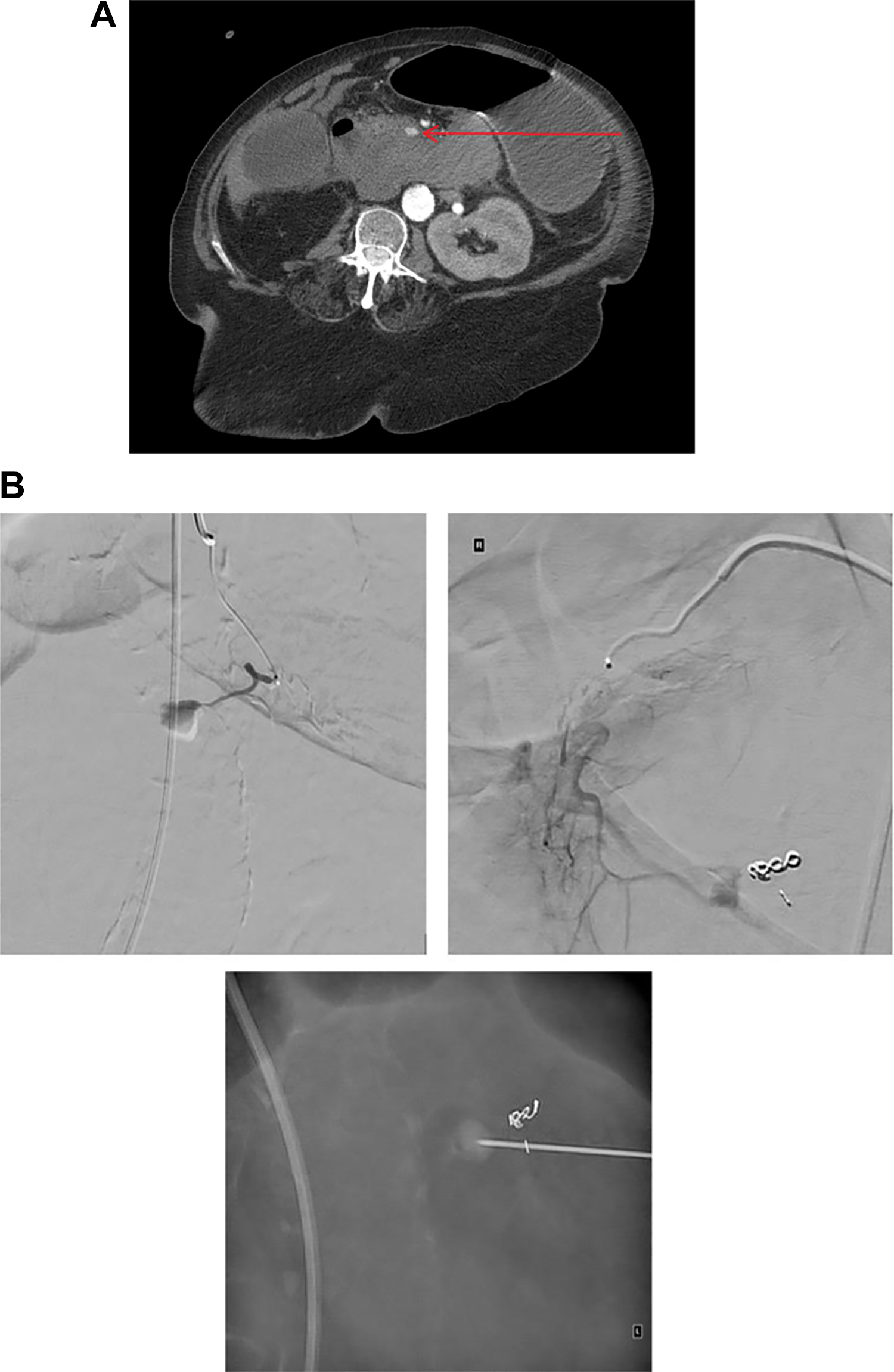
(A) Original computed tomography angiogram showing arterial “blush.” (B) Sequence of angioembolization procedure. 1
Embolization was initially attempted via a femoral approach under general anaesthesia. Under ultrasound guidance, the right common femoral artery was punctured. Coeliac angiography demonstrated a pseudoaneurysm arising from a proximal branch of the SMA. An attempt was made to cannulate the feeding vessel with a microcatheter; however, the position was difficult to maintain.
A Simmons 3 catheter was then used within the SMA for support and a microcatheter was used to cannulate the branch of the SMA that supplied the pseudoaneurysm. This vessel was embolized with a 2-mm straight coil followed by 3 and 2 mm Boston Scientific (Marlborough, Massachusetts)Vert X coils with good occlusion. Repeated coeliac angiography during the embolization demonstrated that there was a further supply from a branch of the gastroduodenal artery.
A 6F up-and-over sheath was used for stability, and a microcatheter was used to attempt to cannulate the branch of the gastroduodenal artery that supplied the pseudoaneurysm. Despite multiple attempts and guide wires, the microcatheter could not be advanced into this vessel.
Angiography from a more proximal position of the microcatheter opacified the pseudoaneurysm. This was used to target the pseudoaneurysm percutaneously using a transabdominal approach. Direct puncture of the pseudoaneurysm was thus performed using a 19-G needle under fluoroscopic guidance (Figure 1B). Multiple projections during injection of contrast to opacify the pseudoaneurysm confirmed the needle tip position. Injection of contrast through the 19-G needle opacified the pseudoaneurysm, which was then directly embolized with Histoacryl and Lipiodol in a ratio of 1:3. In total, 1.5 mL was injected with occlusion of the pseudoaneurysm. Postintervention scans showed complete occlusion.
Following the procedure, the patient was transferred to ICU. The gastric outlet obstruction of the patient was managed with regular nasogastric aspirates and aggressive electrolyte replacement with good clinical response. The patient was successfully transferred 5 days later to a regional hospital for further care.
Discussion
Retroperitoneal haemorrhage is a rare but potentially life-threatening event with an incidence ranging from 0.1% to 0.6% in patients on oral anticoagulation. 2 Clinical presentation is often subtle and varied. Patients with frank retroperitoneal haemorrhage may initially only show very subtle clinical signs, such as tachycardia with hypotension that is only transiently fluid responsive. 2 Single spiral CT and multidetector-row CT provide valuable information on the type, site, extent, and management of retroperitoneal fluid collections and therefore play an important role in diagnosis. 3 Computed tomography angiography may identify a bleeding site and extravasation of contrast, often warranting urgent intervention.
The management of retroperitoneal haematoma is variable and lacks definitive guidelines. Regardless of the aetiology of retroperitoneal haemorrhage, all patients should be monitored in an intensive care or high-dependency setting with close monitoring, intravascular volume resuscitation, blood transfusion, and correction of coagulopathy. 2 Stable patients may often be managed conservatively in this setting, unless the discovery of a focal source of bleeding mandates intervention (eg, a “blush” on CT angiography which may be amenable to embolization).
Direct percutaneous puncture of an intrabdominal pseudoaneurysm is an accepted but infrequently performed technique often due to the difficulty in visualizing the source of haemorrhage. A number of case reports describe percutaneous thrombin injections of splanchnic or splenic artery aneurysms and direct needle puncture with transcatheter N-butyl cyanoacrylate injections of aneurysms of the hepatobiliary system and gastrointestinal (GI) tract. 4 -6 Injection of thrombin into pseudoaneurysms in the groin (usually a complication of femoral arterial punctures) is commonly performed because visualization with ultrasound is possible. In this case, the pseudoaneurysm was visualized as the lesion could be opacified and therefore targeted fluoroscopically.
Percutaneous transabdominal approaches to embolization are uncommonly performed, with Boks et al first describing this technique in 2005. 7 Several studies have found this to be a safe and efficacious approach, including one retrospective study of 30 patients who underwent 33 transabdominal direct sac puncture embolization procedures for type II endoleaks after endovascular aortic aneurysm repair. 8,9
Another retrospective review of 23 patients who underwent 35 embolizations found that transabdominal or translumbar direct sac puncture versus transarterial embolization of type II endoleak with aneurysm sac obliteration were similarly effective for the prevention of aneurysm sac growth, with direct sac puncture yielding significantly shorter fluoroscopic and procedural times. 10
Conclusion
Transarterial embolization via the femoral approach is an accepted standard in the radiological approach to GI tract bleeding, such as from gastroduodenal, left gastric, or splenic arteries. To the best of the authors’ knowledge, there are only few reports of a transabdominal approach in the literature. Our case report supports that when conventional treatment fails, a percutaneous transabdominal approach to SMA haemorrhage can be lifesaving.
Footnotes
Authors’ Note
Verbal consent for publication was obtained for every individual person’s data included in the study.
Declaration of Conflicting Interests
The author(s) declared no potential conflicts of interest with respect to the research, authorship, and/or publication of this article.
Funding
The author(s) received no financial support for the research, authorship, and/or publication of this article.