






Abstract
Objective:
In this retrospective study, we presented the results of Castor single-branched stent-graft in a small series of patients with acute type B aortic syndrome and aberrant right subclavian artery (ARSA).
Methods:
Between January 2019 and November 2019, 5 patients were diagnosed with acute type B aortic syndrome and ARSA (4 patients with intramural hematoma and ARSA, 1 patient with type B aortic dissection and ARSA). All the patients underwent thoracic endovascular aortic repair (TEVAR) using Castor single-branched stent-graft. In-hospital and 3-month outcomes were collected.
Results:
The mean operative time was 116 ± 20.43 minutes (range 90-145). All the TEVAR procedures were successfully performed without conversion to open surgery (100% success rate). All the ARSAs of the 5 patients were revascularized in situ by Castor single-branched stent-grafts. No deaths and complications were observed in the 3-month follow-up. The maximal diameters of diseased aortas in the 4 patients with IMH decreased 3 months after TEVAR. The false lumen in the graft-covered segment was completely thrombosed in the patient with type B aortic dissection.
Conclusions:
Castor single-branched stent-graft may be a good choice in treatment of acute type B aortic syndrome and aberrant right subclavian artery.
Keywords
Introduction
Aberrant right subclavian artery (ARSA), originating from the descending thoracic aorta, is a common variant of the aorta arch. 1 The prevalence of ARSA is reported to be 0.5-1% in the population. 2 Nearly 60% of the ARSA patients coexist with aneurysmal dilation at the level of its origin, also called the Kommerell diverticulum, which may increase the risk of acute aortic dissection.3,4 However, ARSA patients with acute type B aortic syndrome, including type B aortic dissection (TBAD), intramural hematoma (IMH), and penetrating aortic ulcer (PAU), are extremely rare.
Owing to the rarity of coexistence of ARSA and acute type B aortic syndrome, only a few cases have been reported on the treatment methods, mainly including open surgery,5,6 and hybrid operation.7-9 Over the past 20 years, thoracic endovascular aortic repair (TEVAR) has been developed rapidly and becomes more accepted in the treatment of acute type B aortic syndrome for its lower mortality and morbidity. However, the existence of ARSA brings additional challenges for TEVAR in repairing the diseased aorta due to preserving the blood supply from the ARSA to brain and right upper extremity. In recent years, new endovascular procedures were invented, including chimney technique, periscope technique, and fenestration technique, which make it possible to treat this pathology with total endovascular repair. Zhang et al. reported that 8 patients with type B aortic syndrome and ARSA were successfully treated with chimney technique and periscope technique. 2 Herein, we presented the results of Castor single-branched stent-graft in a small series of patients with acute type B aortic syndrome and ARSA.
Materials and Methods
Study Design
This study was conducted according to the Declaration of Helsinki (as revised in 2013). The study protocol was approved by the Ethics Committee of Qilu Hospital (KYLL-2020-096). Informed consent was obtained from all patients. Between January 2019 and November 2019, 5 patients were diagnosed with acute type B aortic syndrome and ARSA (4 patients with IMH and ARSA, 1 patient with TBAD and ARSA). The mean age of these patients was 64.8 ± 7.33 (range, 54-72). Three patients were female and 4 of them had hypertension. Patient characteristics were listed in Table 1.
Clinical Profiles of Patients and CTA Measurements.

N: number; M ± SD: mean and standard deviation; CTA: computed tomography angiography; TBAD: type B aortic dissection; IMH: intramural hematoma; LSA: left subclavian artery; ARSA: aberrant right subclavian artery.
All patients were scanned preoperatively by thin-slice computed tomography (CT). CT images were analyzed by 3-dimensional reconstruction with special software (Figure 1, 3mensio Vacular 10.0, Pie Medical Imaging, Maastricht, Netherlands). Measurements were obtained (Figure 2, A-D), including the angle of aortic arch, aortic diameter at the distal edge of left subclavian artery (LSA), length of zone 2, diameter of LSA ostium, the distance between the distal end of the LSA ostium and the proximal end of ARSA ostium (distance between LSA and ARSA), the diameter of ARSA, the distance between the distal end of LSA and proximal margin of hematoma (distance between LSA and hematoma), and the projection angle of ARSA (Table 1 and Figure 2, E-I). As described previously, angle of aortic arch was defined as the angle along the inner curve at zone 3. 4 The division of zones refers to the definition of “zone” system. 10

A-D, Patient with type B aortic dissection and ARSA. E-H, Patient with type B intramural hematoma and ARSA. A, B, E, and F, Three-dimensional reconstruction. C and G, The ARSA originates from the descending aorta. D and H, Aorta at the level of trachea bifurcation. The red arrows indicated the origin of ARSA.

Measurements and ARSA projection angle. A, The distance between LSA and ARSA. B, The diameters of descending aorta at different levels. C, The distance between LSA and hematoma. D, The diameter of ARSA. E-I, The ARSA projection angles were analyzed by software before operation. The red arrows showed the ARSA. The blue arrow showed the LSA.
TEVAR Procedure
The indications for TEVAR were as follows: aortic rupture, refractory arterial hypertension, pleural effusion, persistent pain, descending aorta diameter > 5.5 cm.
All the patients underwent total TEVAR using Castor single-branched stent-grafts (Microport Medical, Shanghai, China). The structural features of this unibody stent-graft were presented in Figure 3. The lengths of L1 segment vary from 0.5 cm to 3.0 cm, which could adapt to more possible situations. The lengths and diameters of branch stents are also multiple, which could fit more kinds of mid-sized arteries. The mainbody and branch were self-expanding.

A and B, The structure of Castor single-branched stent-graft used in the present study. L1: the length between branch stent-graft and the proximal end of stent mainbody. L2: the length of branch stent-graft. D1: the diameter of branch stent-graft.
The operation details were as follows: initially, a 6-F sheath was inserted percutaneously into the left femoral artery, and a 5-F angiographic pigtail catheter (the first catheter, Figure 4, blue arrow) was led via the sheath to the ascending aorta. Then, angiography was performed to obtain the measurements of the thoracic aorta and confirm the projection angle of ARSA. Afterward, the second 5-F catheter was introduced via ARSA from the right brachial artery to the right femoral artery and it was exteriorized by incising the right femoral artery (Figure 4, green arrow). After that, the third pigtail catheter with a guidewire (Figure 5A, orange arrow) were led from the right femoral artery to the ascending aorta and exchanged for a Lunderquist stiff guidewire (Figure 5B, yellow arrow). Then, the traction wire (Figure 3B and 5C, black arrow) was inserted into the second catheter (Figure 5C, green arrow) and exteriorized from the right brachial artery.

TEVAR procedure (part 1). A, A 5-F angiographic pigtail catheter (the first catheter) was led via the left femoral artery to the ascending aorta. B, The second catheter was introduced via ARSA from the right brachial artery to the right femoral artery and it was exteriorized by incising the right femoral artery. C and D, Angiography at different angles. The blue arrows showed the angiographic pigtail catheter. The green arrows showed the second catheter.

TEVAR procedure (part 2). A and B, The third pigtail catheter with a guidewire were led from the right femoral artery to the ascending aorta and exchanged for a Lunderquist stiff guidewire. C, The Lunderquist stiff was inserted into the maibody of stent-graft, and the traction wire was inserted into the second catheter. D, The branch graft was dragged into the ARSA by pulling the traction wire. E and F, The main body and the branch graft were deployed and angiography was reperformed. The green arrows showed the second catheter. The orange arrow showed the third catheter. The yellow arrow showed the Lunderquist stiff guidewire. The black arrows showed the traction wire.
The mainbody of the single-branched stent-graft was transported to the descending aorta by the stiff guidewire. At the same time, the second catheter combined with the traction wire proceeded by following the main body of the single-branched stent graft.
If necessary, rotate the mainbody to ensure that the second catheter combined with the traction wire will not wrap around the main body sheath.
The projection angle of ARSA, as well as the position of the stent graft, was adjusted according to the measurements of CT angiography (CTA) and the marks, respectively. Then, the outer sheath and the soft sheath were removed and subsequently the branch graft was dragged into the ARSA by pulling the traction wire (Figure 5D). Finally, the mainbody and the branch graft were deployed by pulling the trigger wire and the traction wire, respectively (Figure 5, E, F).
After TEVAR, all the patients were given aspirin (100mg/per day) to prevent thrombus formation in the branch stent.
Outcome Collection
In-hospital outcomes were based on the medical records, including technical success rate of TEVAR, postoperative morbidities, and postoperative mortality. The 3-month follow-up outcomes were obtained by physical examination and CTA. The indicators of outcomes referred to the latest SVS reporting standards. 11 Aortic remodeling was evaluated in the graft-covered segment, the downstream distal thoracic zone, visceral zones (zone 6-8), distal abdominal zone (zone 9).
Categorical factors were expressed by percentage, and continuous measurements were presented by means and standard deviations (SPSS Inc., Chicago, IL).
Results
CT Measurements
The mean angle of the aortic arch was 117.6 ± 13.2°. The distance between the distal end of the LSA ostium and the proximal end of ARSA ostium was 20.98 ± 5.27 mm (range 13.4-28.2 mm). The diameter of ARSA was 10.18 ± 1.71 (range 9.2-13.2 mm). The distance between the distal end of LSA ostium and proximal margin of hematoma was 15.62 ± 3.61 mm (range 10.2-20.1 mm). Additional CT measurements were listed in Table 1. The projection angles of ARSA were presented in Figure 2 (E-I).
Operation Data
The mean operative time was 116 ± 20.43 min (range 90-145). All the TEVAR procedures were successfully performed without conversion to open surgery (100% success rate). The mean length of stent-graft was 200 mm. No endoleaks were observed during operation. Procedural complications, such as stent graft-induced newentry (SINE), branch vessel dissection, vascular injury, retrograde dissection, aortic rupture, were not observed during TEVAR. All the ARSAs were reconstructed in situ by the single-branched stent-grafts (Figure 5, E, F). All the LSAs were preserved without coverage. More detailed information was presented in Table 2.
TEVAR Information.
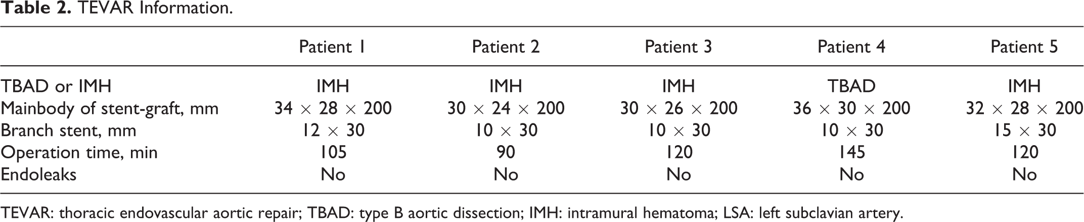
TEVAR: thoracic endovascular aortic repair; TBAD: type B aortic dissection; IMH: intramural hematoma; LSA: left subclavian artery.
In-Hospital Morbidity and Mortality
No in-hospital death was observed in this study. The mean hospital stay was 13.2 ± 6.76 days (range 8-22). No postoperative complications were observed in the 5 patients, including paraplegia, stroke, acute kidney injury, bowel ischemia, aortic rupture, pulselessness of right arm, intermittent claudication of right arm, and cold shoulder feeling.
3-Month Follow-Up Outcomes
All the patients were followed up successfully and underwent physical examination and CTA 3 months after TEVAR (Figure 6). All of them survived. No complications were observed, including paraplegia, stroke, pulselessness of right arm, intermittent claudication of right arm, and cold shoulder feeling. Stent graft-induced newentry was not observed in the 5 patients. Postoperative maximal diameters at different segments for each patient were listed in Table 3. Preoperative maximal diameters were also listed in the table for comparison. The aortic maximal diameters in both of the thoracic and abdominal zones decreased in the 4 patients with IMH 3 months after TEVAR. The aortic hematomas were absorbed (Figure 6, F, G, H and J). The aortic maximal diameters in the graft-covered segment and distal thoracic zone decreased in the TBAD patient. The false lumen in the graft-covered segment was completely thrombosed in the TBAD patient (Figure 6, I). No endoleaks, stent-graft migration and aortic rupture were observed in the 5 patients. All the branch stent-grafts maintained complete patency without thrombus formation and stenosis (Figure 6, K-O).

Three-month postoperative CTA. A-E, Three-dimensional reconstruction of CT. F, G, H and J, The aortic hematomas in the 4 patients with IMH were absorbed. I, The false lumen in the graft-covered segment was completely thrombosed in the TBAD patient. K-O, All the branch stent-grafts maintained complete patency without thrombus formation and stenosis. The red arrows showed the ARSA.
Aortic Remodeling 3 Months After TEVAR.

TBAD: type B aortic dissection; IMH: intramural hematoma; TEVAR: thoracic endovascular aortic repair.
Discussion
Several surgical procedures have been reported to treat patients with acute type B aortic syndrome and ARSA, including replacement of the descending aorta, total arch replacement plus stent elephant trunk, only the stented elephant trunk method, hybrid technique, and TEVAR.7,12 The descending aorta replacement for treating TBAD and ARSA was first reported by DeBakey in 1955. 13 It was not until 1973 that Syme et al. carried out the second descending aorta replacement for managing this pathology. 14 In 2008, a study of 452 patients by Wang et al. revealed that the mortality rate of descending aorta replacement was 6.35% and the early incidence of postoperative complications was 11.1%. 15 High mortality and morbidity rates, as well as the trauma of left thoracic posterior-lateral incision, limit the application of descending aorta replacement for treating thoracic aortic disease. The stented elephant trunk procedure was first applied to repair TBAD and ARSA by Zhu et al. in 2015. 12 Satisfactory early and mid-term outcomes were observed. However, median sternotomy, cardiopulmonary bypass, cardioplegia, and circulatory arrest would cause severe trauma, from which it takes a long time for the patients to recover.
Compared with open surgery, TEVAR was a less invasive technique for the treatment of acute type B aortic syndrome and ARSA. However, it has been shown that ARSA in most patients with this pathology was involved by the dissection or hematoma. Thus, total coverage of ARSA was sometimes inevitably performed to obtain an adequate landing zone.2,4 Total coverage of ARSA may induce ischemia of brain, spinal cord, and right arm, increasing the risks of stroke, paraplegia, pulselessness, intermittent claudication of right arm, and cold shoulder feeling. To date, there are several techniques for the ARSA revascularization during TEVAR, including hybrid technique, fenestration, chimney, and periscope techniques.7,12,16 In Ding er al.’s study, 16 patients with TBAD and ARSA underwent the hybrid procedure. The incidences of type Ia endoleak and branchial plexus injury were 18.8% and 12.5%, respectively. 7 Zhou et al. used chimney and periscope techniques to manage patients with TBAD and ARSA. 4 However, Lindblad et al. reported that the endoleak incidence of chimney technique could reach 11%, and nearly 42% endoleaks required reinterventions. 17 High incidence of type Ia endoleak limits its application.
In the literature, only Dr. zhang reported the use of TEVAR plus fenestration technique in managing a patient with TBAD and ARSA, and more studies are required to figure out the safety of fenestration. 2 Compared to in situ fenestration endograft, we speculated that Castor single-branched stent-graft has several advantages: (1) due to the unibody design, it is more physiological, (2) the procedure is less complicated, (3) the incidence of junction site endoleak between the main body portion and the branch portion is lower. In the study, we successfully used Castor single-branched stent-graft to treat patients with acute type B aortic syndrome and ARSA. All the ARSAs were revascularized in situ by Castor single-branched stent-grafts. No deaths and procedural complications were observed during TEVAR.
To evaluate the aortic remodeling, we referred to the latest SVS reporting standards. 11 Aortic maximal diameters were measured in both of the thoracic and abdominal zones, including the graft-covered segment, the downstream distal thoracic zone, visceral zones, distal abdominal zone. False lumen thrombosis in the TBAD patient was also evaluated. We found that the maximal diameters of diseased aortas decreased in the 4 patients with IMH 3 months after TEVAR. The aortic maximal diameters in the graft-covered segment and distal thoracic zone decreased in the TBAD patient. The false lumen in the graft-covered segment was completely thrombosed in the TBAD patient. All the branch stent-grafts maintained complete patency without thrombus formation and stenosis.
The present study was with several limitations. First, it was a retrospective study and the sample size was small. Second, the long-term follow-up should be performed to further evaluate the efficacy of Castor singe-branched stent-graft in the treatment of acute type B aortic syndrome and ARSA.
Conclusion
The Castor single-branched stent-graft may be a good choice in treatment of acute type B aortic syndrome and aberrant right subclavian artery.
Footnotes
Declaration of Conflicting Interests
The authors declared no potential conflicts of interest with respect to the research, authorship, and/or publication of this article.
Funding
The authors disclosed receipt of the following financial support for the research, authorship, and/or publication of this article: The study was supported by the National Natural Science Foundation of China (81500367).
