






Abstract
Background:
Despite considerable morbid-mortality rates, common femoral endarterectomy is still considered the gold standard for atherosclerotic common femoral artery (CFA) disease. The aim of this study was to demonstrate computed tomography angiography based long-term patency after CFA stent placement and to analyze associated risk factors for restenosis.
Methods:
A retrospective and observational study was carried out in consecutive patients treated with endovascular stent placement in CFA lesions. A clinical follow-up and imaging study was performed using MD-CTA to assess different degrees of in stent restenosis (ISR) and primary, assisted, and secondary patency rates.
Results:
In a 5-year period, 35 extremities were treated in 33 patients with self-expandable nitinol stents. The technical success was 100% without complications related to the procedure. The mean follow-up (FU) was 32.2 months, and 8 limbs were lost. The degree of CFA stenosis was reduced from 79.69 ± 26.47% to 11.23 ± 24.53%. ISR < 20%, 20–70%, and ≥ 70% was evident in 15 (55.6%), 9 (33.3%), and 3 (11.1%) limbs, respectively. Estimated primary, assisted, and secondary patency was 79.5, 96.3, and 96.3%, respectively, after 24 months and 79.5, 96.3, and 96.3%, respectively after 60 months, with a freedom of clinical driven target lesion revascularisation rate of 87.8%.
Conclusion:
Endovascular treatment with self-expandable nitinol stents in CFA lesions had a high technical success rate and was related to few complications. A mild form of intimal hyperplasia was observed in a considerable number of cases. However, long-term patency was high; therefore, CFA stent placement might be a suitable therapeutic alternative in selected patients.
Introduction
Substantial evidence supporting image-based patency for common femoral artery (CFA) stenting is still limited. Common femoral endarterectomy (CFE) is still considered the gold standard for CFA atherosclerotic disease.1,2 Since open surgical treatment is related to a significant morbidity and mortality rate,1,3-5 endovascular techniques have emerged as therapeutic alternatives for this type of atherosclerotic lesions.
In the past, endovascular treatment of the CFA with stent placement was not considered a feasible option, mainly because of the potential risk of stent fracture related to hip joint flection. Future potential arterial access sites could also be compromised by the stent.
In recent years, observational studies evaluating the clinical outcome after CFA stenting have shown good results with low complication rates.1,6,7 More recently, results of a small randomized trial comparing surgery with endovascular stent placement in the CFA support a significant benefit of stenting over surgery with respect to the perioperative morbidity and mortality rate. 2
The aim of this study was to demonstrate computed tomography angiography (CTA) based long-term patency after CFA stent placement and to analyze the associated risk factors for restenosis.
Material and Methods
Patients
The study was approved by the Institutional Review Board. Data of all consecutive patients treated with CFA stents from September 2012 to June 2017 were retrospectively analyzed. All patients signed a written informed consent form. Inclusion criteria were patients with intermittent claudication symptoms non-respondent to optimal medical treatment (Rutherford II & III) or with critical limb ischemia (Rutherford IV–VI). 8
CFA lesions were classified into 4 types based on Azema et al. 9 Type I lesions were those extending from the iliac artery to the CFA; type II were isolated lesions in the CFA; type III affected the CFA and the ostium of the deep or superficial femoral artery, or both; and type IV represented proximal or distal stenosis bypass anastomosis. 9
The interventional procedure was also reviewed to include the utilized endovascular devices and performed techniques to address CFA stent failures and their complications. Technical success was defined as successful recanalization and CFA stent placement with residual stenosis < 30%.
Procedure/ Post-Procedural Protocol
Self-expandable stents were implanted in all patients through a 6Fr contralateral access. Heparin (5000 U) was given in every intervention prior to stent placement. After successful guidewire passage, the lesion was predilated with non-compliant balloon-catheters. The election of the stent device was left to the discretion of each operator. After stent placement, post-dilatation was performed using a balloon-catheter of the same size with respect to the native vessel diameter. Patients were on dual antiplatelet therapy (acetylsalicylic acid 100 mg and clopidogrel 75 mg, daily) before the intervention and for 3 months after the procedure; after that, mono antiplatelet therapy (acetylsalicylic acid 100 mg) was continued indefinitely.
Follow-Up
A clinical follow-up (FU) was carried out at 1, 3, 6, and 12 months, then annually thereafter. The FU period was defined as the time from CFA stent placement to the last multidetector CTA (MD-CTA) or reintervention date available. Clinical improvement was considered closure or reduction in size of the ulcer or at least a 1-point decrease in the Rutherford’s scale, without the need of target lesion revascularisation (TLR).
An ultrasound was used at each FU with the limitation of subsequent acoustic shadow when calcium of the anterior wall was present. MD-CTA was performed using an Adaptive Iterative Dose Reduction 3D (AIDR 3D) protocol. Multiplanar and 3D volume reconstructions were chosen for evaluation of the degree of in-stent restenosis (ISR) and stent fractures. This was performed during FU in a patient when the investigators considered it necessary (Figure 1).

(A) Angiographic image of a CFA chronic total occlusion. (B) Angiographic result after recanalization and CFA stent placement. (C) MD-CTA: multiplanar reconstruction and axial images of the CFA stent 20 months after the procedure. CFA, Common Femoral Artery; MD-CTA, Multidetector Computed Tomography Angiography.
ISR was divided into 5 grades depending on the degree of stenosis of the target lesions: 1 (0–20%), 2 (21–50%), 3 (51–70%), 4 (71–99%) and 5 (occluded). ISR ≥ 70% was considered significant; stent fractures were categorized according to a classification proposed by Jaff et al. 10
Primary patency was defined as a freedom from vessel diameter reduction of more than 50% without any percutaneous or surgical intervention. Assisted patency was considered after endovascular reintervention due to significant ISR, and secondary patency as a stent occlusion. TLR was defined as any procedure for restenosis in patients with new symptoms. Patients were considered lost when there were no more FU studies, whether due to medical consultation absences or death. A clinical FU was not considered a primary endpoint for this analysis, since it can be related to the affection of another vascular territory, in addition to the CFA.
Statistical Analyses
Statistical analyses were performed using SPSS version 15 (SPSS, Inc., Chicago, IL, USA. UU). Categorical variables were presented as the absolute value and percentage. Continuous data were given as the mean ± standard deviation. The relationship between ISR and other variables was evaluated with the chi-square test or the Fisher’s exact test. All values of p < 0.05 were considered significant. A Kaplan-Meier analysis was performed to determine the freedom from TLR, primary, assisted, and secondary patency rates.
Results
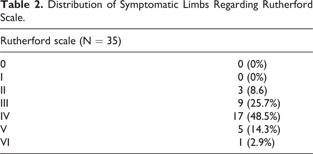
Thirty-five extremities with CFA lesions were treated in 33 patients using self-expandable nitinol stents. Patient- and lesion-related parameters are summarized in Table 1. The mean age was 71.9 ± 10.9 years. The clinical indication of the treatment was intermittent claudication in 12 (34.3%) and critical limb ischemia in 23 (65.7%) extremities (Table 2). The distribution regarding the morphological classification of the CFA lesions is given in Table 1.
Patient- and Lesion-Related Characteristics and Risk Factor Analysis.

Abbreviation: ISR, in-stent restenosis.
Distribution of Symptomatic Limbs Regarding Rutherford Scale.
Additional affected vascular territories were as follows: 11 extremities (31.42%) had associated iliac lesions and 15 (42.85%) had femoro-popliteal lesions. Concomitant iliac artery lesions were treated using the same procedure in 14 (40%) patients and femoro-popliteal artery lesions in 5 (33.3%) patients.
Technical success was achieved in 100% of all cases without any complications related to the treatment or puncture site. No patient required conversion to open surgery. Thirty-five stents were used, of which 32 were uncovered self-expandable nitinol stents (Smart, Cordis Inc, Miami FL; N = 20, and Zilver, Cook Inc, Europe; N = 12) and 3 were covered self-expandable nitinol stents (Viabahn, W.L. Gore & Associates, Flagstaff, Ariz). The only complication was 1 fatal ischemic stroke 48 hours after the procedure. This patient was treated through a contralateral femoral access, with no relation to the procedure or instrumentation of the material.
The mean FU was 32.2 ± 19.1 months. From 35 treated extremities, 8 were lost during FU, 5 did not return for medical consultation, and 3 died due to oncological and cardiovascular diseases.
During FU, 22 (81.48%) patients remained asymptomatic (Rutherford 0) and 3 (11.11%) were symptomatic (Rutherford I) but with significant improvement. The symptomatic patients had associated femoro-popliteal lesions, which were not treated in the same procedure to evaluate clinical progress. From the 6 patients that presented with ulcers before treatment (Rutherford V & VI), all but 1 showed improvement or complete healing. The patient (3.7%) without any clinical improvement continued with medical treatment.
The degree of CFA stenosis was reduced from 79.69 ± 26.47% to 11.23 ± 24.53% at the last CTA-based FU. Significant and clinically relevant ISR (≥ 70%) developed in 3 (11.1%) limbs (stenosis, N = 2; occlusion, N = 1). These patients were symptomatic and were treated by angioplasty at 7, 8, and 20 months from the initial treatment. Non-significant ISR (20–70%) was evident in 9 (33.3%) and no ISR (< 20%) was found in 15 (55.6%) limbs; all of them were asymptomatic. Freedom from different degrees of ISR is given in Figure 2.

Kaplan Meier estimate for different degrees of in-stent restenosis during follow-up in 27 limbs.
The estimated primary, assisted, and secondary patency were 88.9, 96.3, and 96.3%, respectively after 12 months, 79.5, 96.3, and 96.3%, respectively, at 24 months, and 79.5, 96.3, and 96.3%, respectively, at 60 months (Figure 3), with a freedom of clinical driven TLR of 87.8% (Figure 4). There were no stent fractures detectable in the MD-CTA.

Kaplan Meier estimate for primary, assisted and secondary patency rates during follow-up in 27 limbs.

Kaplan Meier estimate for freedom from TLR rate during follow-up in 27 treated limbs. TLR: target lesion revascularization.
An analysis of possible risk factors was performed, comparing patients with and without clinically significant ISR (≥ 70%) in the postoperative period. Patient- and lesion-related parameters were analyzed (Table 1); however, there was no statistical difference in these variables between both groups. It seems that these variables were not associated with the development of ISR and were, therefore, not considered risk factors.
Discussion
Common femoral endarterectomy for CFA atherosclerotic disease has been the gold standard for many years. The CFA is easy to reach for surgical treatment due to its superficial location, and positive long-term patency rates have been reported in many scientific publications.3-5,11,12 However, with a complication rate of 11.1–17%, it is not an innocuous procedure.4,5 Wound infection is one of the most frequent complications and can be considered the Achilles heel of this invasive treatment. Lymph leaks and hematomas are other common complications in this anatomic location. The combined morbidity-mortality rate has been reported at 15%. 3
The endovascular approach is an accepted treatment alternative for peripheral vascular disease in the lower limbs. Different endovascular devices have been used to treat CFA atherosclerotic lesions, such as the balloon catheter, drug-eluting balloon catheter, cutting balloons, and atherectomy devices, 1 all with suboptimal results in heavily calcified plaque. Stavroulakis et al. reported a primary patency rate of 68% after 1 year with a TLR of 25% using conventional balloon catheters. 13
Stent placement has not been considered a viable option for the treatment of the CFA due to several considerations. The anatomy and vessel related features are challenging for stent placement. The hip joint is located near the artery, so the artery is subject to flexion, and the stent is at risk of crushing. This leads to potential stent fractures, which are associated with intimal hyperplasia and increased in-stent restenosis.2,14 In the present study, no stent fractures were detectable during the FU period. Despite concerns related to reduced stent patency in the CFA, clinically significant ISR during FU was evident in only 3 patients in the present study. These results are comparable to CFE in terms of long-term patency but with lower perioperative mortality and morbidity.
The TECCO trial was a randomised study that compared CFE with angioplasty and stent placement in CFA lesions. The study revealed that stenting of the common femoral artery with a self-expanding stent was feasible and associated with a lower rate of general and local complications at 30 days compared with surgery. 2 The study concluded that there was a tendency toward a higher patency rate in favor of endovascular treatment but without any statistical significance.
Lesion characteristics may have an influence on patency rates. Native lesions of the CFA tend to be heavily calcified, which leads to some potential limitations. Severe calcified lesions have a negative impact on lumen gain after endovascular therapy due to an inadequate result after angioplasty or self-expandable stent placement. 15 Severely calcified arteries also predispose patients to stent fractures. In the present series, there were no detectable stent fractures. There are no reports regarding patency after stent placement in heavily calcified CFA lesions; however, stent placement in the superficial femoral artery was interestingly related with a higher restenosis rate but not with a higher TLR rate. 15 In the present study, the amount of calcification in the CFA did not predict the ISR rate (p = 0.98). Furthermore, a trend toward a worse outcome without statistical significance (p = 0.09) was observed in type 3 lesions, and all clinically relevant ISR lesions were classified as type 3 lesions. This finding has been published by Nasr et al. before; however, in their study, they used balloon-expandable stents in this type of lesions. 7 Given the increase in the risk of procedural failure and a trend toward more restenosis and target lesion revascularisation, an alternative treatment strategy, such as CFE, should be considered for type 3 lesions.
Over recent years, stent design improved, and new devices have appeared on the market. Due to unsatisfactory results after angioplasty, these stents have been used to treat elastic recoil, especially in calcified plaques. Elastic recoil plays a major role in the treatment of CFA lesions. Paris et al. reported a treatment on 26 CFAs using angioplasty, in which 100% needed a stent placement. Similarly, in a study by Bilic et al., 66% of the patients needed a stent placement after angioplasty.16,17 Metha et al analyzed 167 patients treated with angioplasty and conventional balloon versus atherectomy with angioplasty and eventual stent for lesions in CFA. They reported that combined primary patency at 42.5 months was 77% for the group of patients treated without stents versus 100% for the group of patients treated with stents. 18
Nitinol self-expandable stents were used in our patients, but stents with increased resistance to compression might improve long-term results. Eccentric and heavily calcified plaques, complex plaque morphology, and biomechanical forces pose a significant challenge to self-expandable nitinol stents. The Supera stent has been used to overcome these problems since the stent is extremely crush resistant. A 6-month cumulative primary patency rate of 100% has been reported with a cumulative freedom from TLR rate of 100%; however, no long-term data are available. 19
The common CFA is frequently used as an arterial access site in endovascular procedures, and a CFA stent placement could potentially compromise future treatment options. Although the puncture through the stent has not been the objective of this study, we observed that there were no complications related to the vascular access site in endovascular procedures up to 8Fr.
In the present study, clinically driven TLR agreed with stenosis greater than 70% with a primary patency rate of 79.5% in the long-term. This clinical study is the only 1 in which patients were followed with MD-CTA. This allows evaluation of the degree of restenosis with a high sensitivity and specificity, since calcium in the anterior wall of the artery is not a limiting factor, as it often is with ultrasound control. This leads us to the conclusion, that intimal hyperplasia at a minor degree might appear in a considerable number of cases; however, clinically relevant restenosis is less frequent with a high primary patency rate in the long-term.
This study has some limitations as it was designed as a retrospective single centre study without a control group or randomization. The number of included patients was relatively small, and the group of patients was heterogeneous.
Conclusion
Endovascular treatment with self-expandable nitinol stents in CFA lesions had a high technical success rate and was related with a low complication rate. A mild form of intimal hyperplasia was observed in a significant number of cases; however, long-term patency was high. Therefore, CFA stent placement might be a suitable therapeutic alternative in selected patients.
Footnotes
Authors’ Note
The study conforms to the guiding principles of the Declaration of Helsinki.
Declaration of Conflicting Interests
The author(s) declared no potential conflicts of interest with respect to the research, authorship, and/or publication of this article.
Funding
The author(s) received no financial support for the research, authorship, and/or publication of this article.
