






Abstract
Purpose:
Nonspecific aortoarteritis (NSAA) is a chronic inflammatory vasculitis involving aorta and its branches. We conducted a study prospectively to compare time resolved magnetic resonance angiography (MRA) with diagnostic digital subtraction angiography (DSA) for the vascular assessment in the patients of NSAA.
Materials and Methods:
Seventeen patients of NSAA were recruited in the study over the period of 3 years. Contrast enhanced MRA using Time-resolved angiography With Interleaved Stochastic Trajectories (TWIST) sequence and diagnostic DSA were performed in these 17 patients.
Results:
The majority of the patients were young (median age was 25 years, range 8 to 46 years) and 11 patients were females. Erythrocyte sedimentation rate (ESR) was elevated in 9 patients and C-reactive protein (CRP) was elevated in 6 patients. Most commonly involved vessels in our patients were right renal artery (14 patients), abdominal aorta (12 patients) and left renal artery (11 patients). Left and right subclavian arteres were involved in 10 and 6 patients respectively. The sensitivity and specificity of time-resolved MRA using TWIST sequence is 100% as compared to DSA in the assessment of major vessels such as aorta, arch vessels, celiac artery and superior mesenteric artery. However, the sensitivity and specificity of time resolved MRA in the evaluation of renal arteries and vertebral arteries were 100%, 71.4% and 85.7%, 33.3% respectively. No significant association of MRI contrast enhancement with erythrocyte sedimentation rate (p = 1.00) and C-reactive protein (p = 0.600).
Conclusion:
Time resolved MRA images obtained using TWIST sequence were as qualitative as DSA images and can noninvasively evaluate the vascular involvement in NSAA patients.
Keywords
Introduction
Nonspecific aortoarteritis (NSAA) is a rare inflammatory vasculitis, characterized by granulomatous pan-arteritis of the aorta, its branches and pulmonary arteries.1-3 It may cause cardiovascular complications including congestive cardiac failure, pulmonary hypertension, stroke and aortic complications. Doppler ultrasonography, computed tomography (CT), magnetic resonance imaging (MRI) and fluoro deoxy glucose (FDG)-positron emission tomography (PET) are used to diagnose and evaluate the patients of NSAA. 3
The diagnosis of NSAA depends on clinical features, radiologic changes, and biochemical findings. The diagnosis of NSAA depend on the following criteria: (1) clinical complaints caused by ischemic changes involving the central nervous system, kidneys, or upper limbs; (2) fever, reduced or absent pulses, audible bruits, and fundoscopic findings; and (3) increased erythrocyte sedimentation rate (ESR) and C-reactive protein (CRP) levels.1-3 Imaging and angiographic changes suggesting NSAA vary from minimal intimal irregularity to complete occlusion in the involved vessels. Disease involving the aorta and/or at least 2 medium-sized arterial branches is required for diagnosis.
The most useful markers for the diagnosis of NSAA are erythrocyte sedimentation rate (ESR) and C-reactive protein (CRP). These markers are used to assess disease activity and follow-up. Early disease may have a waxing and waning course that may finally lead to fibrotic occlusion of vessels. The early non-specific clinical inflammatory features may progress to a vascular inflammatory stage in 80% of patients.1-3 Late clinical features are likely due to ischemic changes caused by arterial involvement. Disease recurrence commonly occurs in new arterial territories, with the presence of active and quiescent disease.
MR angiography comprehensively evaluates arterial stenosis and dilatation. It noninvasively evaluates vascular lesions without radiation exposure. It can demonstrate vessel wall thickening and vessel wall enhancement. We have assessed the time-resolved magnetic resonance angiography (MRA) using the TWIST (Time-resolved angiography With Interleaved Stochastic Trajectories) sequence in the evaluation of NSAA. TWIST is a time-resolved 3D MRA technique, which is having a high temporal and spatial resolution. It has the advantage of acquisition of angiographic images in multiple phases with low dose contrast agent without the need for breath-hold during the scan. It has certain disadvantages in the form of a large volume of acquired data with a poor temporal resolution of small branches.
To the best of our knowledge, there is no study in the literature, which used time-resolved MRA using TWIST sequence for the vascular assessment in the patients of NSAA. We conducted this prospective study with the aim to assess the efficacy of time-resolved MRA using TWIST sequence in the assessment of vascular involvement in NSAA patients.
Materials and Methods
This prospective study was conducted between 2017 and 2019 after institional ethical committee approval. The patients of NSAA presenting to the Departments of Cardiology and Cardiovascular Radiology and Endovascular Interventions, All India Institute of Medical Sciences, Delhi, India were included in the study. All the patients were diagnosed with NSAA as per American College of Radiology (ACR) criteria. Informed consent was taken from all patients.
Seventeen patients with NSAA, who underwent both time-resolved MRA using TWIST sequence and digital subtraction angiography (DSA) were included in the study. Informed consent was obtained in all the patients. Doppler evaluation was done for all the patients, following which time-resolved MRA using TWIST sequence was done. Laboratory investigations such as erythrocyte sedimentation rate (ESR), C-reactive protein (CRP) were performed in all patients. All the MRA were done on 1.5 Tesla MRI scanner (Aera; Siemens, Erlangen, Germany).
TWIST MRA was performed after administration of 0.01 ml/kg of gadoterate meglumine (DOTAREM) with a flow rate of 3 ml/sec. This is followed by post-contrast T1 weighted imaging for the assessment of vessel wall thickness and contrast enhancement. DSA with or without endovascular interventions were performed in all 17 patients.
TWIST MRA images were analyzed as compared to DSA images and their performance was analyzed. The wall thickness of the vessel was assessed in the axial sections of post-contrast T1 weighted sequence. The images of TWIST and DSA were analyzed individually by 2 radiologists, who were separate from the study and having experience of more than 10 years. The sensitivity and specificity of both imaging modalities were based on the expert opinion of the scoring radiologists.
The image quality was assessed using the following scores
Image quality is poor and not of diagnostic quality
Image quality is optimum to make the diagnosis, but inadequate for treatment planning
Image quality is good for the diagnosis as well as treatment planning
Statistical Analysis
To describe the role of time-resolved MRA using TWIST sequence in the vascular assessment of patients of Nonspecific Aorto-arteritis, the data were summarized and analyzed using statistical package for social sciences (SPSS version 24.0) or STATA(version 14) software. Data were expressed as mean ± SD or number and percentage as appropriate for qualitative and quantitative variables.
Results
The median patient age in our study was 25 years (range 8 to 46 years) and about 11 were females. Other patient details are summarized in Table 1. The most common presentation of patients was with diminished or absent pulses with limb pulse difference (15 patients) followed by hypertension (14 patients). Eleven patients complained of dyspnea. Four patients presented with syncope and palpitation. Claudication was seen in 2 patients. Two patients had complaints of fever. Hemiparesis, carotid tenderness, and vision disturbance were seen in 1 patient each. The clinical features are represented in Table 2.
Patient Demographics.

Clinical Features.
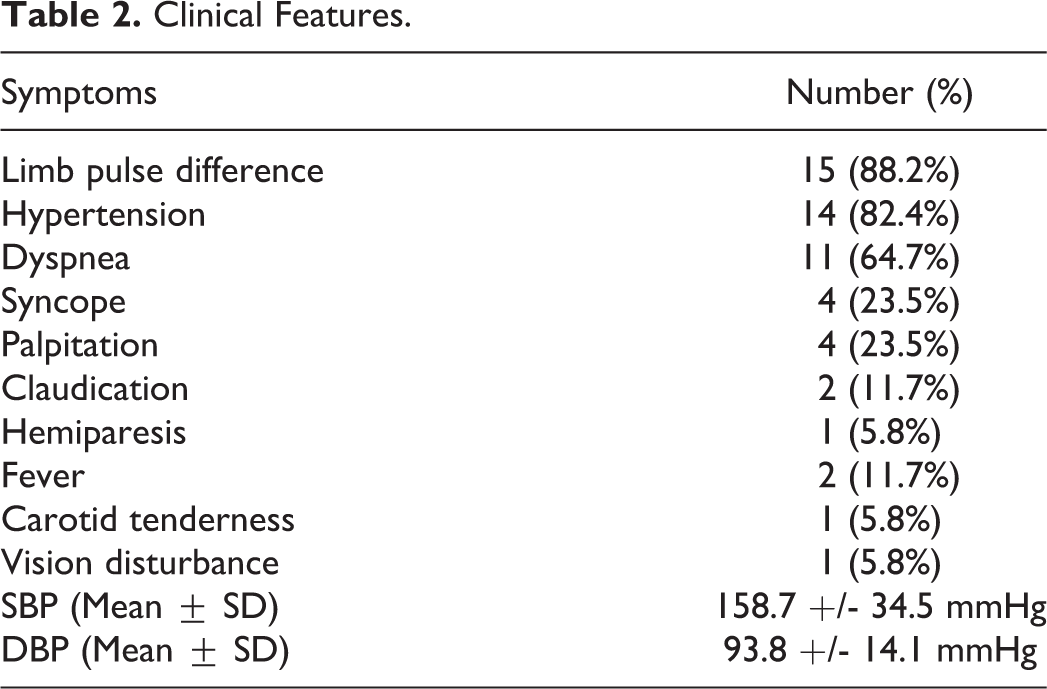
The mean systolic blood pressure was 158.7 +/- 34.5 mmHg. The mean diastolic blood pressure was 93.8 ± 14.1 mm Hg. ESR was elevated in 9 patients (52.9%) and CRP was elevated in 6 patients (35.3%). The average ESR and CRP levels are 25.9 ± 11.6 mm/hr and 10.4 ± 22.4mg/L respectively. Seven out of 17 patients (41.2%) were on steroid therapy. Three patients were additionally receiving immunosuppressants. All the 14 patients having hypertension were on antihypertensive drugs.
Correlation Between TWIST MR Angiogram and DSA Findings
Both time-resolved MRA using TWIST sequence and DSA were performed in 17 patients of NSAA. Among these, 15 patients underwent endovascular interventions. Patient data and imaging features were compared for both MRA and DSA findings in all these patients. DSA was considered the gold standard for the evaluation of the arterial system.
Vascular Involvement
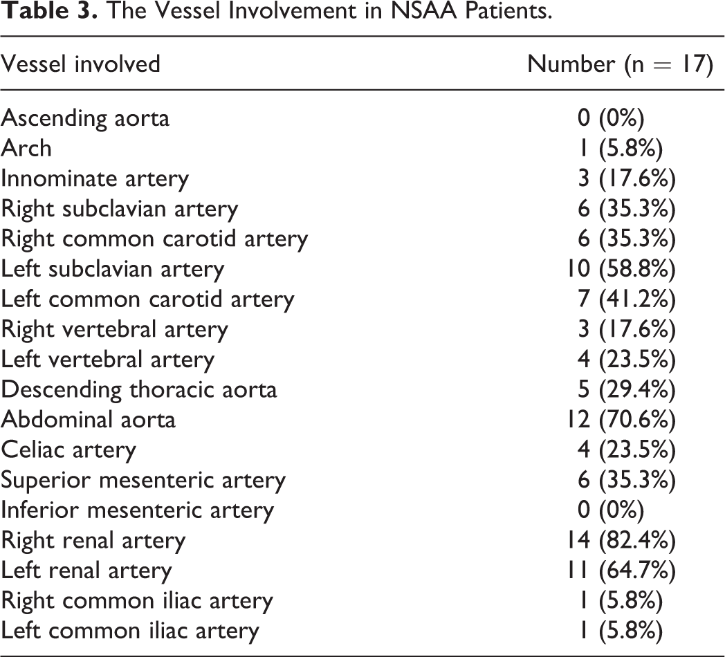
The incidence of involved vessels in our patients (n = 17) is depicted in Table 3. Common arteries involved in our patients are the right renal artery (82.4%), abdominal aorta (70.6%), and left renal artery (64.7%). The left subclavian artery was involved in 10 (58.8%) patients and the right subclavian artery in 6 (35.3%) patients. Ascending aorta and inferior mesenteric arteries were not involved in any of the patients.
The Vessel Involvement in NSAA Patients.
Quality of Images in TWIST and DSA Angiography
The images of time resolved MRA using TWIST sequence were compared with standard DSA images. The involved arteries were correctly identified in both the TWIST angiography images and DSA in all the patients. The sensitivity and specificity of TWIST angiography was 100% when compared to DSA for the detection of the lesions involving the aorta, arch vessels, celiac artery, and superior mesenteric artery. However, the sensitivity and specificity of TWIST MRA for the evaluation of renal arteries are 100% and 71.4% respectively. The sensitivity and specificity of vertebral artery (VA) assessment were 85.7% and 33.3% respectively.
All the involved arteries were scored 3 (good quality) on both TWIST and DSA images except for small vessels such as renal artery branches and vertebral arteries (Figures 1–6). Renal arteries were scored 2 as compared to DSA (3) in all the patients. The vertebral arteries are correctly assessed (score 3) when they are of good size (in 16 patients). In 1 patient, the quality of VA image obtained through TWIST scored 2.

TWIST MRA (A) and DSA (B, C) images of a 17 year old male patient of NSAA showing post-ostial stenosis of bilateral subclavian arteries.

TWIST MRA (A) and DSA (B) images of a 23 year old female patient of NSAA showing occluded bilateral common carotid arteries and Left subclavian artery. Tight ostio-proximal stenosis of right subclavian artery.

TWIST MRA (A) and DSA (B) images of a 11 year old male patient of NSAA showing diffuse disease with aneurysmal dilatation of suprarenal aorta and bilateral renal artery occlusion.

TWIST MRA (A) and DSA (B) images of a 15 year old female patient of NSAA showing diffuse disease of abdominal aorta with significant ostio-proximal stenosis of bilateral renal arteries. The spatial resolution of renal arteries and its branches in TWIST image is lesser than DSA.

TWIST MRA (A) and DSA (B) images of a 8 year old female patient of NSAA showing tight stenosis of peri-celiac abdominal aorta with post stenotic dilatation. Small bridging collaterals at stenosed segment is better appreciated in DSA.

TWIST MRA (A) and DSA (B, C) images of a 25 year old female patient of NSAA showing diffuse dilative disease in descending thoracic aorta and innominate artery. Diffuse stenosis seen injuxta-diaphragmatic and perirenal aorta.
Vessel Wall Morphology and Disease Activity
The average vessel wall thickening on MRA in our patients is 2.65 ± 0.94 mm. The vessel wall enhancement was seen in 5 patients (29.4%). However there was no significant association between vessel wall thickness and contrast enhancement or disease activity (p-value = 0.224). No significant association of MRI contrast enhancement with ESR (p-value = 1.00) and CRP values (p-value = 0.600).
Discussion
In the present study, females were more commonly involved than males in our study, which well supports the other studies of NSAA.1-3 The most common presenting symptom in our study was pulse difference in limbs (88.2%) followed by hypertension (82.4%) and dyspnea (64.7%). There was no correlation between serum ESR/ CRP levels and contrast enhancement of the vessel wall. Evidence of ongoing activity could not be related to elevated ESR or CRP.
Doppler ultrasonography determines vessel wall thickening and estimates blood flow dynamics across the lesions. It is ideal for follow-up assessment of the renal and carotid endovascular treatment. Carotid ultrasound evaluates the concentric thickening of common/internal carotid arteries and helps in follow-up in NSAA patients. 1 However, it is operator dependant and thoraco-abdominal aorta, and its branches will not be properly evaluated due to poor visualization. CT angiography can also demonstrate the vascular lesions and is useful for surveying the lesions. 1 CT showing diffuse wall thickening of the aorta or its major branches in febrile patients should raise the suspicion of NSAA. Delayed scans are important in the demonstration of vessel wall enhancement. Radiation exposure is one of the main concerns in the evaluation of young patients.
Digital Subtraction Angiography (DSA) is usually performed with the intent to treat the lesions. DSA can be used for diagnosis and endovascular treatment of NSAA. 4 But it is not useful for vessel wall imaging. Early treatment should be initiated to prevent permanent vascular damage and end-organ injuries. The treatment includes steroids, immunosuppressive medication with aggressive management of hypertension.
Surgical repair has been the standard therapy for treating vascular lesions. Even after bypass surgery, the disease may progress involving proximal or distal to anastomotic sites resulting in graft failure. 5 With recent advances, endovascular treatment is gaining a lot of attention in the management of NSAA, despite high rates of restenosis/occlusion. The endovascular treatment (angioplasty and stenting) for chronic inactive lesions show the success of revascularization with better safety. Surgery or endovascular treatment should ideally be avoided in the active stage of disease except in life-threatening conditions. 6
Comprehensive cardiovascular MRI in NSAA provides detailed cardiovascular anatomical information. Some of the previous studies have looked at the diagnostic accuracy of MRA with conventional angiography. Yamada et al conducted a study to assess the diagnostic accuracy of 3D MRA in NSAA and found out that the sensitivity and specificity of 100% for the diagnosis of NSAA. 7 Kumar et al analyzed the role of 3D TOF (time of flight) MRA found out an accuracy of 86% for the detection of the aortic arch and arch vessels and 92% for the detection of the abdominal aorta and visceral branch involvement. 8
Recently, advances in MRI sequences in the form of time-resolved MR angiography further added value in the comprehensive dynamic evaluation of vascular involvement. TWIST MRA is one of the methods of time-resolved dynamic MR angiography study which is based on the alternating sampling of the 2 regions of K space. The peripheral region of K space is less sampled compared to the central region. The main advantage is of this sequence is accurate timing of MRI acquisition is not needed as the multiple measurements are acquired in quick succession. We can simply trigger the sequence and run the sequence till the complete washout of the contrast through vessels.
TWIST MRA is a time-resolved 3D MRA technique having a high temporal and spatial resolution. It has the advantage of the acquisition of angiographic images in multiple phases with low dose contrast agent without the need for breath-hold during the scan. It has certain disadvantages in the form of the large volume of acquired data with a poor temporal resolution of small branches. It is commonly used, where the flow is fast or in conditions of unpredictable flow dynamics. This can be very useful in those uncooperative patients, issues of breath-holding, and children. In NSAA patients who often present with dyspnea, this sequence can be very useful.
Few of the studies also looked at the role of TWIST MRA in various other conditions. Jin et al assessed the role of TWIST MR angiography to evaluate the vascular invasion in musculoskeletal tumors of lower extremities and found out that it reliably delineates the vascular involvement. 9 Armstrong et al evaluated the role of TWIST MRA in helping to image the complex central venous accesses and concluded that it can reliably identify the candidate sites for the venous catheter insertion. 10 Korn et al found double bolus injection TWIST-MRA helps the evaluation of cervical arteries with the elimination of venous contamination. 11 Kinner et al found that TWIST MRA can assess the entire peripheral vascular system without any timing issues and venous contamination. 12
In our study, TWIST images were as sensitive and specific as DSA in identifying all the involved major vascular lesions except for renal and small-sized arteries. However, DSA could demonstrate the renal artery branches better than the TWIST angiography images. Hence TWIST MRA can be utilized for the diagnosis and pre-intervention assessment of all the involved major branches of the aorta. However, for the accurate assessment of renal arteries, vertebral arteries, and small caliber arteries, DSA scored better over the TWIST MRA images. Hence, the pre-treatment assessment of involved vessels can be reliably done in most of the vascular lesions on TWIST MRA images instead of subjecting patients to invasive DSA except for the small caliber vessels.
As the NSAA patients usually younger and anxious patients, symptomatic patients with an inability to breath-hold for a longer duration, new MRA sequences such as TWIST angiography can be utilized in these patients. To the best of our knowledge, this is the first prospective study that has used time-resolved MRA using TWIST sequence for the vascular assessment of NSAA patients and found that it could provide us reliable information regarding vascular involvement.
The sample size of our study was limited as our inclusion criteria were strict to include only those NSAA patients who underwent both the examinations. The number of patients (n = 17) in our study is relatively small to make a strong recommendation. However, we expect that our study can serve as a prototype for more research to assess the role of TWIST MRA in the assessment of vascular involvement in NSAA patients.
Conclusion
Time-resolved MRA using TWIST sequence images were as qualitative as DSA images. TWIST MRA can be used for comprehensive vascular evaluation in patients with NSAA. TWIST MRA can be very useful in uncooperative patients and those facing breath-holding difficulty. It provides dynamic vascular assessment and helps to evaluate the arterial system (such as the aorta and its major branches except for the small-sized vessels) without much venous contamination. It has the potential to replace existing conventional MRA in the evaluation of NSAA patients.
Footnotes
Authors’ Note
The abstract of this study has been presented at Society of Interventional Radiology 2020 conference and same abstract has been published as abstract no 698 in Journal of Vascular and Interventional Radiology, 2020. This is part study of original ethical approval by Institute Ethics Committee, All India Institute of Medical Sciences, Delhi, India—IECPG-448/29.11.2017.
Declaration of Conflicting Interests
The author(s) declared no potential conflicts of interest with respect to the research, authorship, and/or publication of this article.
Funding
The author(s) received no financial support for the research, authorship, and/or publication of this article.