
Abstract
Mycotic subclavian aneurysms are rare, and their presence typically mandates urgent repair due to the associated high risk of rupture and mortality. A multi-disciplinary team effort is of utmost importance in ensuring favorable results. In this case report, we present a 79-year-old male with a rapidly enlarging mycotic left subclavian artery aneurysm secondary to a retrosternal abscess and left sternoclavicular septic arthritis, who underwent aneurysmal exclusion, a left carotid-left axillary bypass and pectoralis muscle flap coverage with a good outcome.
Introduction
Mycotic aneurysms involving the subclavian artery are uncommon and are associated with significant morbidity and mortality, mainly due to aneurysm rupture. These aneurysms may occur due to primary infection of the artery or from secondary bacterial seeding of an aneurysmal subclavian artery. Mycotic aneurysms may also be complications of local bony and soft tissue infections, infective endocarditis, or bacterial and fungal bloodstream infections.
The open approach is the traditional method of repair for these aneurysms, however, successful management using the endovascular or combined (hybrid) approach have also been described in some case reports.1-4 Most surgeons however favor the open repair whenever feasible for adequate source control, especially in patients with limited or no co-morbidities. 5 This case report describes our technique for repairing a proximal mycotic left subclavian aneurysm and also highlights the importance of early management and a multi-disciplinary team approach.
Consent for publication was obtained from the patient.
Case Report A 79-year-old male with a past medical history of hypertension and type 2 diabetes mellitus presented to the emergency department with a 1-week history of left shoulder pain and fever, as well as altered mental status for two days. Physical examination revealed a lethargic elderly male, with point tenderness focal to the left sternoclavicular joint. His admission white cell count was 18 000 cells/cm.
3
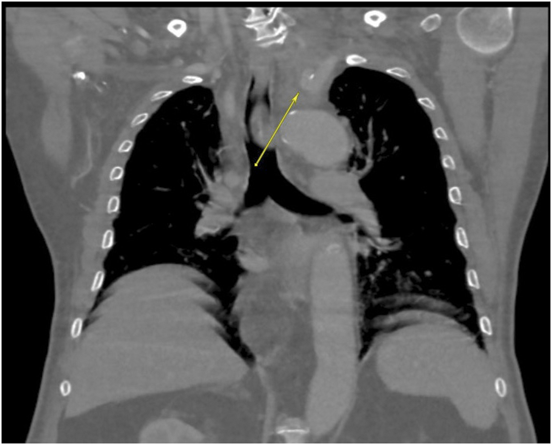
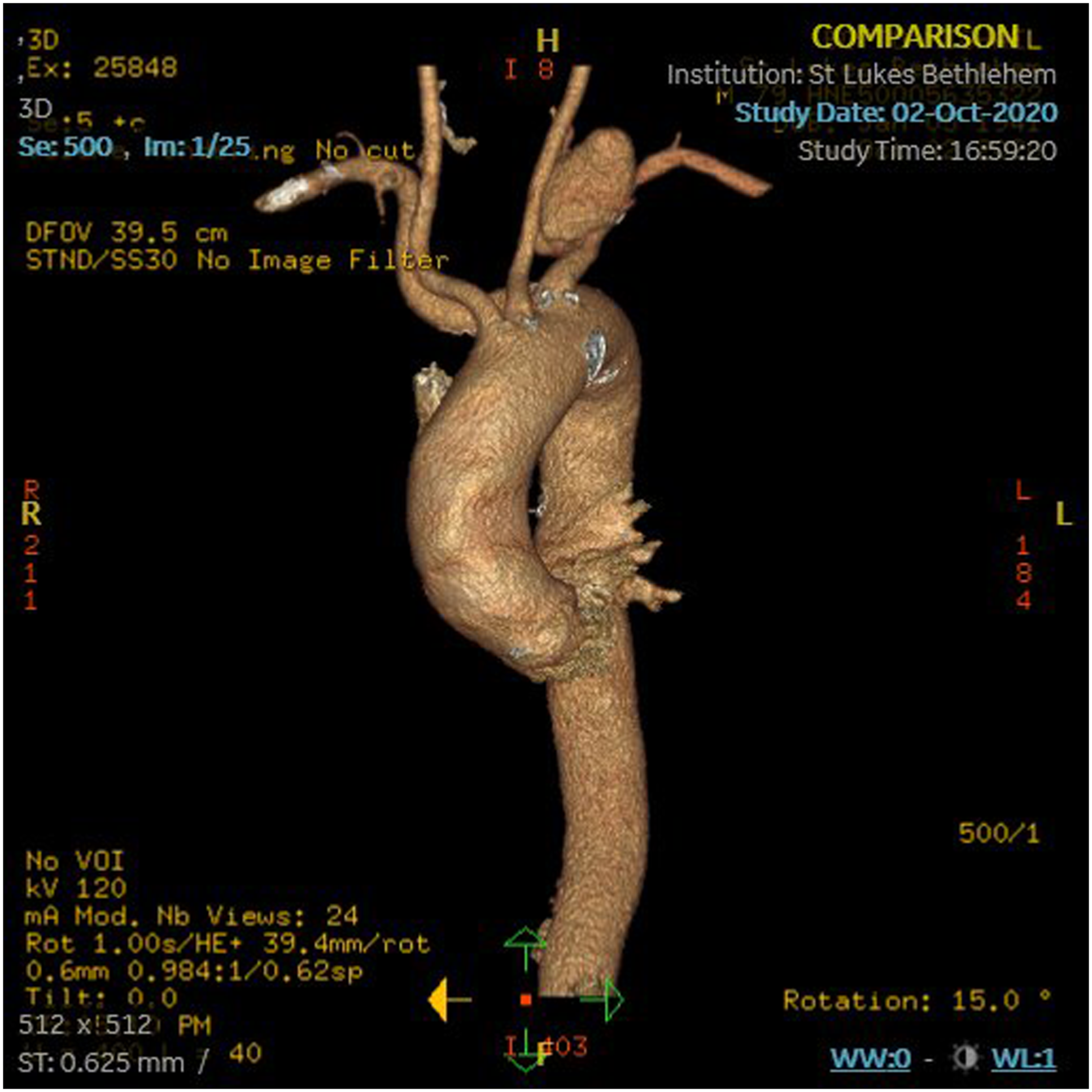
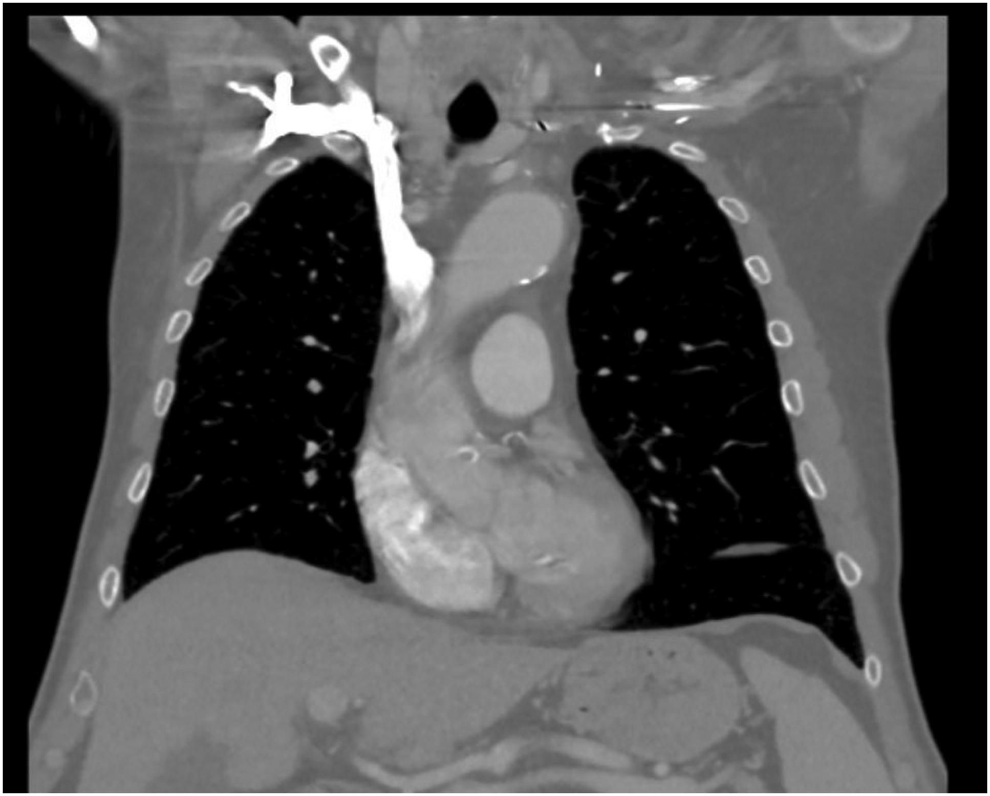
He had a contrast-enhanced chest computed tomography (CT) scan which showed a 9 cm x 7 cm x 2.8 cm retrosternal abscess with involvement of the left sternoclavicular joint (Figure 1). A 1.4 cm x .9 cm left subclavian artery aneurysm was also noted on the imaging study (Figure 2). He was emergently taken to the operating room for a retrosternal debridement and washout during which the clavicular head and the medial third of the clavicle were excised. No intervention was performed on the left subclavian aneurysm at this time. His wound was managed with negative pressure therapy and he was taken back to the operating room twice over the course of 4 days, with no evidence of residual gross infection. He was initially placed on vancomycin which was narrowed to cefazolin following the growth of methicillin-sensitive Chest CT (axial) showing the retrosternal abscess (arrow). Chest CT (coronal) showing initial small left subclavian mycotic aneurysm. The plastic surgery team was then consulted for wound closure of his complex chest wall wound, and a chest CT angiogram with 3-dimensional reconstruction (CTA) was ordered as part of his pre-operative work-up. The CTA revealed that the left subclavian artery aneurysm now measured 4 cm x 4 cm (Figure 3). Also noted on the CTA was an aberrant right subclavian artery arising from the aortic arch distal to the origin of left subclavian artery (Figure 3). The cardio-thoracic, vascular, and plastic surgery services promptly intervened via an open approach due to the rapid short interval growth in aneurysm size and substantial risk of rupture. He was deemed at moderate operative risk and informed consent was obtained. Chest CTA with 3-dimensional reconstruction showing the enlarged left subclavian mycotic aneurysm. Operative exposure was obtained through a median sternotomy and extended into the left 4th intercostal space, then further extended laterally to the anterior axillary line. The innominate, left common carotid and the left subclavian arteries were mobilized and controlled proximally at their origins. An incision was made along the clavicle and the middle third of the clavicle was resected, providing exposure and control of the left axillary artery. A separate longitudinal cervical incision was made to isolate the left common carotid artery, and a left common carotid-to-left axillary artery bypass was performed using autologous reversed great saphenous vein tunneled through grossly non-infected tissue. The left subclavian artery was then divided at its origin with a stapler and the left axillary artery was mobilized proximally to the aneurysmal portion of the left subclavian artery. The subclavian artery was divided just distal to the mycotic aneurysm sac. Direct access to the sac was challenging due to surrounding inflammatory changes and this was facilitated by division of the thoracic duct, the left internal jugular vein and the left recurrent laryngeal nerve with sparing of the left phrenic nerve. Upon opening the sac, brisk bleeding from numerous side branches (left vertebral and thoraco-acromial arteries) was noted and these branches were oversewn from within the sac. Blood loss at this stage was significant and we elected to debride the sac and place mediastinal and chest drains. A pectoralis major flap was subsequently performed to cover the exposed vasculature and close the dead space, with local tissue advancement skin flaps for definitive wound closure. He was transferred to the intensive care unit post-operatively and was successfully extubated the following day. He developed hoarseness post-operatively but passed a speech and swallow evaluation and tolerated soft diet. He had excellent perfusion to his left upper extremity with a palpable radial pulse. He was placed on a short course of steroids following laryngoscopy findings of left hemi-laryngeal edema and left vocal fold paralysis. He was discharged home on post-operative day 12 on lifelong, suppressive antibiotics (oral cephalexin). He underwent an outpatient left vocal fold injection and vocal cord medialization by the otorhinolaryngology team. One month post-discharge, he was doing well. He healed all his incisions and had excellent left upper extremity perfusion. He also resumed his full pre-morbid physical activities. He continued to have an excellent post-operative course at his 8 months post-discharge visit, with imaging studies showing a patent bypass with left axillary artery perfusion (Figure 4). Chest CT at 8 months follow-up showing perfusion of the left axillary artery.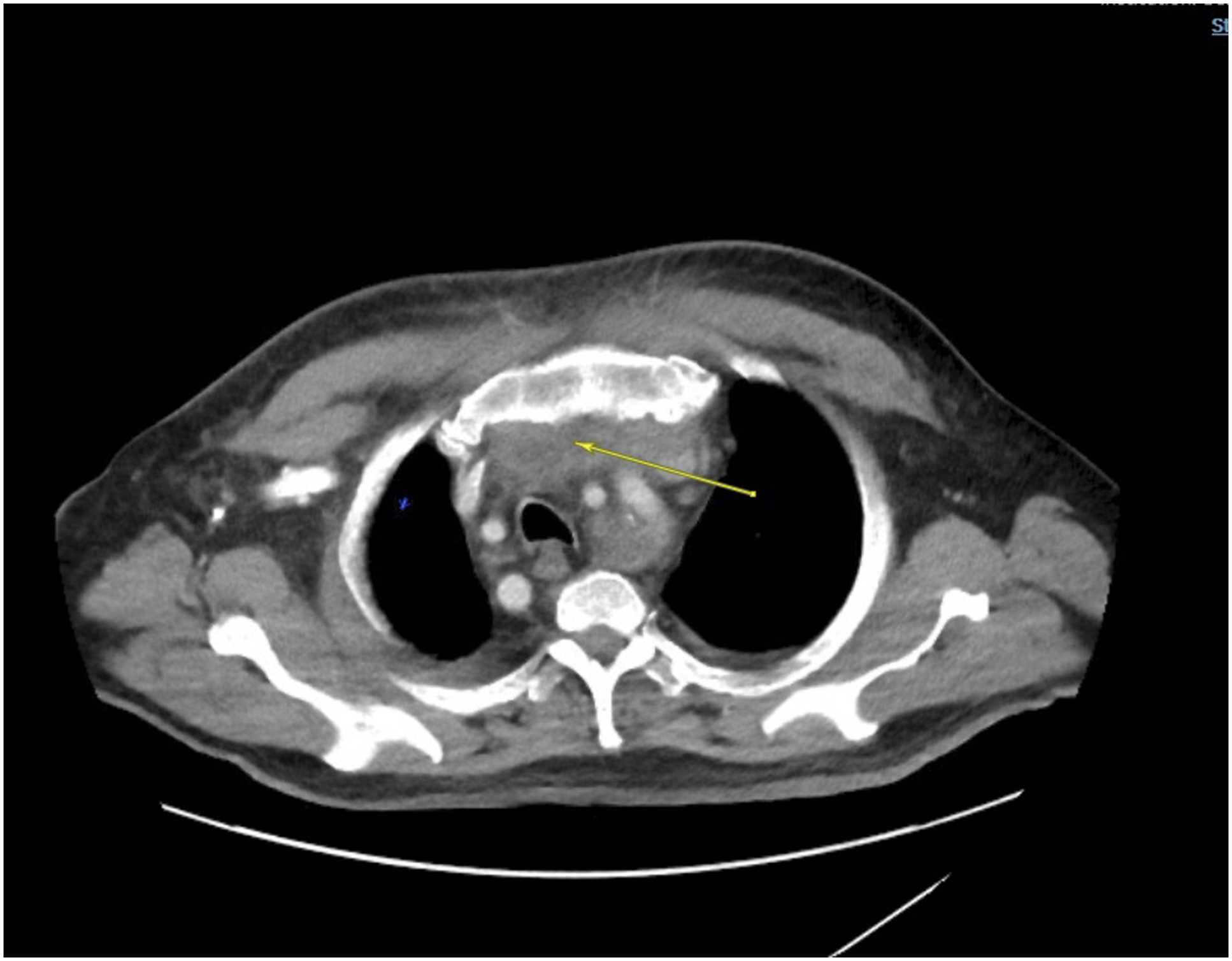
Comment
Mycotic aneurysms of the subclavian artery are extremely rare and have only accounted for less than 5% of all documented subclavian aneurysms over the past 3 decades. 6 They are often technically challenging to treat, and the open repairs, especially in poor surgical candidates, can be associated with significant morbidity and mortality. This case was particularly unique in several ways; the first being that no obvious inciting cause of the retrosternal abscess was found. He also had a significant interval increase in the aneurysm size (3 cm growth in 2 weeks), thus, highlighting the fact that infected aneurysms can grow drastically in short periods of time. In terms of operative technique, we chose a left axillary-to-left carotid bypass as it afforded us the least infected plane for tissue dissection as well as for our conduit path. The choice of an autologous great saphenous vein conduit was to reduce the risk of post-operative graft infection as we were working in a contaminated surgical field. Similar patency rates for autologous and antibiotic-soaked prosthetic grafts have also been reported. 6 Cryo-preserved allografts have also been used successfully in the absence of suitable autologous veins or prosthetic grafts. 7 The endovascular technique for repair has been described in some series either as a sole treatment strategy or as a bridge to open repair in high risk surgical patients or in patients presenting with ruptured aneurysms.8-10 The goals of open repair are complete aneurysmal sac excision, debridement of non-viable surrounding tissues, and upper extremity revascularization. 11 Complete sac excision was not done in our case as dissection and sac exposure was extremely difficult which would inevitably have prolonged the operative time in the face of already increasing transfusion requirements. This prompted our decision to widely open the sac, debride it, and leave a drain in place.
In conclusion, this case also illustrates the importance of a team approach in the successful management of mycotic aneurysms. The patient presented had a good outcome despite his advanced age and medical co-morbidities, a direct result of the early diagnosis and the combined efforts of the multiple disciplines directly involved in his care.
Footnotes
Declaration of Conflicting Interests
The author(s) declared no potential conflicts of interest with respect to the research, authorship, and/or publication of this article.
Funding
The author(s) received no financial support for the research, authorship, and/or publication of this article.