






Abstract
Purpose:
To report a case of delayed splenic rupture after percutaneous transsplenic portal vein stent deployment.
Case Report:
A 72-year-old male patient presented at a medical center with abdominal pain and reduced liver function according to laboratory tests. Due to a history of right hemihepatectomy and left portal vein occlusion, the percutaneous transhepatic approach was considered inappropriate. Instead, percutaneous transsplenic access was selected as a suitable procedure for portal vein catheterization. Eight days following the procedure, the patient developed abdominal pain, and a computed tomography scan showed a small splenic pseudoaneurysm that was underappreciated at the time. Patient suffered acute splenic rupture 32 days post-procedure. Subsequent embolization was performed, achieving complete hemostasis.
Conclusion:
The transsplenic approach should be considered when the transhepatic or transjugular approach is unfeasible or difficult to implement. A careful plugging of the puncture tract is necessary to prevent or minimize hemorrhage from the splenic access tract. In addition, careful serial follow-up computed tomography should be used to evaluate the splenic puncture tract.
Introduction
The successful endovascular treatment of portal vein (PV) occlusion or portal hypertensive varices can be achieved via various access methods, such as transvenous (via internal jugular vein), transhepatic, and transsplenic approaches. Transjugular access is commonly adopted for a transjugular intrahepatic portosystemic shunt, whereas percutaneous transhepatic PV access has been widely used to treat PV stenosis. However, these approaches may be unfeasible in the PV system in some cases related to perihepatic ascites, main PV occlusion, or non-visualized intrahepatic PV.1,2 In such cases, the percutaneous transsplenic approach is a suitable choice. Procedure-related major bleeding complications have been rarely reported after percutaneous transsplenic access.2-4 We report a case of delayed splenic rupture after applying the percutaneous transsplenic approach to treat PV occlusion and highlight the importance of serial follow-up imaging.
Case Report
A 72-year-old male presented at our medical center with abdominal pain and worsening liver function, as verified by laboratory tests. The patient was diagnosed with extrahepatic cholangiocarcinoma and underwent a right hemihepatectomy with hepaticojejunostomy. A contrast-enhanced computed tomography (CT) scan of the abdomen obtained 9 days postoperatively showed evidence of complete thrombosis of the main PV and proximal left PV. Due to the right hemihepatectomy and PV occlusion, a percutaneous transhepatic approach was considered inappropriate. Instead, a percutaneous transsplenic access was selected as a less risky approach because the patient presented a splenomegaly of 13 cm. Because of the large size of spleen, the location of an intrasplenic vein branch can be accurately veiwed using ultrasonography.
The segmental splenic vein was punctured with a 21G micropuncture needle under both sonographic and fluoroscopic guidance. Successful puncture was achieved after a single penetration of the splenic capsule. After insertion of a 6F sheath, selective portography showed a heavy burden of thrombosis and narrow post-stenotic left PV branches. A trial of pharmacomechanical thrombolysis was unsuccessful due to the chronic thrombus. After the subsequent pharmacomechanical thrombolysis and balloon angioplasty of the PV and preceding segments, major resolution of thrombus from the main PV to the left PV was observed. A discrepancy of the PV caliber at the level of left PV confluence was also observed. Therefore, after insertion of a 7F sheath, PV stent placement was performed. After completing the procedure, the sheath was carefully removed, and contrast was injected through the sheath to confirm its tip position within the splenic vein or transparenchymal puncture tract. The contrast medium extending through the puncture tract to the perisplenic space allowed us to determine the position of the sheath within the puncture tract and specify the distance between the sheath tip and splenic capsule. After positioning one 3 mmx5 cm 0.035-inch coil (Meyer; Cook Medical, Bloomington, IN, USA) at the intended closure site, the coil was pushed using a 0.035-inch guide wire while fixing the coil in position. Then, the sheath was completely removed by injection of n-butyl-2-cyanoacrylate (NBCA) glue (Histoacryl, B Braun, Melsungen, Germany) and a lipiodol mixture (ratio 1:2) throughout the puncture tract in the splenic parenchyma, splenic capsule, and abdominal wall (Figure 1).

A, Fluoroscopic image and (B) transverse contrast-enhanced CT image show successful embolization of percutaneous transsplenic tract by a coil (white arrow) and NBCA glue (black arrows). Note the NBCA glue along the puncture tract in splenic parenchyma, splenic capsule, and abdominal wall.
The patient received postoperative antibiotics (intravenous cefazolin) to treat any post-procedural infection. The patient complained of abdominal pain on day 8 after the procedure and underwent a CT examination. The examination showed a small pseudoaneurysm in the splenic parenchyma, which was underappreciated at the time. On day 23 after the procedure, another CT examination was performed due to persistent abdominal pain. In addition to the small pseudoaneurysm, a small amount of fluid collection around the pseudoaneurysm was now present. On day 32 after the procedure, the patient complained again of persistent abdominal pain and laboratory exam revealed a drop in serum hemoglobin level. The ruptured pseudoaneurysm was revealed on CT images (Figure 2). Splenic angiography showed contrast medium extravasation in the spleen around the previous puncture site. Subsequent embolization using polyvinyl alcohol particles (Bearing; Merit Medical, South Jordan, UT, USA) was performed (Figure 3), finally achieving complete exclusion of the bleeding focus. After 2 months of follow-up, the patient recovered with patent PV and satisfactory liver function.
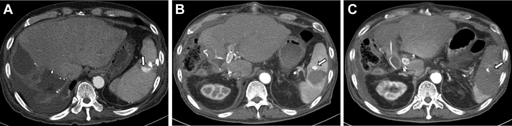
Serial transverse enhanced CT follow-up images. A, CT scan 8 days after procedure shows a small splenic pseudoaneurysm near the previous transsplenic puncture site (arrow). B, CT scan 23 days after procedure shows persistence of splenic pseudoaneurysm (arrow) with a newly developed intrasplenic hematoma. C, CT scan 32 days after procedure shows splenic rupture with peritoneal hematoma (arrow).

A, Selective angiogram of splenic artery reveals contrast medium extravasation near the previous transsplenic puncture site (arrows), which was embolized with polyvinyl alcohol particles. B, Completion angiogram shows no residual contrast medium extravasation.
Discussion
The spleen can be useful to achieve an alternative percutaneous approach for endovascular PV treatment, especially when transhepatic or transjugular access is inappropriate. Indeed, transsplenic access in PV procedures provides a straight path to the PV. Several studies have reported the feasibility and safety of the transsplenic approach for PV intervention.3,5-7 Although the transsplenic approach provides effective access, the associated bleeding risk remains controversial. The fundamental complication of splenic vein catheterization is bleeding from the splenic puncture tract. Bleeding is more likely to occur in thrombocytopenia, splenomegaly, and portal hypertension.1,8
Successful access to the intraparenchymal splenic vein is key to effective transsplenic access. However, the tortuous course of the splenic vein may prevent catheter advancement, and more bleeding complications may occur compared with other access types.4,7,8 Therefore, the puncture route should be carefully planned regarding aspects such as the site, direction, depth, and intrasplenic vein branches. 2 Several studies have reported successful embolization of the transsplenic tract using Gelfoam and NBCA glue or a combination of Gelfoam and coils,2,4,8 but bleeding complications may occur in some patients. In contrast, no major bleeding complications have been reported after embolization of the transsplenic route by using a combination of coils and NBCA glue.1,5
The spleen is a hypervascular organ that communicates with the parenchymal tissue through complex channels. Some studies have analyzed mechanisms for delayed splenic hemorrhage, such as the direct action of fracture edges on the splenic capsule and parenchyma, possibly resulting from patient mobilization and producing delayed laceration.9,10 Other studies have suggested that delayed splenic rupture may be mainly related to inadequate radiological investigations. 9 Unfortunately, our patient had a delayed splenic rupture after the procedure despite successful transsplenic tract embolization combining coils and NBCA glue. On the early follow-up CT image, a small splenic pseudoaneurysm was observed. If the pseudoaneurysm had been noticed earlier, splenic hemorrhage may have been avoided. Therefore, clear recommendations should be set for both short- and long-term follow-up imaging of the splenic tract. We recommend obtaining a CT scan 1 week post-procedure. Contrast-enhanced ultrasonography can also be helpful for tract evaluation, but its usefulness is limited due to metallic artifacts.
Conclusion
Although various studies have evaluated the safety of the percutaneous transsplenic approach to the portal vein, we propose that this approach should be considered when transhepatic or transjugular access is unfeasible or difficult. To prevent or minimize hemorrhage from the splenic access tract, the puncture tract should be carefully plugged with embolic material. Most importantly, serial surveillance follow-up CT or contrast-enhanced ultrasonography is mandatory to assess the splenic puncture tract and rule out bleeding complications which can result in splenic hemorrhage.
Footnotes
Authors’ Contributions
HJH: Acquisition and analysis of the work, Drafted the work. KJH: Conception of the work and substantively revised it. LKH: Writing—review & editing. PS: Writing—review & editing. All authors have checked the authorship to a submitted version and agreed to the author list and contributions.
Consent for Publication
Written informed consent was obtained from the patient for publication of this case report and any accompanying images.
Declaration of Conflicting Interests
The author(s) declared no potential conflicts of interest with respect to the research, authorship, and/or publication of this article.
Ethical Approval
All procedures performed in human participants were in accordance with the Declaration of Helsinki and approved by the Institutional Ethical Committee (Gachon University Gil Medical Center, GFIRB2020-331).
Funding
The author(s) received no financial support for the research, authorship, and/or publication of this article.

Informed Consent
Informed consent was obtained from the individual included in the study.