
Abstract
High-flow priapism is an uncommon condition typically resulting from penile or perineal trauma, due to laceration of cavernosal artery. We present a case of 24-year-old male who presented with post-traumatic painless priapism. Ultrasound showed hematoma with arterio-cavernosal fistula. On CT Angiogram, the cavernosal artery was seen arising from accessory pudendal artery, which arose from inferior epigastric artery (IEA), branch of external iliac artery (EIA). Catheter angiogram of EIA showed fistulous communication at the base of the penis from a branch of IEA. Selective embolisation of the artery was done using 33% glue (n-butyl cyanoacrylate). Post embolisation, no residual filling of the fistula and partial detumescence of penis was noted. Transarterial embolisation is usually preferred as first line of management in high-flow fistulous priapisms.
Keywords
Introduction
Priapism is an uncommon medical condition characterized by prolonged tumescence or erection of the penis for more than 4 hours unrelated to sexual stimulus or desire. 1 Clinically it is classified into low-flow priapism (veno-occlusive or ischaemic) and high-flow priapism (arterial or non-ischaemic). Its identification is important as lack of timely treatment can result in persistent erectile dysfunction due to irreversible corporal fibrosis. High-flow type usually follows perineal or penile trauma with disruption of cavernosal artery, and resulting in arterio-cavernosal fistula. 2 The goal of management in arterial priapism is to achieve detumescence by obliterating the fistula. Here we present a case of high-flow priapism with unique anatomical variation of origin of the cavernosal artery from accessory pudendal artery which was successfully managed by endovascular embolization.
Case Report
A 24-year-old male came with complaints of persistent painless penile erection for 14 days. He had history of road traffic accident 3 weeks prior to erection, with perineal trauma. There was no obvious swelling at the perineum or bleeding from the penis at the time of trauma. Treatment with local ice bag pressure was attempted, but no improvement was noted. On local examination, penis showed grade III rigidity; Erectile hardness score 3 (Supplementary file 1). There was no evidence of penile tenderness, perineal tenderness or testicular swelling.
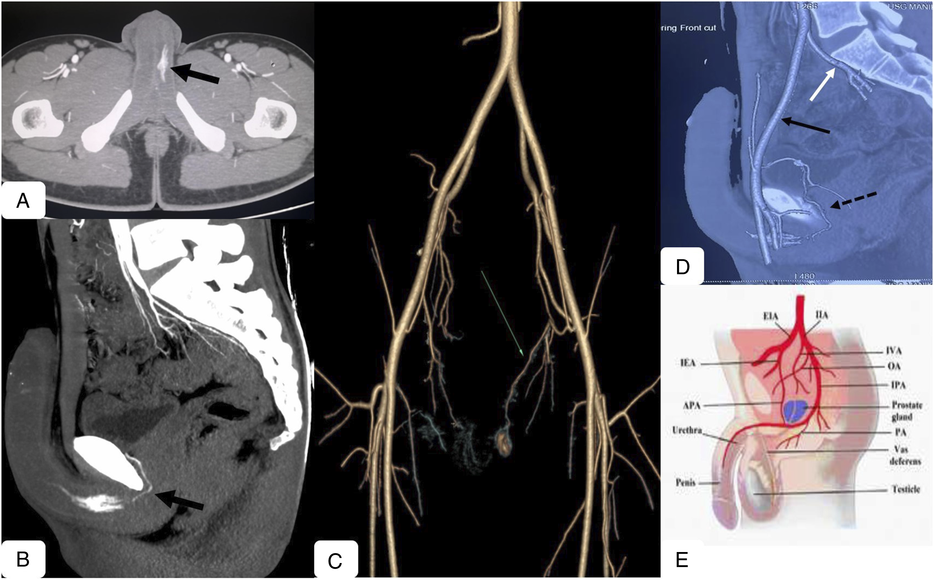
Penile Doppler (PD) was done which showed turbulent high velocity low resistance flow in left cavernosal artery (CA), whereas the right CA showed normal high resistance flow. Hypoechoic areas were noted in body of bilateral corpora cavernosa, suggestive of hematoma (Figure 1). CT Angiogram (CTA) showed extravasation of intravenous contrast from the CA into the venous sinusoids. Normally, CA arises from internal pudendal artery (IPA) which is a branch of internal iliac artery (IIA). But in our patient, the CA did not arise from the IIA. The fistula was seen from the branch of inferior epigastric artery (IEA), from external iliac artery (EIA). Based on literature search, this branch of IEA was found out to be accessory pudendal artery (APA) (Figure 2). Ultrasound images of 24-year-old male presenting with painless penile erection and high-flow arterio-cavernosal fistula. (a) B-mode image showing hematoma (*) in the body of bilateral corpora cavernosa; (b) Color Doppler image showing aliasing within the cavernosal artery and likely site of arterio-cavernosal fistula; (c) Spectral Doppler image showing high velocity (PSV-100 cm/s) low resistance flow in left cavernosal artery; and (d) low velocity high resistance flow (normal flow) in right cavernosal artery. Contrast Tomographic (CT) images of 24-year-old male presenting with painless penile erection and high-flow arterio-cavernosal fistula. (a) Axial CT Angiogram image in arterial phase showing clustering of intravenous contrast in the body of left corpora cavernosa (arrow); (b) Sagittal CT Angiogram image showing contrast extravasation at the base of the penis (arrow); (c) Volume Rendered Image showing the fistula arising from accessory pudendal artery (APA), a branch of inferior epigastric artery (IEA), from left external iliac artery (EIA) [arrow]; (d) Sagittal maximum intensity projection (MIP) image showing cavernosal artery arising from APA (dashed black arrow), branch of IEA from EIA (black arrow), [white arrow - internal iliac artery]; (e) Schematic image showing APA arising from IEA (Image courtesy: Thai CT et al. Pelvic magnetic resonance imaging angioanatomy of the arterial blood supply to the penis in suspected prostate cancer patients. Eur J Radiol. 2015 May; 84 (5):823-7).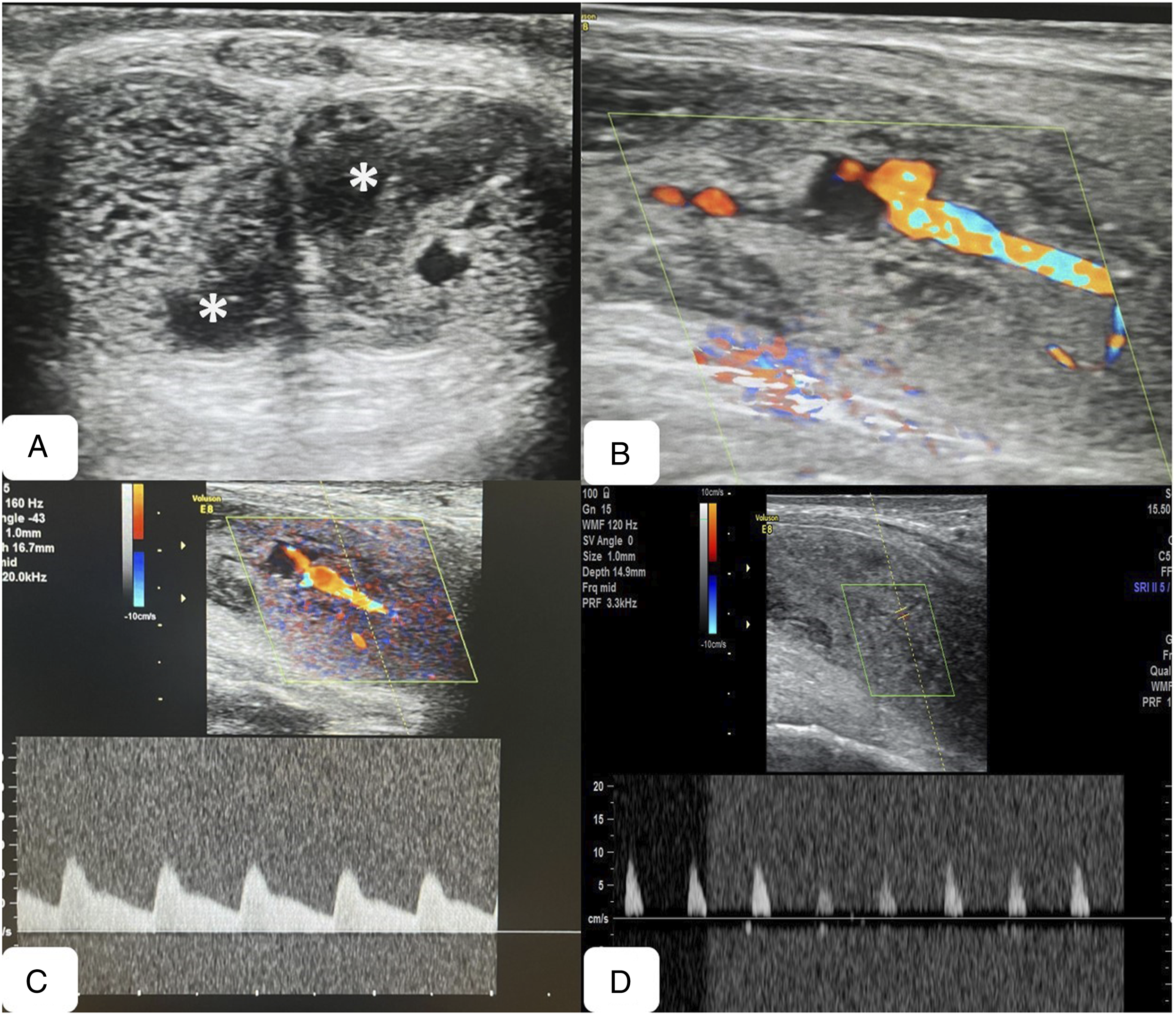
The patient was planned for transarterial embolisation (TAE) to occlude the fistula. Under local anaesthesia, through right transfemoral approach, left iliac angiograms were performed. There was no fistula seen from IIA angiogram. EIA angiogram showed fistulous communication at the base of the penis from a branch of IEA. Selective cannulation of the artery was done using microcatheter and the fistula embolised with using 33% glue (n-butyl cyanoacrylate; NBCA). Post embolisation, no residual filling of the fistula was noted. Immediately post procedure, partial detumescence of penis was noted. PD performed 48 hours later showed low velocity monophasic flow in the left CA (Figure 3). On follow up after 3 months, the patient did not have complaints of erectile dysfunction. Digital Subtraction Angiographic (DSA) images of 24-year-old male undergoing embolisation for high-flow arterio-cavernosal fistula. (a) Left external iliac artery (EIA) angiogram image showing the fistula arising from a branch of inferior epigastric artery (IEA) - Accessory Pudendal artery (APA), supplying the fistula at the base of the penis; (b) Selective micro-catheterisation of APA delineating the fistula; (c) Embolisation of the branch of APA and fistula using 33% glue; (d) Post embolisation image showing complete obliteration of fistula with normal filling of proximal APA and IEA.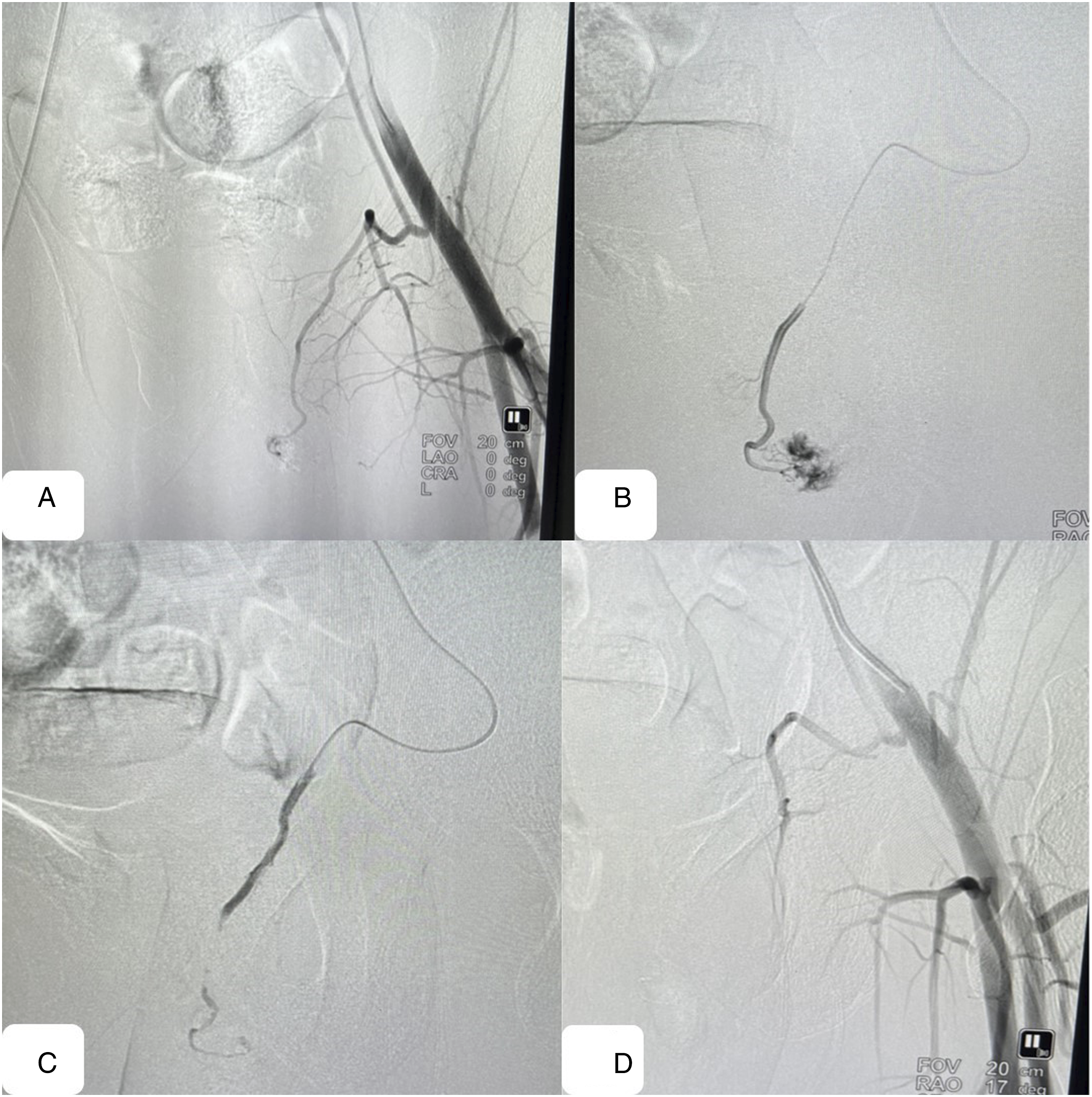
Discussion
Blunt perineal trauma is the most common cause for high-flow priapism, especially in young adults. Trauma causes laceration of the cavernosal arteries with direct communication between the arterial blood and cavernosal sinusoids, resulting in arterio-cavernosal fistula. The turbulent flow due to the fistula stimulates the release of endothelium derived nitric oxide resulting in trabecular dilatation and persistent erection. 3 Unlike ischemic priapism, there is no sinusoidal thrombosis or infarction, and hence arterial priapism is usually painless. Arterial priapism can occur immediately following trauma or can occur after delay of few days due to initial spasm of the injured vessel. 4
The cavernosal artery arises from penile artery which in around 50-60% of the cases, originates from internal pudendal artery (IPA), a branch of internal iliac artery (IIA). In 35-40% cases, the penile artery in addition to arising from IPA, can also arise from accessory pudendal artery (APA). In 2-5%, penile artery can solely arise from APA. APA is located within peri-prostatic region, running caudally towards the anterior perineum. 5 The origin of APA is varied, with most commonly arising from either obturator or inferior vesical arteries from IIA. In around 6-10%, APA can arise from inferior epigastric artery (IEA), a branch of external iliac artery (EIA). 6 Our case was unique in the aspect that left cavernosal artery was seen arising from APA which in turn arose from IEA. There was no cavernosal supply from left IPA/IIA. The main clinical significance of such an uncommon variation lies in the fact that inadvertent injury to IEA during inguinal surgeries or dialysis catheter placements, can lead to erectile dysfunction.5,6
Arterial priapism being non-ischemic, conservative management in the form of ice and compressive perineal dressing can be tried as an alternative option. The rationale behind medical management is to cause vasospasm of the inured vessel with subsequent thrombosis and spontaneous detumescence. 7 However, excessive arterial inflow can lead to hemodynamic alterations causing damage to cavernosal smooth muscle resulting in secondary irreversible corporal fibrosis and eventually erectile dysfunction. For these reasons, endovascular embolisation is usually preferred as first line of management in high-flow arterio-cavernosal fistulous priapisms. 8 Selective transarterial embolisation (TAE) has a reported detumescence rate of almost 90-100% with recurrence rates of 6-20% in literature.7-10 The various embolic agents which can used for TAE include microcoils, polyvinyl alcohol (PVA) particles, n-butyl cyanoacrylate (NBCA; glue), gel-foam and autologous blood clot.8,10 The choice of embolic agent usually depends upon the expertise of the interventionist and doesn’t significantly alter the treatment results or penile erection. Erectile dysfunction post embolisation can be seen in 10-20% of the patients. 10 Surgical treatment consists of resection of arterio-cavernosal fistula or ligation of pudendal/cavernosal artery.1,8 However, they are associated with more chances of erectile dysfunction. All these patients with arterio-cavernosal fistulas who undergo treatment should be followed up at regular intervals to detect any recurrence or occurrence of erectile dysfunction.
Conclusion
High-flow priapism is an uncommon condition caused due to penile or perineal blunt trauma. TAE is relatively safe and effective procedure and preferred treatment for arterio-sinusoidal fistula following traumatic priapism. The origin of cavernosal artery can be varied and have to be kept in mind prior to embolisation.
Supplemental Material
Supplemental Material - Post-Traumatic High-Flow Priapism: Accessory Pudendal Origin of Cavernosal Artery From External Iliac Artery - A Case Report
Supplemental Material for Post-Traumatic High-Flow Priapism: Accessory Pudendal Origin of Cavernosal Artery From External Iliac Artery - A Case Report by Karan Visanji Dedhia, DNB, S. Vignesh, MD, PDCC, T. Mukuntharajan, DMRD, N. Karunakaran, DMRD, DNB(RD), and Ganesh Rajagopal, DMRD, DNB(RD), FRCR in Vascular and Endovascular Surgery
Footnotes
Acknowledgments
I would like to thank the Department of Urology for referring the case.
Declaration of Conflicting Interests
The author(s) declared no potential conflicts of interest with respect to the research, authorship, and/or publication of this article.
Funding
The author(s) received no financial support for the research, authorship, and/or publication of this article.
Ethical Approval
Patient’s informed consent was obtained during the procedure. Care has been taken not to disclose the patient’s identity directly or indirectly in any form. Institutional ethical committee approval was obtained for this study.
Supplemental Material
Supplemental material for this article is available online.
References
Supplementary Material
Please find the following supplemental material available below.
For Open Access articles published under a Creative Commons License, all supplemental material carries the same license as the article it is associated with.
For non-Open Access articles published, all supplemental material carries a non-exclusive license, and permission requests for re-use of supplemental material or any part of supplemental material shall be sent directly to the copyright owner as specified in the copyright notice associated with the article.