
Abstract
Venous stasis ulcers are nonhealing lesions due to venous hypertension secondary to valvular dysfunction or deep venous outflow obstruction. We describe a case of a 71-year-old male with a history of polycythemia vera, secondary myelofibrosis, and massive splenomegaly up to 38 cm who presented with chronic, perimalleolar venous stasis ulcers and pain on the left lower extremity. CT showed significant compression of the left common iliac vein due to mass effect from the spleen. He was managed medically while being evaluated for partial splenic artery embolization but expired due to other chronic conditions before any intervention could be performed. Partial splenic artery embolization may be considered as a treatment option for patients with symptomatic iliac vein compression due to massive splenomegaly secondary to myelofibrosis, as long as extramedullary hematopoiesis is not compromised.
Introduction
Venous stasis ulcers are open lesions, often located in the gaiter region of the lower extremity, that do not heal on their own. In many Western countries, these ulcers represent a common, chronic problem, with an estimated prevalence of 1% to 3%, which increases in patient populations over 65 years of age, and is the prevailing type of lower extremity ulcer.1-3 Though the exact pathogenic steps are still under investigation, the typical pathophysiology of venous ulcers is venous hypertension, which can occur secondary to superficial, communicating, or deep vein valvular dysfunction, deep venous outflow obstruction, and muscle dysfunction.1,3 The present report describes a case of nonhealing venous ulcers on the left lower extremity, which were thought to be caused by compression of the left common iliac vein (LCIV) due to massive splenomegaly from secondary myelofibrosis. To our knowledge, this specific cause of deep venous outflow obstruction has not yet been described in the literature.
Case Report
A 71-year-old male with a history of polycythemia vera and secondary myelofibrosis presented to the vascular surgery clinic with chronic, nonhealing venous stasis ulcers (Figure 1) and pain in the left lower extremity (LLE). At the time of presentation, he had been followed by wound care for 2 years and had presented to the Emergency Department several times because of his ulcers. Physical examination was significant for a massive, firm, palpable spleen that extended into the pelvis as well as left medial and lateral perimalleolar ulcers. Reflux studies on the left lower extremity showed no evidence of acute or chronic deep venous thrombosis, no evidence of venous valvular incompetence in the deep venous system, and no evidence of acute or chronic superficial vein thrombosis, though there was evidence of superficial vein incompetence along the distal thigh great saphenous vein and mid-calf short saphenous vein. Medial and lateral views of nonhealing perimalleolar ulcers on the left lower extremity.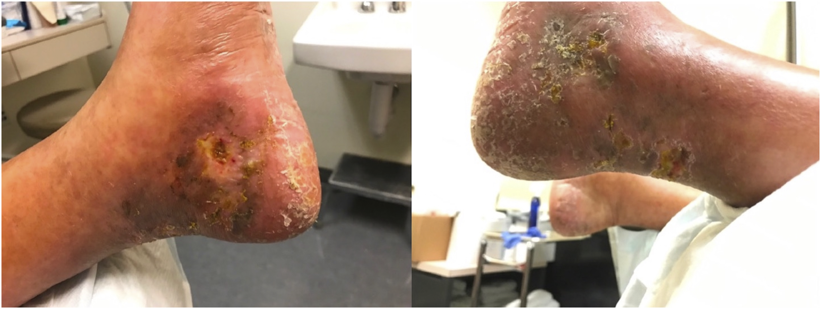
The patient was diagnosed with polycythemia vera 30 years prior and had been undergoing treatment with hydroxyurea during most of that time, except for a period between June 2020 and February 2021. He developed secondary myelofibrosis with marked splenomegaly up to 38 cm which was observed on multiple abdominal ultrasounds and CT scans in the 10 years leading up to presentation. A left upper back mass biopsy and bone marrow biopsy, both performed 2 years prior, confirmed extramedullary hematopoiesis and markedly fibrotic marrow with atypical megakaryocytic hyperplasia consistent with persistent myeloproliferative neoplasm, respectively. Further review of the abdominal CT demonstrated significant compression of the proximal left common iliac vein, likely by the distal aorta due to mass effect from the spleen, resulting in venous stasis of the left lower extremity (Figure 2). Coronal and axial views of patient’s CT scan of the thoracic, abdominal, and pelvic cavities. The spleen measured up to 38 cm from the superior to inferior poles. The black arrow indicates the area of compression of the left common iliac vein.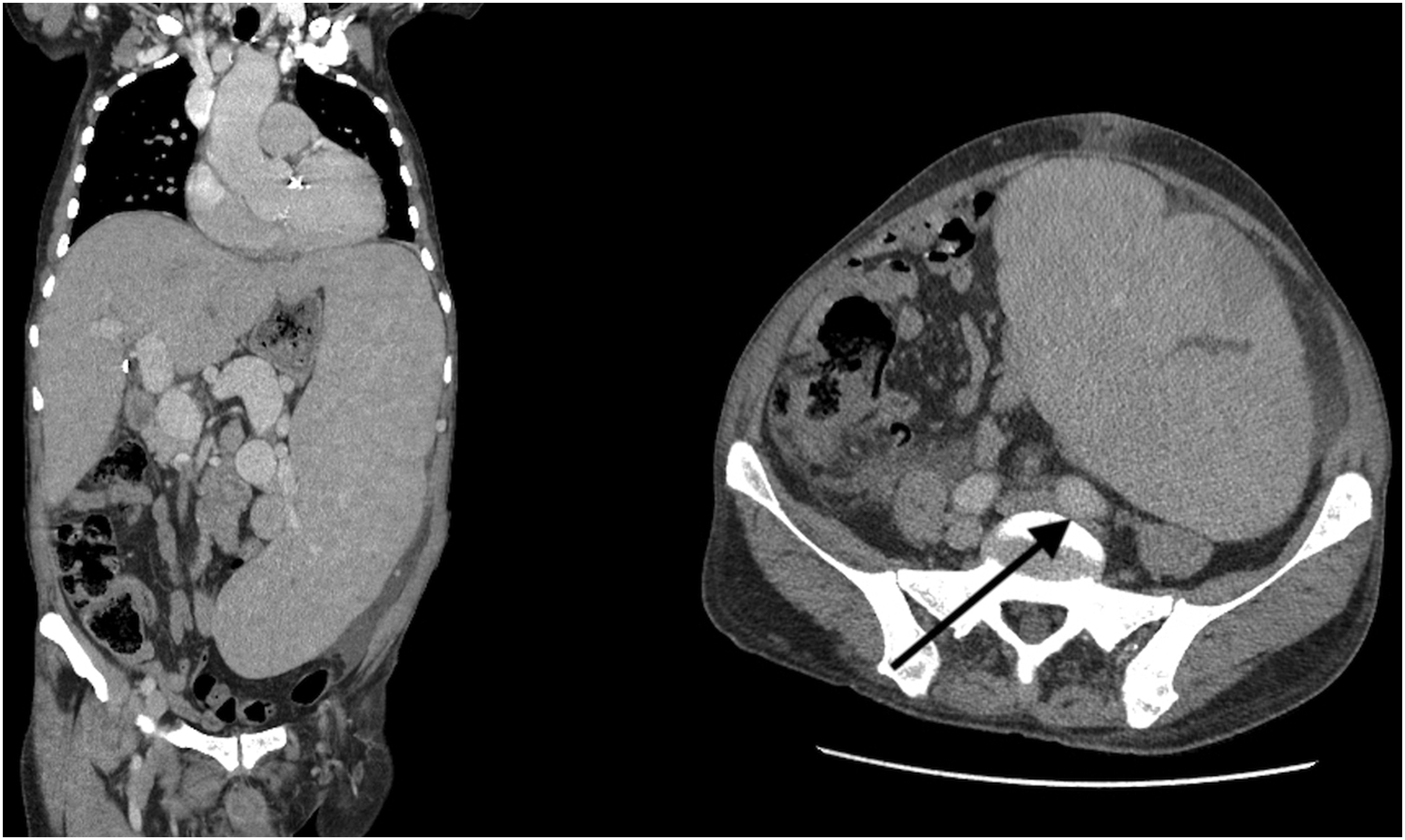
The patient was treated with leg elevation, 30-40 mmHg compression with Unna boot therapy, wound care, and discontinuation of hydroxyurea. He was subsequently evaluated for splenectomy, but the team eventually decided against splenectomy after consultation with hematology cautioned that the spleen was a significant source of hematopoiesis due to the patient’s myelofibrosis. At a multidisciplinary conference involving vascular surgery, interventional radiology, and hematology/oncology, the option of partial splenic artery embolization was presented as a means of shrinking the spleen, thereby relieving the compression of the left common iliac vein, while simultaneously preserving hematopoiesis by preventing the removal or total embolization of the entire spleen. Intravascular ultrasound and venous stenting were considered for this patient but was ultimately decided against due to concerns that the mass effect from the spleen would compromise the integrity of the stent. Though the patient agreed to this plan, he expired due to his other chronic conditions before any intervention could be performed. The patient expired at a skilled nursing facility 3 days after being discharged in stable condition from the hospital where he was admitted for septic shock secondary to a lower respiratory tract infection and bilateral lower extremity cellulitis.
Discussion
This report documents an unusual patient case of left lower extremity venous stasis ulcers in the context of secondary myelofibrosis resulting in massive splenomegaly and left common iliac vein (LCIV) compression. Though LCIV compression is a common cause of nonhealing ulcers on the left side, this is most often due to compression via the right common iliac artery (RCIA), known as May-Thurner Syndrome (MTS). The presentation of MTS is often asymptomatic, though clinical symptoms can include venous thromboses and unilateral left lower extremity edema, pain, heaviness, venous claudication, hyperpigmentation, varicosities, and venous ulcers.4,5 50% stenosis of the iliac vein can be present in up to 37% of patients with healed and active venous ulcers, 6 implying that LCIV compression may be a relatively common contributor in many cases of venous ulcers. The Clinical-Etiology-Anatomy-Physiology (CEAP) classification system for describing patients with chronic venous disorders was updated in 2020 to allow for further characterization of secondary vascular disease with the addition of the Esi (chronic venous disorder secondary to an intravenous etiology) and Ese (chronic venous disorder secondary to an extravenous etiology) classifications; this case described in this report would fall into Ese category. 7
May-Thurner Syndrome is most often attributed to compression via the RCIA, but other pelvic structures have also been identified to cause LCIV compression, including the left common iliac artery, uterine leiomyoma, prominent bony structure, and aneurysms on either the LCIA or RCIA.4,8-10 Though there is significant variation in the exact etiology that causes MTS, many symptomatic patients present with the same or similar symptoms, 4 and previous case reports have reported success with endovascular stent placements.8,9,11 However, to our knowledge, LCIV compression secondary to massive splenomegaly has not been previously reported in the literature and is a unique presentation that draws special consideration for treatment.
Splenomegaly due to extramedullary hematopoiesis is one of the most characteristic features of myelofibrosis12,13 and can be treated through either pharmacologic or interventional options. JAK1/2 inhibitors are often the mainstay of therapy, though patients can become refractory to treatment over time.12,13 Our patient was initially pharmacologically managed with fedratinib, but did not tolerate the medication well over time and white blood cell counts continued to trend upwards. Nonmedical management options include splenectomy, splenic irradiation, and partial splenic artery embolization (PSE) for patients who are refractory or intolerant to pharmacological therapy. In the present case, PSE was deemed the safest intervention after bone marrow biopsy showed fibrotic marrow, suggesting that the spleen likely played an important role in the patient’s hematopoiesis. A previous case report demonstrated favorable outcomes with preoperative PSE followed by laparoscopically-assisted splenectomy in 2 patients with myelofibrosis and massive splenomegaly. 14 However, there is a general paucity of literature in utilization of PSE for splenomegaly in the context of myelofibrosis specifically. 13 Further, a large single-center study of 982 obstructive venous outflow lesions in 870 patients undergoing iliac stents found that relieving the obstruction healed venous ulcers in 58% of patients. 15 Together, the lack of proven treatment options identified in the literature and the knowledge that stenting other similar types of venous obstruction resolves only 58% of venous ulcers explain the rationale of first proceeding with relief of the LCIV external compression. PSE would likely be most appropriate in a patient who can safely undergo the procedure without jeopardizing hematopoiesis.
Another important factor to consider in our patient case, and in similar cases of ulcers in the context of polycythemia vera and secondary myelofibrosis, is hydroxyurea (HU) as a possible iatrogenic etiology of venous ulcers, which has been documented previously in the literature.16-24 The patient’s usage of hydroxyurea is a possible confounder in his clinical picture and explains the reasoning, in part, that splenomegaly was not pursued, along with the understanding of the spleen’s role in hematopoiesis. HU-induced ulcers occur in approximately 9% of patients on high-dose, long-term HU therapy for myeloproliferative disease 25 and typically present with characteristic clinical features that include occurrence in patients on long-term HU treatment of at least 1 g/day17,22,23 and perimalleolar localization.19,21-24 Though this pattern of presentation is similar to that of our patient, it should be noted, however, that in nearly all cases reported in the literature, the most distinguishing characteristic of HU-induced ulcers was that the ulcers promptly healed upon cessation of hydroxyurea.17,19,21-25 In our patient, there was a period of approximately 8 months between June 2020 and February 2021 where he discontinued hydroxyurea, yet his nonhealing ulcers persisted. Because of this, we postulate that iliac vein compression remains the most likely explanation for the chronic venous ulcers, though the etiology may indeed be multifactorial.
Conclusion
Myelofibrosis with massive splenomegaly can result in LCIV compression. Our case report documents a patient with symptoms of MTS – including LLE venous stasis ulcers, pain, and edema – most likely due to LCIV compression via splenomegaly. PSE should be considered as a potential treatment strategy, with caution, in patients with venous ulcers secondary to myelofibrosis and splenomegaly in which splenectomy is not a treatment option due to splenic extramedullary hematopoiesis.
Footnotes
Declaration of Conflicting Interests
The authors declared no potential conflicts of interest with respect to the research, authorship, or publication of this article.
Funding
The author(s) received no financial support for the research, authorship, and/or publication of this article.