
Abstract
AN0128 is a boron-containing compound with antibacterial and anti-inflammatory properties. To test its potential effectiveness in treating periodontal disease, we induced experimental periodontitis in the rat by placing ligatures and assessed the impact of AN0128 and positive and negative controls by micro-CT and histologic measurements. The formation of an inflammatory infiltrate was measured in hematoxylin-and-eosin-stained sections. Daily application of AN0128 (1%) compared with controls reduced bone loss by 38 to 44% (P < 0.05), while vehicle alone had no effect (P > 0.05). The reduction in bone loss with AN0128 was similar to that achieved with a NSAID, ketorolac, and Total toothpaste containing triclosan. AN0128 also reduced the level of gingival inflammation 42% compared with the ligature only (P < 0.05), whereas vehicle alone had no effect (P > 0.05). The results indicate that AN0128 significantly reduces the formation of an inflammatory infiltrate and reduces bone loss, measured histologically and by micro-CT.
INTRODUCTION
Periodontal diseases are induced by bacterial biofilms that stimulate a host response in gingival connective tissue that leads to destruction and bone loss (Rosan and Lamont, 2000; Graves and Cochran, 2003; Haffajee and Socransky, 2006). Periodontitis occurs in approximately 15 to 25% of the US population and is the most common cause of tooth loss in adults in the United States (Borrell et al., 2005). A critical event in the initiation of periodontal disease is the colonization of the tooth by bacteria and bacterial invasion of connective tissue (Sanavi et al., 1985; Holt et al., 1988; Listgarten and Loomer, 2003; Haffajee et al., 2006). This leads to stimulation of the host, resulting in activation of innate and acquired immune responses (Kornman, 1999; Graves and Cochran, 2003; Teng, 2003). Cause-and-effect relationships between periodontal disease and initiating factors have been established by the use of specific inhibitors, which, taken together, have indicated that the production of prostaglandins, interleukin-1, tumor necrosis factor, and RANK ligand is likely to be an important trigger in the disease process (Williams et al., 1985; Assuma et al., 1998; Teng et al., 2000; Delima et al., 2002).
Several different models of periodontal disease have been used to establish bacteria as a critical early initiator. In rat models, placement of a ligature around the teeth causes the formation of a pathogenic biofilm, gingival inflammation, and bone loss (Rovin et al., 1966; Kenworthy and Baverel, 1981; Liu et al., 2006). In contrast, ligatures placed around the teeth of germ-free rats have little effect on gingival inflammation and do not induce periodontal bone loss (Rovin et al., 1966). Furthermore, topical application of an antibacterial agent, chlorhexidine, and treatment with antibiotics reduce loss of bone, supporting the role of bacteria in initiating destruction in this model (Weiner et al., 1979; Kenworthy and Baverel, 1981). In contrast, increasing Gram-negative bacterial burden enhances osteoclastogenesis and bone resorption (Samejima et al., 1990).
Borinic acid quinoline esters are a recently identified class of new antibacterial compounds (Benkovic et al., 2005). One of these compounds, AN0128, is currently undergoing clinical trials for the treatment of atopic dermatitis, which is associated with Staphylococcus aureus colonization of the skin (Baker et al., 2006). To test the potential effectiveness of AN0128 as a treatment for periodontal disease, we applied AN0128 topically in a rat ligature model of experimental periodontitis.
MATERIALS & METHODS
Animals
All experimental procedures involving animals were approved by the Institutional Animal Care and Use Committees at the Boston University Medical Center. Twelve-week-old male Sprague-Dawley rats (weighing from 275 to 300 g each) were purchased from Charles River Laboratories (Wilmington, MA, USA). In the first set of experiments, rats were divided into 5 treatment groups (n = 6): 1% AN0128 in 40% Transcutol P, 40% PBS, and 20% ethanol vehicle (Anacor Pharmaceuticals, Palo Alto, CA, USA); 0.5% ketorolac tromethamine ophthalmic solution (Allergan, Inc., Irvine, CA, USA); vehicle alone; ligature only; and no ligature. Ketorolac was used as the positive control, since it has been reported to reduce periodontal bone loss (Jeffcoat et al., 1995). Cotton thread ligatures were placed around the cervix of the right and left second maxillary molars. The experimental drug and positive and negative controls were applied locally to the second maxillary molars with a tuberculin syringe once daily. In the second set of experiments, treatment groups consisted of Total toothpaste (0.3% triclosan, 2% copolymer, and 0.243% sodium fluoride; Colgate-Palmolive, New York, NY, USA), 5% AN0128, or vehicle applied daily via a cotton-tip applicator. (AN0128 is available from Anacor Pharmaceuticals upon request.) Total toothpaste was used as a positive control, since it has been shown to reduce the progression of periodontal bone loss in susceptible persons in human clinical trials (Rosling et al., 1997). The experiments were carried out with small groups of animals so that, in any individual experiment, n = 2 or n = 3. The results from the different "rounds" of experiments were then combined so that, in the end, n = 6 per group. In no case were all of the animals treated at a single time.
Specimen Preparation
Animals were euthanized by CO2 overdose, and the intact jaws and associated structures were fixed for 72 hrs in cold 4% paraformaldehyde. The right and left maxillary jaws were then dissected free and examined by micro-CT (microCT-40, Scanco Medical AG, Bassersdorf, Switzerland), after which they were decalcified by incubation in cold Immunocal (Decal, Congers, NY, USA) for approximately 12 days, with solution changed daily, then embedded in paraffin, and sectioned in the mesiodistal plane at 5 microns. Hematoxylin-and-eosin-stained sections at a midpoint in the buccal-palatal direction were identified by visible root canal systems in adjacent teeth. In most cases, 2 tissue sections were examined (on the right and left sides of the maxillary right and left second molars of each animal) for inflammation and loss of bone.
Micro-CT analysis
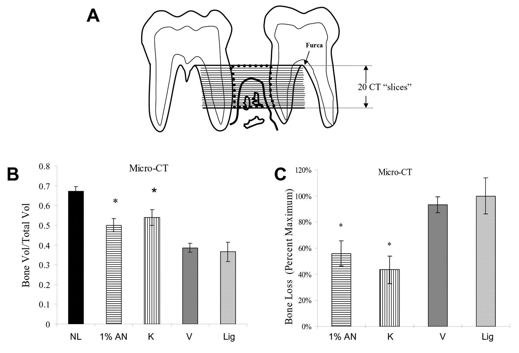
The sagittal plane of each specimen was set parallel to the x-ray beam axis. The specimens were scanned at a resolution of 16 μm in all 3 spatial dimensions. To facilitate reproducible analysis, the interdental area between the first and second and second and third molars was examined, starting coronally by a line drawn between adjacent molar furcations, continuing in an apical direction for 20 continuous scan slices. The outer walls of the scan were determined by the root surface. The results represent the residual bone volume (BV) per total volume (TV) for 20 slices. The latter includes the PDL space so that the [BV/TV x 100] was always less than 100%.
Histomorphometric Analysis of Hematoxylin-and-eosin-stained Sections
Histomorphometric analysis was performed with Image ProPlus software (Media Cybernetics, Silver Spring, MD, USA). The bone area was assessed by the amount of bone coronal to a line 1 mm below the adjoining cemento-enamel junction in the mid-interproximal region extending coronally to a line 0.2 mm apical to the cemento-enamel junction, bordered laterally by the root surfaces. The total area included the PDL space and was used to calculate (bone area/total area) x 100. In most cases, the mesial and distal sides of the right and left second maxillary molars were assessed, providing 4 measurements from which the value was calculated for a given animal, which was the unit of measurement. An inflammatory infiltrate in the gingival connective tissue was assessed at 1000x magnification and scored according to the following criteria: 0 - no inflammation; 0.5 - slight inflammation limited to the epithelium; 1 - slight inflammation in connective tissue near the epithelium, with most fields having approximately 2 to 4 inflammatory cells in this area per field; 2 - moderate inflammation in connective tissue, with most fields having approximately 5 to 10 inflammatory cells per field; and 3 - severe inflammation in connective tissue consistent with an abscess. Gingival inflammation was assessed between the first and second molars and second and third molars. The mean value was then established for a given animal. Two examiners were calibrated before analysis. All data were analyzed by a blinded examiner, and the majority of sections were examined by a second blinded independent examiner, whose results were similar to those of the first examiner.
Statistical Analysis
The individual rat was chosen as the unit of analysis. Results are presented as the mean of 6 specimens ± SEM. Differences in bone parameters between and among groups were determined by ANOVA with Scheffé’s post hoc test with significance at the 0.05 level. Differences in gingival inflammation values between and among groups were determined by the Wilcoxon rank-sum test for multiple comparisons, significant at the 0.05 level.
RESULTS
Histologic analysis demonstrated that placement of ligatures caused a 57% decrease in bone area (BA/TA) compared with the ’no ligature’ group (Fig. 1A). When rats were treated with 1% AN0128, the bone area was 50% higher than in ’ligature only’ rats, and, for ketorolac, the bone area was 46% greater (P < 0.05). There was no difference between vehicle-treated and ’ligature only’ rats, and no difference between AN0128- and ketorolac-treated animals (P > 0.05). When expressed in terms of bone loss, treatment with AN0128 resulted in a 38% decrease in bone loss compared with a 37% decrease in bone loss when ketorolac was used, both of which were significant (P < 0.05) (Fig. 1B).
When the same specimens were assessed by micro-CT analysis, the placement of ligatures caused a 46% decrease in bone volume compared with that in rats without ligatures. When rats were treated with AN0128 (1%), the bone volume was 35% higher and, with ketorolac, was 45% more than in ’ligature only’ animals. Treatment with vehicle alone did not significantly change bone volume (P > 0.05) (Fig. 2A). There was no significant difference between results with AN0128 and ketorolac (P > 0.05). When the data are expressed in terms of bone loss, treatment with 1% AN0128 reduced bone loss by 44%, and ketorolac reduced it by 56% (Fig. 2B) compared with bone loss in the ’ligature only’ group. Vehicle alone had no effect (P > 0.05).
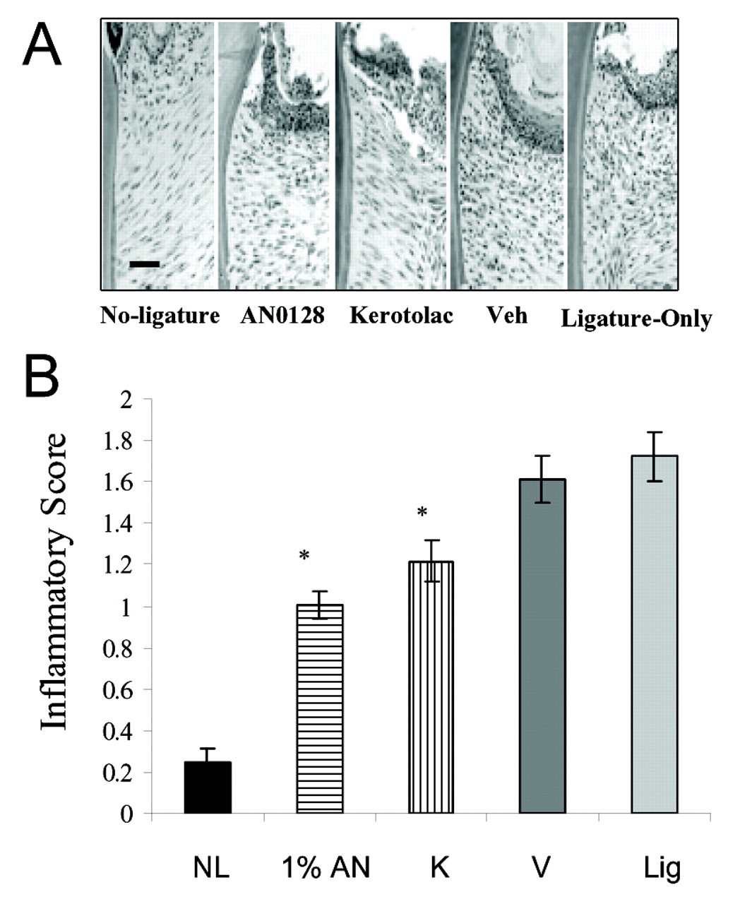
To assess the impact of AN0128 on the inflammatory response in the gingiva, we examined the relative numbers of PMNs and mononuclear cells in histologic sections, using a scale from 0 to 3 (Fig. 3). There was very little inflammation in the absence of ligatures, while placement of ligatures induced a moderate level of inflammation. When rats were treated with AN0128, there was a 42% reduction in the level of inflammation, which was similar to that in the ketorolac group (P > 0.05). Results with AN0128 and ketorolac were statistically significant (P < 0.05).
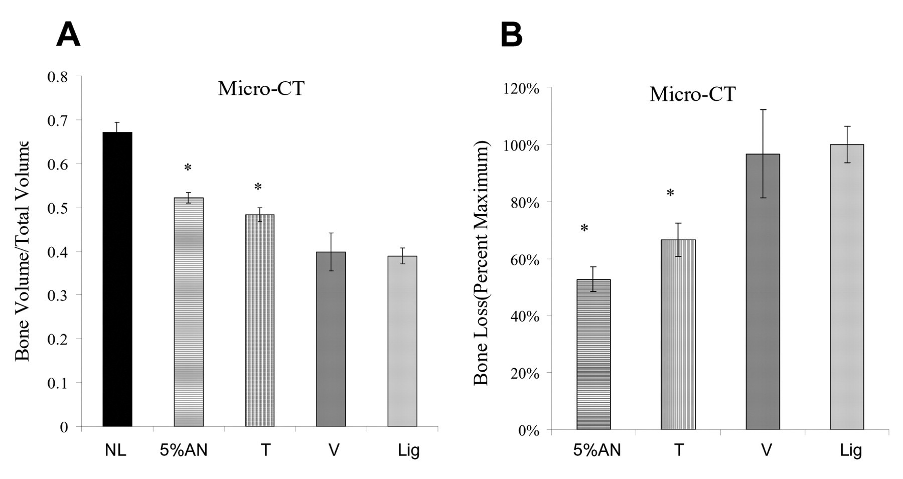
We carried out micro-CT experiments to compare a higher concentration of AN0128 (5%) with Total toothpaste. The induction of experimental periodontitis reduced bone volume by 42%, very similar to the amount produced with the first set of animals. Treatment with 5% AN0128 resulted in a 33% increase, and Total toothpaste produced a 24% increase in bone volume, compared with ’ligature only’ animals with periodontal bone loss (P < 0.05), while vehicle alone had no effect (P > 0.05) (Fig. 4A). When the data were expressed as bone loss, application of AN0128 resulted in a 47% decrease, compared with a 33% decrease in bone loss when Total toothpaste was used. There was no reduction in bone loss in vehicle-treated rats, and there was no significant difference between applications of Total and AN0128 (P > 0.05).
DISCUSSION
Studies presented here found that animals treated topically with the boron-containing compound AN0128 had significantly less bone loss and a smaller inflammatory infiltrate than did either vehicle-treated or ’ligature only’ rats. Histologically, there was a 50% improvement in bone area. This compares favorably with results obtained with tetracycline or chlorhexidine, in which there was a 33% or 40% increase, respectively, in alveolar bone in the treatment groups (Leonard and Mandel, 1979; Weiner et al., 1979). Furthermore, AN0128 reduced the inflammatory infiltrate by 42% compared with treated controls. This is consistent with a 30% decrease in vascular leakiness reported with an inducible nitric oxide synthase inhibitor, mercaptoethylguanidine, and a 55% decrease in infiltrating inflammatory cells in tetracycline-treated animals (Weiner et al., 1979; Lohinai et al., 1998). Thus, AN0128 applied topically has efficacy that is similar to that of other biologically effective treatments, including antibiotics.
Two doses of AN0128, 1% and 5%, were tested. When measured by micro-CT, both doses of AN0128 increased bone volume by a similar amount, 33 to 35%, compared with the ’ligature only’ rats with experimental periodontitis, while vehicle alone had no effect. When the micro-CT data were expressed as bone loss, treatment with AN0128 caused a 44 to 47% reduction, demonstrating that both doses were effective. There was no significant difference between the two doses tested. AN0128 (5%) was a suspension with less than 2% in actual solution. This may account for the lack of dose response. Furthermore, both positive controls, ketorolac and Total toothpaste, significantly inhibited periodontal bone loss. This is the first report that Total toothpaste inhibits bone loss in an animal model, while it has previously been shown to reduce bone loss in human clinical studies (Rosling et al., 1997). AN0128 had an efficacy similar to that of both of these positive controls.
The rat ligature model of experimental periodontitis has been used for several years, with most studies measuring bone loss by optical methods. We used both histologic and micro-CT analysis to assess the impact of AN0128 and two different positive controls. In both cases, the same analytical approach was taken, whereby the amount of bone present was assessed within a space defined by the walls of the dental root. This took into account discrepancies in bone width caused by various interdental spaces. When maximum bone loss was compared (’ligature only’ group) with minimal bone loss (no ligature), histologic analysis indicated that there was a 2.3-fold difference in bone area. In contrast, micro-CT indicated a 1.8-fold difference in the first set of animals and a 1.7-fold difference in the second set of animals. Thus, there is very good reproducibility in the micro-CT analysis and close agreement with another analytical approach, histologic assessment. The reliability of micro-CT analysis is consistent with results reported recently (Wilensky et al., 2005).
AN0128 is both anti-inflammatory and anti-bacterial. AN0128 has shown in vitro activity against several bacteria associated with periodontal disease, namely, Prevotella intermedia, Porphyromonas gingivalis, Eubacterium nodatum, and Treponema denticola (data not shown). In addition, AN0128 has shown inhibition of LPS-induced TNF-α release from human monocytes, and thus this compound exhibits both antibacterial and anti-inflammatory activity (Baker et al., 2006). The decrease in the inflammatory infiltrate noted could be from either antibacterial or anti-inflammatory pathways. AN0128 has shown activity against some bacteria associated with periodontal disease, Prevotella intermedia, Porphyromonas gingivalis, Eubacterium nodatum, and Treponema denticola, with minimum inhibitory concentrations of < 0.5 μ g/mL (data not shown). AN0128 also has an IC50 against LPS-induced TNFα release from human monocytes in the low micromolar range (Baker et al., 2006). It is known that penetration of AN0128 into the skin results in concentrations well above the aforementioned MIC and IC50 values (Baker et al., 2006). Since a detailed microbiologic assessment was beyond the scope of the present manuscript, we cannot determine which parameter was most significantly affected. Regardless of mechanism, AN0128 performed as well as the NSAID ketorolac and Total toothpaste in a rat model of periodontitis.
1% AN-0128 reduced alveolar bone loss assessed histologically. We induced periodontal bone loss by placing ligatures around the right and left second maxillary molars for 7 days. The molars were treated by daily topical application of 1% AN-0128 (1% AN), vehicle alone (V), or positive control, ketorolac (K). One control group had no ligatures (NL), and another had ligatures placed but no treatment (Lig). 1% AN-0128 reduced alveolar bone loss assessed by micro-CT. Periodontal bone loss was induced by placing ligatures around the right and left second maxillary molars for 7 days. The molars were treated by daily topical application of 1% AN-0128 (1% AN), vehicle alone (V), or positive control, ketorolac (K). One control group had no ligatures (NL), and another had ligatures placed but no treatment (Lig). AN-0128 reduces inflammation in experimental periodontitis. Periodontal bone loss was induced by placement of ligatures around the second maxillary molars. The molars were treated by daily topical application of 1% AN-0128 (1% AN), vehicle alone (V), or positive control, ketorolac (K). One control group had no ligatures (NL), and another had ligatures placed but no treatment (Lig). AN-0128 reduces alveolar bone loss. We induced periodontitis by placing ligatures around the right and left second molars for 7 days. Molar teeth were treated with 5% AN-0128 (5% AN), vehicle alone (V), or Total toothpaste (T) on a daily basis. One control group had no ligatures (NL), and another had ligatures placed but no treatment (Lig). Bone volume 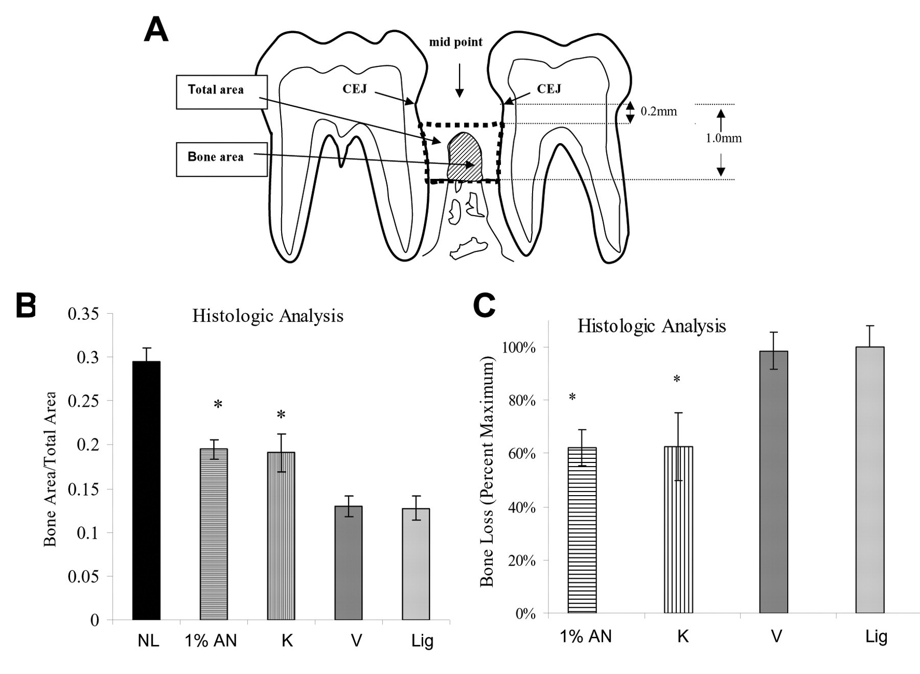
Footnotes
Notes
The review of this paper was overseen by the Editor-in-Chief, and, to avoid any potential conflict of interest, the Associate Editor for Critical Reviews was not involved in the review process.
Acknowledgements
This work was supported by a grant from Anacor Pharmaceuticals, Inc. No consultant fees were paid. We thank Dr. Elise Morgan for assistance with the micro-CT analysis and Alicia Ruff for help in preparing this manuscript.