
Abstract
Much controversy exists regarding the origin of hemangiomas. Most investigators believe them to be hamartomas. These lesions have been found virtually in all the organs in human body, but the head and neck area appear to be involved more frequently than other regions. Hemangiomas are rarely found in the salivary glands. The identification of salivary gland hemangiomas requires a minimum of ultrasound examination along with color Doppler. One such report of hemangioma of the parotid gland is reported, which was diagnosed with the aid of color Doppler ultrasound examination.
Case Report
A 43-year-old male patient reported for routine dental checkup. Extra-oral examination revealed a solitary well-defined swelling on the right side of the face, below the lobe of the ear and behind the angle of the mandible, corresponding to the tail of the parotid gland, measuring approximately 2 × 2.2 cm in size. The skin over the swelling appeared intact and healthy. The mass was firm in consistency, nontender, with no visible or palpable pulsations (Figure 1). The left parotid and the submandibular glands were not enlarged or palpable. The oral mucosa appeared adequately hydrated and normal. The patient revealed that the swelling started insidiously 6 years previously and had progressed during a period of 6 months to its present size. Ever since, the swelling had remained static, and the patient was not concerned about esthetics.
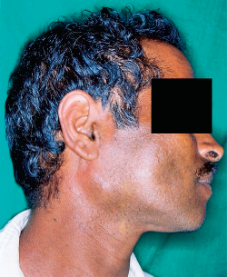
Photograph showing swelling in the right parotid region.
A rotated posteroanterior radiograph of the skull failed to reveal any abnormalities. Fine-needle aspiration yielded a copious amount of frank blood and cytology revealed components of normal blood. This finding prompted an ultrasonographic examination of the swelling, which revealed a well-defined, hypoechoic, intraparotid mass with sinusoidal spaces that measured 2.8 × 1.4 cm in size (Figure 2). No calculi were noted. A color Doppler study revealed the mass to be of vascular origin and further showed a feeder artery into the mass (Figure 3).
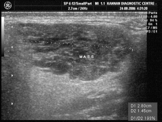
Well-defined, hypoechoic, intraparotid mass with sinusoidal spaces measuring 2.8 × 1.4 cm in size.
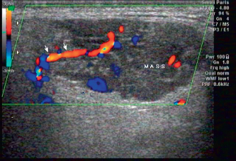
Color Doppler showing the mass with the feeder artery.
On the basis of the clinical findings, fine-needle aspiration cytology (FNAC), and color Doppler ultrasound findings, a final diagnosis of intraparotid hemangioma was made. The patient declined any additional studies, such as magnetic resonance imaging or angiography, and declined treatment, citing the asymptomatic nature of the mass. The patient is currently under follow-up, and no clinical changes have been noted in the swelling.
Discussion
Hemangiomas are tumors of vascular origin and constitute 7% of all benign tumors of the human body. 1 The head and neck appear to be the favored sites for the occurrence of these tumors because approximately 60% of them are found in this region. 2 Salivary gland tumors are relatively rare, and approximately 80% occur in the parotid salivary gland. Also, approximately 80% of the salivary gland tumors are benign. 3 Most of the salivary gland tumors are ectodermal in origin, and mesenchymal tumors are rare. Chuang et al 4 assessed the mesenchymal intraparotid tumors and found the prevalence was 1.4%. Approximately 30% of these mesenchymal tumors were hemangiomas.
Hemangiomas of the salivary glands are quite uncommon. Most salivary gland hemangiomas are found in the parotid gland. 4 Parotid hemangiomas occur commonly in early childhood or infancy and are extremely rare in adults.4,5 Approximately 90% of adult salivary hemangiomas are found in persons younger than 40 years of age. It is currently believed that hemangiomas are congenital lesions and remain quiescent within the gland and go undetected. Unknown causes are said to induce sudden growth or pain, and diagnosis of the lesion follows the symptoms. 4 In the present case, the patient was 43 years of age. Precise epidemiologic data depicting the prevalence of parotid hemangioma in adults are lacking. Salivary hemangiomas, similar to hemangiomas elsewhere in the body, are found most commonly in female patients. The left-side salivary glands appear to a favored site for their occurrence. In the case being reported, the lesion was noted on the right side.
Clinically they present as a slow-growing, soft to firm, mobile, painless mass. Acute hemorrhage or thrombosis within can give rise to symptoms of pain. 5 FNAC has been performed in isolated cases of parotid hemangioma. The diagnosis of a hemangioma through FNAC depends on the quantity of the aspirate obtained. 6 In the present case, FNAC in fact raised the suspicion of the lesion being entirely vascular leading to further imaging studies.
Imaging of salivary gland hemangiomas consist of plain film radiography, ultrasonography, color Doppler, computed tomography, magnetic resonance imaging, nuclear scan, and angiography. Plain films demonstrate phleboliths, when present.4,6 Ultrasound examination demonstrates a heterogeneous, hypoechoic mass with sinusoidal spaces. 4 Color Doppler studies show color flow within the lesion, perfusion and the feeder artery. 5 All of the aforementioned ultrasonic and color Doppler findings were noted in the present case. Angiographic studies are indicated to delineate the exact vascular anatomy prior to definitive therapy such as embolization. 5 Computed tomography dynamic scanning will show a tumor mass with enhancing quality akin to blood vessels, depending on the blood flow through hemangioma. 6 On magnetic resonance imaging, a T2, hyper-intense signal will be noted with signal voids and is the hallmark of hemangioma. 5
Regarding the imaging studies for parotid hemangiomas, it is suggested that a confident diagnosis of hemangioma can be established on a high-resolution ultrasonography, and conventional angiography is not routinely necessary for diagnosis 5 A careful monitoring of salivary hemangiomas is necessary because a few congenital hemangiomas undergoing malignant transformation are reported. 7 Therapy for salivary hemangiomas includes conventional surgery, laser therapy, cryotherapy, ligation, or embolization of the vessel or intralesional corticosteroid administration. 4
Conclusion
Intrasalivary gland hemangiomas are extremely rare and, when occurring in the parotid gland, may simulate any other salivary gland neoplasm. For an efficient and swift diagnosis, appropriate imaging studies are e ssential, which includes the basic and most important modality, the color Doppler ultrasound. Long-term follow-up is warranted because the malignant transformation of benign hemangioma to an angiosarcoma is a possibility.