
Abstract
Objective:
The study aimed to investigate the ultrasonographic venous abnormalities in CEAP C1 patients and explore the relationship between venous abnormalities, vessel diameters, and patients’ baseline characteristics.
Methods:
We prospectively collected data from patients with lower limb chronic venous disease, CEAP C1 classification, who underwent Doppler ultrasound at our institution between December 2022 and May 2023. Demographic data, including age, sex, weight, height, underlying non-communicable diseases (NCDs), including diabetes, hypertension, and dyslipidemia, long-standing hours per day, and vessel diameters, were collected. Patients were divided into reflux and non-reflux groups. The association between venous abnormalities, vessel diameters, and patients’ baseline characteristics was analyzed by using correlation tests.
Results:
There were 94 participants, mean 44.9 (+ 13.4 SD) years. Eighty-eight (93.6%) were female. Eleven participants had 1 symptomatic limb; thus, a total of 177 limbs were assessed. In the 177 limbs assessed, refluxes were found in 25 (14.1%) limbs, including 2 (1.1%) limbs in deep veins, 11 (6.2%) limbs in superficial veins, 8 (4.5%) limbs in perforators, and 4 (2.3%) limbs in both great saphenous veins (GSV) and perforators. Perforator diameter ≥1.8 mm had a significant relationship with reflux (p = .017), while GSV diameter and patients’ baseline characteristics had no relationship with venous reflux. No thrombosis or short saphenous vein abnormalities were shown in this study.
Conclusions:
Venous abnormalities were found in 14.1% of our CEAP C1 limbs. All were refluxes. Almost all of the refluxes occurred in superficial veins and/or perforators. There were no thromboses or abnormalities in the short saphenous vein. Perforator diameter ≥1.8 mm had a significant relationship with reflux (p = .017). Short saphenous vein examinations and the augmentation test in perforator veins with a diameter less than 1.8 mm can be omitted from the screening protocol for CEAP C1 patients to reduce examination time.
Introduction
Chronic venous disease (CVD) has been defined as “(any)morphological and functional abnormalities of the venous system of long duration manifest either by symptoms and/or signs indicating the need for investigation and/or care.” 1 Chronic venous disease is a prevalent condition with a significant impact on global healthcare, affecting approximately 60% to 80% of the population. 2 The majority of cases are in the early stages, as classified by the Clinical, Etiology, Anatomy, and Pathology (CEAP) system. 1 The Edinburgh Vein Study, which surveyed 1566 subjects, found that CEAP C1 was present in 90.1% of the population. 3 Similarly, in the American Venous Forum’s National Venous Screening Program, which assessed 5814 limbs, CEAP C0–1 was identified in 59% of cases. 4 More advanced stages of the disease include varicose veins (VVs), observed in 25% of individuals, and chronic venous insufficiency (CVI), found in 5%. 2 Females have a higher risk of developing CVD compared to males. Additional risk factors include age >65 years, overweight status, parity, prolonged standing, and a positive family history. 2 ,5-9 Furthermore, previous studies have indicated that ethnicity is a contributing factor in the development of CVD. 5 ,10-13
Doppler ultrasound (DUS) is widely used to assess venous anatomy, detect venous abnormalities that contribute to CVD, such as valve incompetence, and exclude underlying conditions, such as deep vein thrombosis. However, DUS requires specialized sonographers, is time-consuming, and is labor-intensive. A routine DUS protocol for diagnosing varicose veins involves at least 12 venous segments, excluding perforators. 14 Traditionally, the primary indication for DUS was in patients classified as CEAP C2–6.15,16
CEAP C1, in particular, represents mild venous abnormalities such as telangiectasias and reticular veins without overt symptoms of venous insufficiency. 2 Although a lower CEAP clinical stage suggests milder disease, it does not necessarily correlate with fewer symptoms. 17 Recently, a new recommendation from the Surgery Committee has suggested the use of DUS in CEAP C1 patients. 1 The presence of perforator incompetence and reflux in patients classified as CEAP C1 remains a subject of ongoing research, particularly regarding its role in disease progression. Patients with a CEAP C1 classification may exhibit subtle hemodynamic changes that predispose them to disease progression. Recent studies suggest that subclinical reflux may be detectable in CEAP C1 patients through DUS, 18 raising the possibility that reflux in superficial veins, combined with perforator incompetence, may contribute to the eventual development of symptomatic varicose veins.
Recognizing the prevalence of venous abnormalities requiring surgical intervention in this patient group is crucial, as minimal symptoms and signs may lead to delayed diagnosis and treatment. However, most existing studies have focused on more clinically severe stages of the disease. 19
This study aimed to investigate the prevalence of lower limb venous abnormalities in CEAP C1 Thai patients. Additionally, the relationship between venous abnormalities, vessel diameters, and patients’ baseline characteristics was examined.
Materials and Methods
Participants
This was a descriptive study conducted following the Helsinki Declaration and approved by our Ethics Committee (Approval No. IRB1-096/2565). Informed consent was obtained from all participants.
Data were prospectively collected from Thai patients with telangiectasia or reticular veins in the lower limbs, corresponding to the CEAP C1 classification, 1 who underwent lower limb venous DUS at our institution between December 2022 and May 2023. Demographic data, including age, sex, weight, and height, were recorded. Information on underlying non-communicable diseases (NCDs), including diabetes, hypertension, and dyslipidemia, as well as daily prolonged standing hours, the affected side of lower limb CVD, and vessel diameters, was also documented. Body mass index (BMI) was calculated by dividing weight (kg) by height squared (m²).
Patients were excluded from the study if they had CVD classified as higher than CEAP C1, a history of prior lower limb venous surgery, recent venous thrombosis (within 1 month before the examination), arteriovenous malformations, or tumors.
The calculation of sample size employed a formula designed for estimating population proportions,
There were 94 participants with a mean age of 44.9 years (±13.4 SD). The majority were female (88 participants, 93.6%). Eleven participants had 1 symptomatic limb, resulting in a total of 177 limbs assessed. The baseline characteristics of the patients are presented in Table 1.
Patient’s Baseline Characteristics.
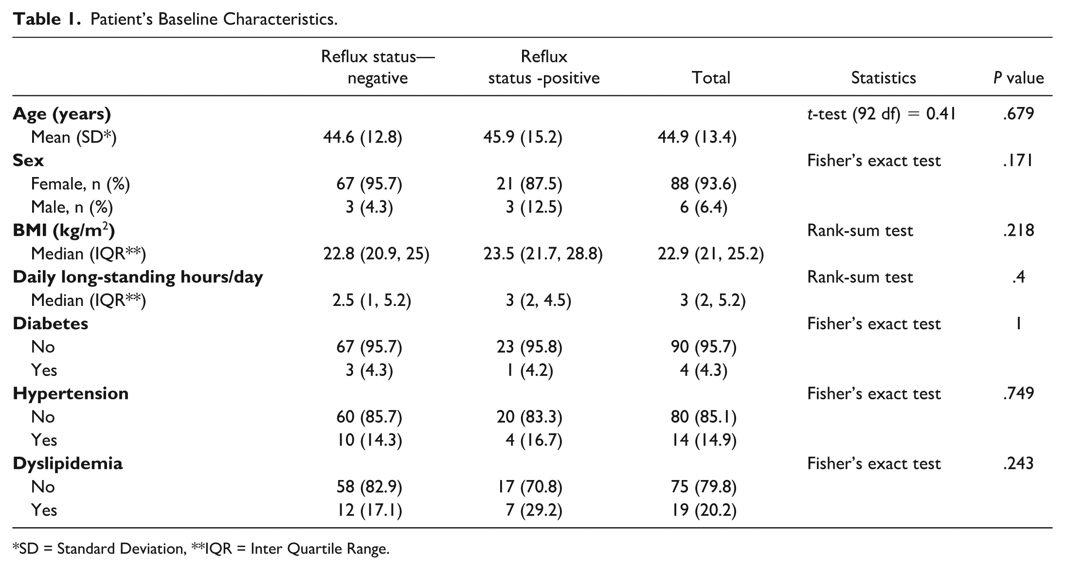
SD = Standard Deviation, **IQR = Inter Quartile Range.
Equipment and Study Protocol
A Logic E10 (GE HealthCare, United States) and a Toshiba Aplio 500, (Canon Medical Systems Corporation, Japan) ultrasound scanners, equipped with 7 to 12 MHz linear-array transducers, were used. B-mode imaging was utilized for detecting thrombosis and measuring vessel diameters. B-mode with manual compression of the veins was performed to detect thrombosis. Color Doppler and spectral Doppler modes were employed to assess reflux. In spectral Doppler analysis, the sample volume gate was adjusted to cover the entire lumen diameter, and the angle of insonation did not exceed 60 degrees. The lumen diameter was measured from the inner wall to the inner wall. Three radiologists with varying levels of experience (1, 6, and 30 years) performed all examinations. Deep venous system examinations were conducted in the 30-degree reverse Trendelenburg position, while the superficial venous system, as well as thigh and medial calf perforators, were examined in the standing upright position. 20 Valsalva maneuvers and distal augmentations were performed during thigh assessments, whereas only distal augmentations were conducted for the leg. Distal augmentations were performed manually by assistants. 15 A cut-off value of reverse blood flow of greater than 1 second in deep veins and greater than 0.5 seconds in superficial veins and perforators was considered reflux. 19 The examination levels were determined per the standard departmental protocol. The details of the assessment are provided in Supplemental Table 1. Tributary veins were not assessed. Perforators were traced from superficial to deep veins, and their diameters were measured at the level where they penetrated the deep fascia.
Statistical Analysis
Data were analyzed using the R program. Normally distributed continuous variables were expressed as the mean ± standard deviation (SD), while non-normally distributed continuous variables were presented as the median and range. Categorized variables were expressed as numbers and percentages. Patient’s characteristics and the average vessel diameters between the non-reflux group and the reflux group were compared. The association between venous abnormalities and patient’s parameters, including age, sex, BMI, underlying NCDs, daily prolonged standing hours, and vessel diameters, was analyzed by performing correlation tests. P-values less than .05 were indicative of statistical significance.
Results
Among the 94 participants, 25 participants (26.6%) had reflux, including 2 participants (2.1%) in the deep veins, 11 participants (11.7%) in the superficial veins, 8 participants (8.5%) in the perforators, and 4 participants (4.3%) in both the superficial veins and perforators.
Among the 177 limbs assessed, reflux was detected in 25 limbs (14.1%), including 2 limbs (1.1%) in the deep veins, 11 limbs (6.2%) in the great saphenous veins (GSV), 8 limbs (4.5%) in the perforators, and 4 limbs (2.3%) in both the superficial veins and perforators. No cases of thrombosis were observed in this study.
No significant associations were found between reflux and age, sex, BMI, underlying NCDs, or daily prolonged standing lifestyle.
Deep Venous System
There were 2 limbs (1.1%) with reflux. In the first limb, reflux was observed along the common femoral vein to the popliteal vein, while in the second limb, reflux was present only in the popliteal vein. Neither limb exhibited associated abnormalities in the superficial venous system.
Superficial Venous System
Reflux was detected in 15 limbs (8.5%), with a total of 29 reflux locations in the GSV. Fifty-three percent of limbs exhibited reflux at a single location per limb. The number of reflux locations per limb is illustrated in Figure 1A. No reflux was detected at either the saphenofemoral junction or the saphenopopliteal junction.
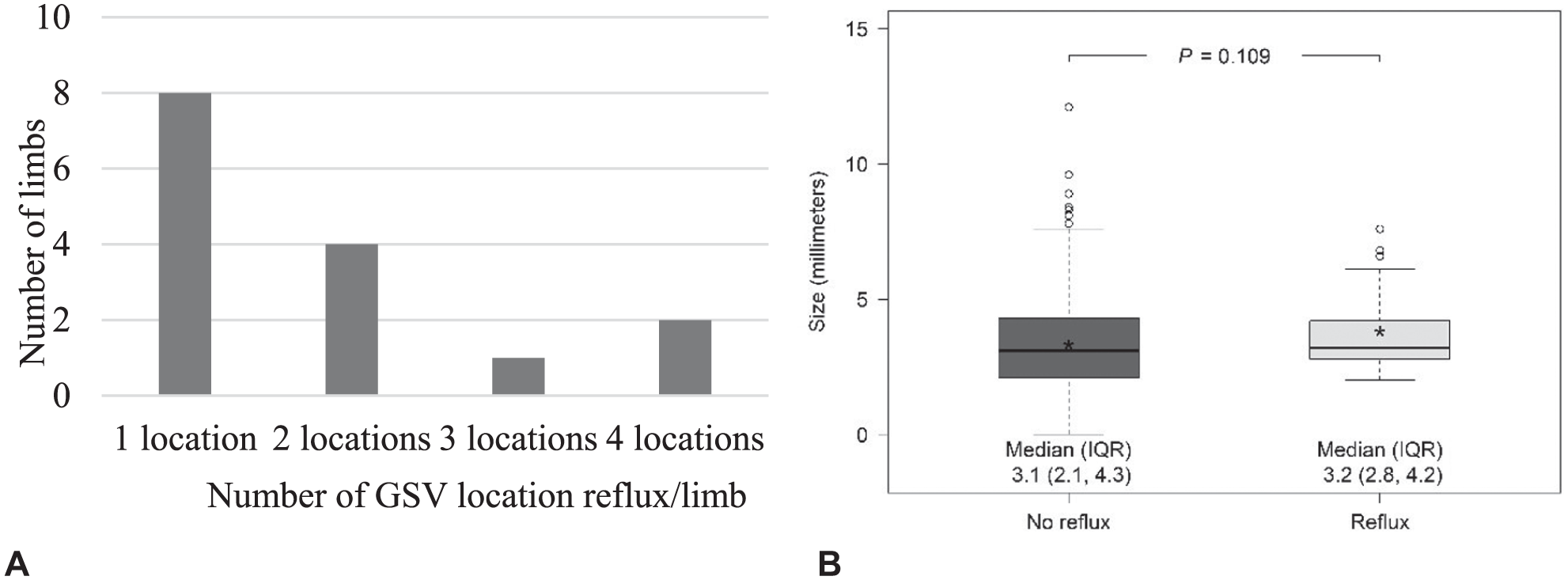
(A) The number of GSV reflux locations per limb. (B) Difference in great saphenous vein diameters between the non-reflux and reflux groups. GSV = great saphenous vein.
The GSV locations and diameters detected by ultrasound, along with the prevalence of reflux, are presented in Table 2. No significant association was found between GSV diameters and reflux (Figure 1B). Additionally, no abnormalities in the short saphenous vein (SSV) were observed in this study.
GSV Locations and Diameters (mm) Detected by Ultrasound, Along With the Prevalence of Reflux (N = 177).
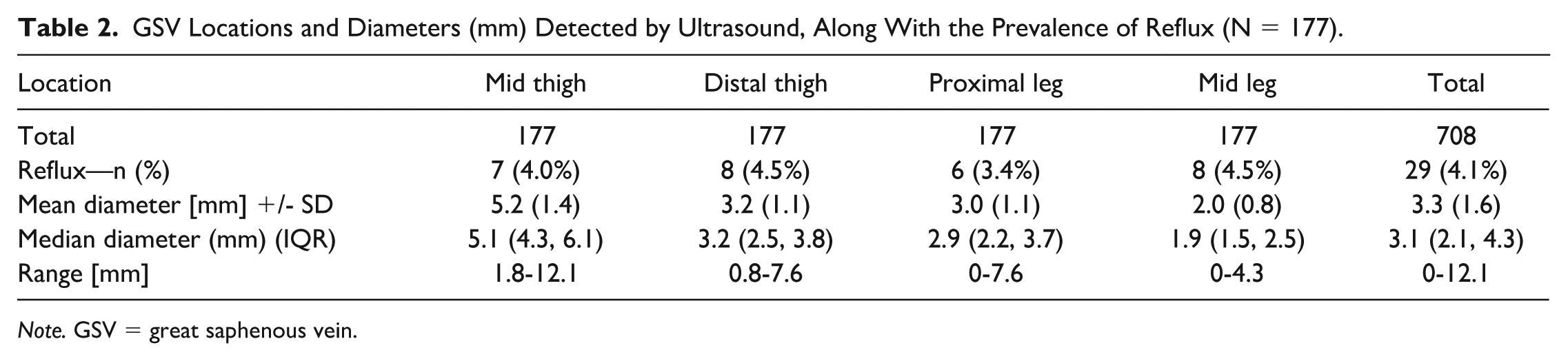
Note. GSV = great saphenous vein.
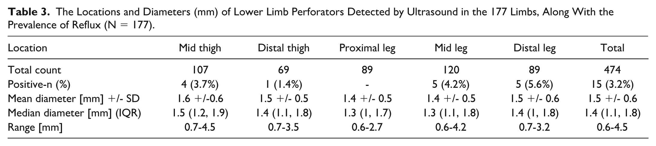
Perforators
Perforators were detected in 474 locations in 177 limbs. The number of perforators found per limb is presented in Figure 2A, with 2 perforators per limb being the most common (34%). Perforator reflux was observed in 12 limbs (6.8%), with reflux occurring in 15 locations. All locations were individually identified as abnormal, except for the proximal leg, which showed no abnormalities in this study. The locations and diameters of lower limb perforators detected by ultrasound in the 177 limbs, along with the prevalence of reflux, are presented in Table 3. A perforator diameter of ≥ 1.8 mm was significantly associated with reflux (P = .017) (Figure 2B).
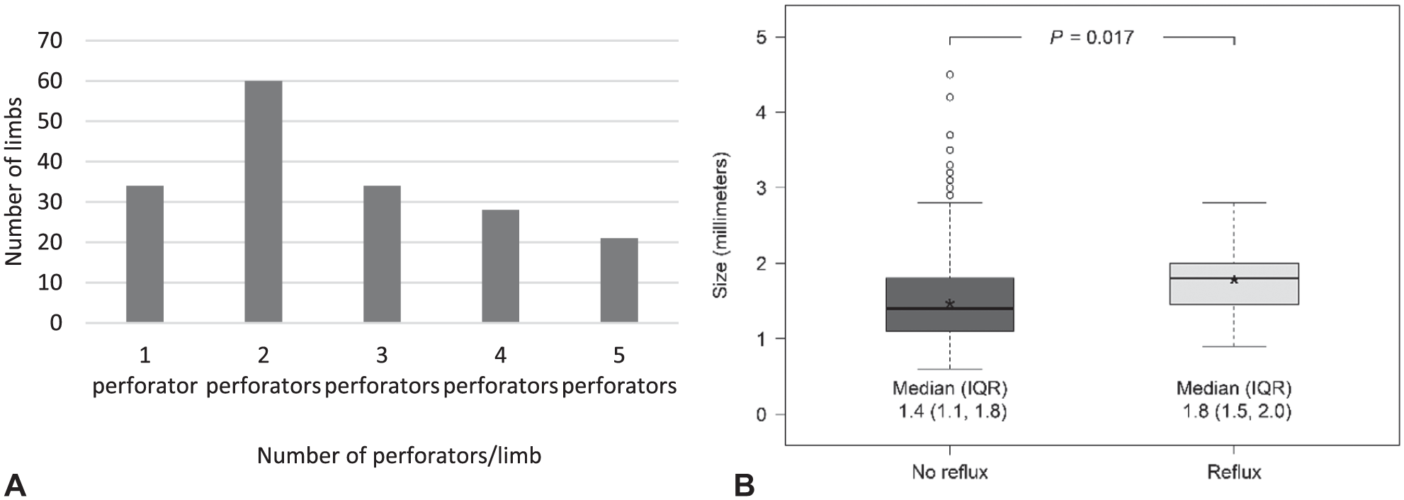
(A) The number of perforators found per limb. (B) Difference in perforator diameters between the non-reflux and reflux groups (P = .017).
The Locations and Diameters (mm) of Lower Limb Perforators Detected by Ultrasound in the 177 Limbs, Along With the Prevalence of Reflux (N = 177).
Discussion
The prevalence of lower limb venous abnormalities in CEAP C1 patients in the current study was 26.6% on a per-patient basis but 14.1% on a per-limb basis. The prevalence on a per-patient basis was nearly twice that on a per-limb basis. The use of paired data from the same subject, which can lead to differences in statistical results, has been a subject of debate. 21 However, most lower limb venous studies utilize a per-limb basis for prevalence calculations.
In the 177 limbs assessed in our study, reflux was detected in 25 limbs (14.1%), in contrast to the study by Engelhorn et al, 16 which examined 269 CEAP C1 women’s limbs and found a higher prevalence of GSV reflux (44%). This discrepancy may be explained by differences in ethnicity. 5 ,10-13 In an Asian male study by Sam et al, 10 a lower prevalence of reflux (7.4%) was found in 122 CEAP C0-1 limbs. In contrast, studies by Panpikoon et al 6 and Hong KP 22 in CEAP C1 limbs reported reflux in the GSV in 23.1% and 40.5%, respectively. These higher prevalences may be attributed to the older mean age in those studies, which was higher than our study’s mean age of 39 ± 11 years.
In the study by Panpikoon et al, 6 the authors suggested omitting SSV and perforator vein examinations to reduce examination time, as they observed a low prevalence of reflux (0.3% and 4.4%, respectively). These findings are consistent with our results, where no SSV reflux was detected, and the prevalence of perforator reflux was only 6.8%. Similarly, Engelhorn et al 16 also recommended detecting only GSV reflux in the screening protocol for CEAP C1 patients, citing similar reasons. In our department, a full study protocol requires approximately 10 to 20 minutes per patient.
This study revealed no significant relationship between GSV diameters and reflux. The diameters in the reflux group and non-reflux group were 3.2 mm versus 3.1 mm, respectively. This is consistent with the study by Kim et al, 23 which also found no significant relationship between GSV diameters and reflux in the thigh, except in the lower thigh segment, where a statistical significance was observed. They suggested that the cutoff value for the lower thigh GSV diameter associated with reflux was 5 mm (P = .025). In contrast, a study involving CEAP C0-3 Thai patients found a significant relationship between GSV diameters and reflux, although they were unable to determine an accurate cutoff diameter. 19 The main reason for this discrepancy is that the study included more advanced stages of disease than our study, where CEAP C1 patients constituted only 7.6% of the sample.
A significant finding in this study was that perforator diameters ≥1.8 mm were significantly associated with reflux (P = .017), which is consistent with previous studies.24,25 Hill et al 20 studied perforator diameters in 20 normal subjects and reported a mean diameter of 1.3 ± 0.6 mm. We observed 1 to 2 perforators per limb, which accounted for 53% of our population, in agreement with a previous study that reported 1 perforator per limb in CEAP C1 patients. 26 Sandri et al 24 reported 56.4% of perforators were incompetent in CEAP C3-4 limbs. The lower number of perforators found in this study may be attributed to the fact that our participants were in the early stage of disease, with only CEAP C1 classification. This is supported by prior studies that demonstrated an increase in the number of perforators with higher CEAP classifications.25,26 Not all dilated perforators are indicative of disease.
Although numerous studies have shown that age, sex, and BMI are important risk factors for VVs,5-7 we found no significant relationship between these factors and reflux, which is consistent with some other studies.10,12,19,23,25,27 Some studies have reported that diabetes and hypertension are risk factors for CVD,7,28,29 but our study did not reveal a significant association. The small number of positive patients in our study may have influenced this result.
Data from a previous study revealed that prolonged standing or walking at work was significantly related to hospitalization for varicose veins in both men and women; however, the study did not specify the number of hours involved. 8 A study among nurses found that prolonged standing 3 hours or more was positively associated with varicose veins. 9 In contrast, our study did not demonstrate a relationship between long-standing hours and CEAP C1 patients, which is consistent with the Edinburgh study. 27 The difference in hours between the positive and negative groups in the current study was not statistically significant (3 hours vs 2.5 hours). These conflicting results may be due to difficulties in obtaining valid data.
The prevalence of deep venous system reflux (1.1%) in our study was low, which is consistent with the Edinburgh study (2.6%). 27 Deep vein insufficiency (DVI) in our study was an isolated finding, without association with the superficial venous system, in contrast to the Korean study, which demonstrated 10.2% of DVI, all of which were accompanied by saphenous vein insufficiency. 22 The majority of DVI in that study occurred in CEAP C2-6 limbs, but the presence of DVI was not related to the CEAP clinical class. This aligns with the findings of the Edinburgh study, which suggested that isolated DVI was not associated with the development of varicose veins. 27
The strength of our study lies in the fact that all superficial veins and perforators were examined in the standing position, as reflux may be missed in other positions.30,31 In our experience, in some cases, we have observed that reflux is absent when examined in the reverse Trendelenburg position but becomes immediately evident when the patient is examined in the standing position.
The current study has several limitations. It was a single-center design, and we detected only 5 clinically significant perforators. 32 While other perforators may have shown reflux, they were not clinically significant. Additionally, non-saphenous superficial veins in the anterior, lateral, and posterior aspects of the thigh were not investigated. Due to the small number of positive findings, the correlations and comparisons between vessel size, reflux, and perforator evaluation may not have had sufficient statistical power to support definitive conclusions. The significance of DVI was not explored due to the small number of positive patients in our study. Further studies with larger samples would provide more comprehensive information. Because our study included only CEAP C1 patients—most of whom were female and all of whom were of Thai ethnicity—the findings cannot be generalized to all patients with venous insufficiency. Before implementing screening protocols, a study with a more balanced gender distribution is necessary. We also did not assess intra- or inter-observer reliability, and patients’ symptoms were not analyzed in this study.
Conclusion
Venous abnormalities were found in 14.1% of our CEAP C1 limbs, all of which were refluxes. Ninety-eight percent of these refluxes occurred in the superficial venous system and/or perforators. There were no instances of thromboses or short saphenous vein abnormalities. A perforator diameter ≥1.8 mm showed a significant relationship with reflux (P = .017), while GSV diameter, age, sex, BMI, underlying NCDs, and daily long-standing lifestyle showed no relationship with venous reflux. Short saphenous vein examinations can be omitted from the screening protocol for CEAP C1 patients, and the augmentation test is unnecessary in perforator veins with a diameter of less than 1.8 mm to reduce examination time.
Supplemental Material
sj-docx-1-jvu-10.1177_15443167261421212 – Supplemental material for Ultrasonographic Venous Abnormalities in CEAP C1 Patients
Supplemental material, sj-docx-1-jvu-10.1177_15443167261421212 for Ultrasonographic Venous Abnormalities in CEAP C1 Patients by Trakarn Chaivanit, Sornsupha Limchareon, Teetawat Jearsirikul, Watanya Jaidee, Thanchanok Nanthanasub and Sorawat Ketkaewmanee in Journal for Vascular Ultrasound
Footnotes
Acknowledgements
The authors would like to thank Keattichai Keeratitanont, MD, for contributing to the revision of the statistical writing.
Declaration of Conflicting Interests
The authors declared no potential conflicts of interest with respect to the research, authorship, and/or publication of this article.
Funding
The authors disclosed receipt of the following financial support for the research, authorship, and/or publication of this article: This work was funded by the Faculty of Medicine, Burapha University. The faculty had no involvement in the study design or collection, analysis, and interpretation of data. The faculty was not involved in the decision to submit the manuscript for publication.
Ethical Considerations
This study was conducted following the Helsinki Declaration and approved by our University Ethics Committee (Approval No. IRB1-096/2565).
Consent to Participate
Written informed consent was obtained from all participants.
Consent for Publication
Informed consent for publication was provided by the participants.
Data Availability
The datasets generated during and/or analyzed during the current study are available from the corresponding author on reasonable request.
Supplemental Material
Supplemental material for this article is available online.
References
Supplementary Material
Please find the following supplemental material available below.
For Open Access articles published under a Creative Commons License, all supplemental material carries the same license as the article it is associated with.
For non-Open Access articles published, all supplemental material carries a non-exclusive license, and permission requests for re-use of supplemental material or any part of supplemental material shall be sent directly to the copyright owner as specified in the copyright notice associated with the article.