
Abstract
The quantification of differences in alpha electroencephalograph (EEG) activity between the eyes-closed and eyes-open resting conditions could be used as a measure of resting state arousal. The objective of this study was to evaluate the contribution of EEG alpha reactivity on opening the eyes, to the neurophysiology of children with attention-deficit hyperactivity disorder (ADHD). Thirty-eight children with ADHD were assessed using quantitative EEG (qEEG) analysis of absolute band power at rest, with eyes open and closed. Alpha reactivity index was calculated on opening the eyes, defined from the relationship between the absolute powers in the respective bands in the periods with the eyes open and closed. EEG data of 38 sex- and age-matched controls, with no neurological or psychiatric problems, were collected for comparison. There was a significant reduction in absolute alpha power at all electrodes for both ADHD and control groups with eyes open, indicating an increase in the arousal level. However, the alpha reactivity index was greater, corresponding to less reactivity, in the frontal regions of the children with ADHD (P < .01). Such a finding suggests alterations in arousal mechanisms in ADHD. This research suggests that alpha reactivity on opening the eyes, allied with other variables from the qEEG, may improve diagnostic accuracy in ADHD.
Introduction
A diagnosis of ADHD is based on the Diagnostic and Statistical Manual of Mental Disorders (Fourth Edition Text Revision {DSM-IV-TR}) criteria, 1 of which the basic characteristic is the persistence of a standard of lack of attention and/or hyperactivity impulsivity, more frequent, and serious than that typically observed in individuals at an equivalent developmental level. Currently, there is no laboratory examination that can serve as a marker for ADHD. Thus, the assessment procedure should consist of a review of the medical, developmental and family histories, as well as an examination of intellectual function and academic performance.
There is wide agreement that ADHD occurs as the result of a dysfunction in the central nervous system, but the mechanisms are still not well known. Zentall and Zentall 2 raised the hypothesis that ADHD symptoms, such as restlessness and hyperactivity, could be associated with chronic underarousal. However, a number of researchers have suggested different mechanisms by which there would be a compromise of arousal, alertness and activation in ADHD.3–5
Since it is functional, simple and accessible, EEG has been widely used to study normal cognitive mechanisms, as well as the nature of brain dysfunction in ADHD. The most frequent alterations in ADHD are greater interhemispheric coherence in the theta and delta bands, less in the alpha band, 6 increased absolute and relative theta and delta,7–11 and lower relative alpha and beta.8,10,12,13 Absolute theta/beta power ratio showed high discriminatory power between ADHD and controls.14,15
Recent studies have tried to characterize concepts of arousal and activation in normal individuals from physiology and neural correlations. By using measurement of electrochemical skin conductance (SCL), the classical correlation of arousal level, it was shown that during eyes-closed resting the arousal level was negatively correlated with the alpha brain electrical activity (8-12 Hz range), and that opening the eyes was associated with an increase in SCL and reduction in alpha activity.16,17 Thus, the quantification of alpha activity could be used as a measure of resting-state arousal under eyes-closed and eyes-open conditions.
In a recent work, Barry et al 18 failed to encounter a significant correlation between theta/beta ratio and SCL, contradicting the supposed linkage between this index and arousal in ADHD. Instead, they found a negative correlation between SCL and alpha activity, suggesting its importance as a marker of arousal.
The objective of the present work was to evaluate the relationship between alpha activity, with eyes open and closed, in ADHD. Although a state of hypoarousal is considered to be an important mechanism in ADHD, no research of alpha reactivity on opening the eyes in children could be found.
Materials and Methods
Thirty-eight school children, aged 8 to 11, diagnosed with ADHD according to DSM-IV-TR, and referred by the outpatients’ departments of the Infancy & Adolescence Psychiatry Department and the Pediatric & Neurological Pediatric Department (HMCP PUC-Campinas), were studied. Thirty-eight children, with no neurological or psychiatric problems, comprised the sex- and age-matched control group, which was also matched for maternal scholastic level. All the children were free of medication at the time of testing, and those taking methylphenidate were taken off this medication for at least 12 hours prior to the assessment, longer than its serum and behavioral half-lives. 19 The following procedures were carried out: medical history, psychiatric evaluation, neurological examination; Conner Parent and Teacher Rating Scales; Child Behavior Checklist 4-18, Wechsler Intelligence Scale for Children; and digital and quantitative EEG (qEEG).
EEG
EEG was recorded with a resolution of 12 bits, 0.5 and 35 Hz filters, and 200 samples/second, using the BrainNet BNT 36 equipment (EMSA Equipamentos Médicos), with impedance maintained below 10 kΩ. The examination was carried out with the patients lying on their backs in a silent environment with low-intensity lighting. The electrodes were placed according to the International 10-20 System, with the use of an additional 2 electrodes placed 1 cm below (left side) and above (right side) the external angle of the eyelid, with the objective of evaluating eye movements. The data referring to the electrodes Fp1, Fp2, F8, and F7 were not computed due to frequent contamination by artifacts related to eye movements. The interconnected ear lobe electrodes served as the reference. The data were recorded while awake during about 5 periods, alternating 2 minutes with the eyes closed and 2 minutes with the eyes open. The individual was stimulated to remain awake when symptoms of somnolence appeared. Stretches of somnolence or sleep were excluded from the quantitative analysis.
For the qEEG analyses, 18 to 26 of artifact-free epochs, each lasting 2.56 seconds, were selected under 2 conditions; awake but resting (eyes closed) and awake (eyes open). After applying Fast Fourier Transform, the absolute powers (μV2/Hz) were studied in the following frequency bands: delta (up to 3.9 Hz), theta (4.9-7.8 Hz), alpha (8.2-12.5 Hz), and beta (12.9-36.3 Hz). To obtain a normal distribution, the values for absolute power were substituted by their logarithms.
The absolute power averages were calculated for the centrotemporal regions (C4, C3, T4, T3, T6, and T5), occipital regions (O1 and O2), and frontal regions (F3 and F4).
Alpha reactivity index, calculated on opening the eyes, was defined as the ratio between the absolute alpha power with eyes open and eyes closed. The greater the reduction in alpha activity with eyes open (alpha reactivity), the smaller the value of this index.
The formula for the alpha index is as follows:

Ethical aspects
The Ethics Commission for Research with Human Beings of PUC-Campinas, Brazil, approved the project, and the participants signed an informed consent.
Results
Table 1 shows the data for age, sex, maternal scholastic level, and school grade of the 38 children with ADHD and of the control group (CG). Table 2 shows the mean obtained for the logarithms of the absolute alpha powers, with eyes closed and open, by location of electrodes and by groups. There was no statistically significant difference between the ADHD and CG for absolute alpha power, whether eyes were closed or open (T test, P > .05, values for P not presented in table).
Data for Age, Gender, School Grade, and Maternal Scholastic Level for the ADHD and Control Groups.a
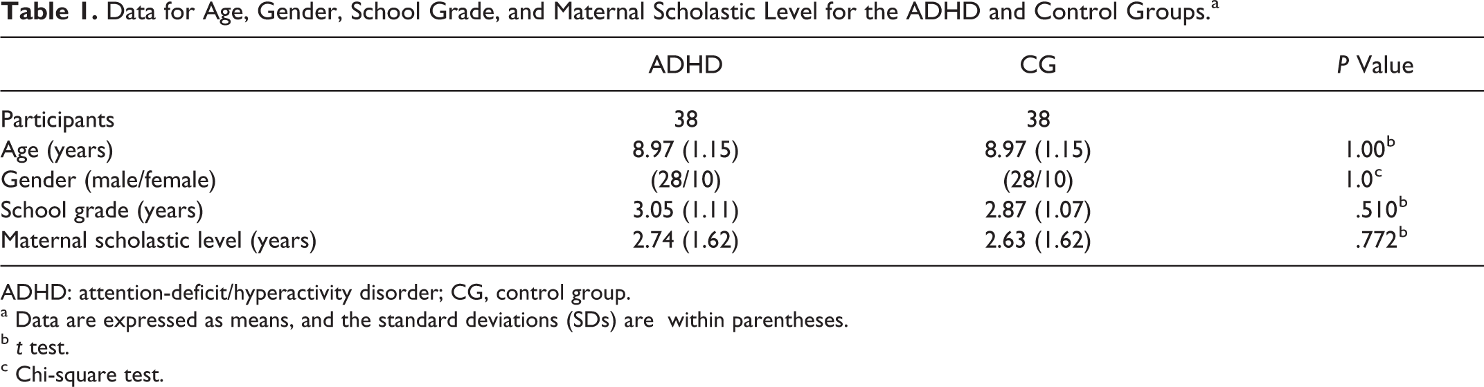
ADHD: attention-deficit/hyperactivity disorder; CG, control group.
a Data are expressed as means, and the standard deviations (SDs) are within parentheses.
b t test.
c Chi-square test.
EEG Absolute Alpha Power With the Eyes Both Closed and Open for the ADHD and Control Groups.
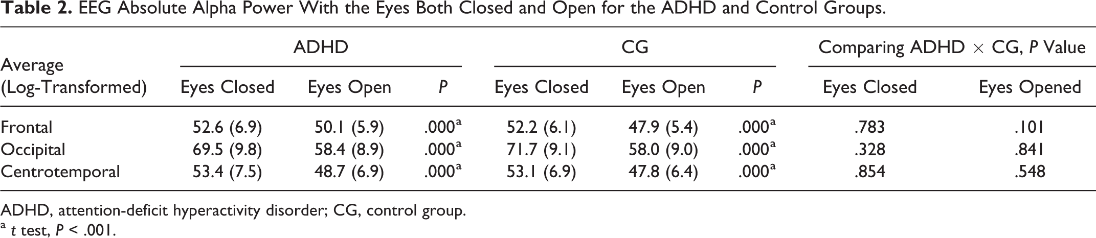
ADHD, attention-deficit hyperactivity disorder; CG, control group.
a t test, P < .001.
A decrease in the absolute alpha power occurred with the eyes open for all the regions studied, with a significance level below .001. Table 3 shows the alpha reactivity indices obtained at the various locations of the electrodes for ADHD and CG. The alpha indices were greater (less reactivity) for ADHD, but statistically significant only in the frontal regions (T test, P < .01).
Alpha Reactivity Indices on the qEEG for the ADHD and Control Groups.
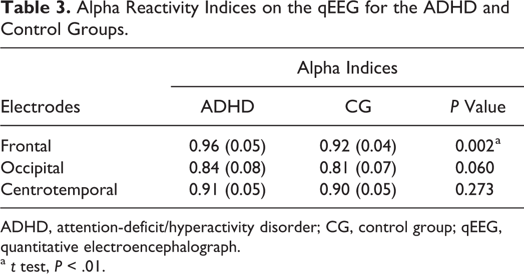
ADHD, attention-deficit/hyperactivity disorder; CG, control group; qEEG, quantitative electroencephalograph.
a t test, P < .01.
Discussion
No significant differences were found between ADHD and CG in absolute alpha power, in agreement with the literature, since increases in absolute power occur predominantly in delta and theta bands7–11 and are expressed in a relevant way in the theta/beta index.14,15
Opening of the eyes, from the eyes-closed resting state, is classical practice in conventional EEG. 20 It results in desynchronization of electrical brain activity, and the alpha rhythm is reduced, giving rise to rapid rhythms. Alpha reactivity to visual stimuli is well known; however, it is not exclusively related to visual information, since it has connections with memory and cognition in general. 21 This is traditionally evaluated by visual analysis of the EEG, although with the advances in computer technology, it may be the object of multiple quantitative analyses of qEEG.
Alpha reactivity can be quantified by indices such as the alpha power index, obtained by dividing the values obtained eyes open by the corresponding values obtained eyes closed. In qEEG studies, a reduction in alpha power reactivity has been found in diverse clinical conditions such as age-related cognitive decline 22 and Alzheimer disease23,24 and has been considered as an indicator of brain disorder. 24
In the present study, a significant reduction in absolute alpha power was observed with eyes open compared with eyes closed for both ADHD and CG, and for all the derivations, providing evidence of clear reactivity of the alpha rhythm, as expected in this age group.25,26 Such alpha desynchronization to visual input could occur due to widespread communication of cortical and thalamocortical interactions, in order to aid information processing.21,27 On comparing the reactivity indexes between ADHD and CG in the present research, values were found to be higher (less alpha reactivity) in the ADHD group at the various electrode positions, although the difference was only statistically significant in frontal regions.
Since quantification of alpha activity can be used as a measure of resting-state arousal under both eyes-closed and eyes-open conditions,16–18 the findings of the present research suggest alterations in the state of alertness of the children with ADHD. This finding in children is original in the use of EEG, since the authors have no knowledge of a similar study. Mechanisms connected to cortical hypoarousal were reported by Lubar 28 as being due to the increase in theta activity, decrease in beta, and eventually in alpha activities. This suggestion is supported by research with skin conductance tests, 29 regional cerebral blood flow and positron emission tomography. 30
van Dongen-Boomsma et al 31 found a greater reduction in alpha power from eyes closed to eyes open in adults with ADHD as compared with controls, which appears inconsistent with the results of the present study. However, there are relevant differences between the findings of EEG at rest in children with ADHD as compared to those of adults with ADHD, since in the latter the data are scarce, disparate, and less consistent, 31–33 suggesting neurophysiologic modifications during the development of the individuals into adult life. Earlier research on ADHD was directed essentially at evaluating brain activation, that is, making recordings during different tasks. The researchers did not search for a characterization of the state of arousal.15,32–37 The fact that in the present study the difference in alpha reactivity was only observed in a significant way in the frontal regions is supported by various other evidences of dysfunction in the frontal cortex. In the study of ADHD in children, alterations in coherence6,38,39 and an increase in theta activity 40 in the frontal regions were found. Frontal alterations have been found in studies with positron emission tomography 39 and single photon emission computed tomography. 41
In summary, elements suggesting alterations in the arousal mechanisms that probably involve the frontal regions were found in the present research. On the other hand, this research opens opportunities for the use of alpha reactivity on opening the eyes, obtained using different evaluation methods, 42 and allied to other variables of the qEEG, with the objective of providing an increment in diagnostic accuracy of ADHD.
Footnotes
Declaration of Conflicting Interests
The authors declared no potential conflicts of interest with respect to the research, authorship, and/or publication of this article.
Funding
The authors received no financial support for the research, authorship, and/or publication of this article.