
Abstract
The aim of this study was to determine the efficacy of tongue somatosensory-evoked potentials (tSSEPs) in evaluation of afferent trigeminal pathway in patients with early multiple sclerosis (MS). The tSSEP was performed on 10 healthy volunteers and 10 patients with the first symptom of MS. Data were compared between the groups, and tSSEP findings of patients with MS were correlated with clinical and magnetic resonance imaging (MRI) data. Among 10 patients, 2 (20%) had clinically evident involvement of the brainstem, 5 (50%) had brainstem lesions on brain MRI, while 9 (90%) had prolonged latencies on tSSEP. Of the 8 patients with no clinical evidence of brainstem pathology, 7 (88%) had prolonged latencies/conduction block on tSSEP. Patients had statistically significant prolongation of N1, P1, and N2 latencies for stimulation of the right side and N2 latency for stimulation of the left side compared to healthy controls. The tSSEP is an efficient method for evaluating the afferent trigeminal pathway in patients with early MS. This study provides evidence that lesions of the afferent trigeminal pathway are more frequently found by tSSEP than by clinical examination or MRI.
Introduction
MS is a chronic disorder of the central nervous system, characterized by autoimmune inflammation, demyelination, and axonal damage. Traditionally, diagnosis of MS was based on neurologic evaluation, MRI, evoked potentials (EPs), and cerebrospinal fluid (CSF) analysis. Although EP and CSF analyses are no longer part of the revised McDonald criteria, 1 they are still useful, and have an important role in satisfying the “no better explanation for the clinical presentation” criteria. 2 Characteristic neurophysiological findings in MS are prolonged latencies in visual, acoustic, somatosensory EP, and vestibular-evoked myogenic potentials. 3,4
Trigeminal somatosensory EP was used in the mapping of somatosensory cortex 5,6 , as well as in diagnosis of clinical and subclinical trigeminal impairment. 7
tSSEPs are a diagnostic method, which by measuring latencies, and/or conductional blocks of applied electrical stimuli, evaluate the integrity of afferent trigeminal pathways. The neuroanatomical basis of tSSEP is based on the transmission of stimuli through lingual and mandibular nerves to the trigeminal nuclei in the brainstem. From the brainstem, the stimulus is transmitted through the trigeminal lemniscus on the opposite side to the thalamus, and finally the somatosensory cortex. Central or peripheral lesions of the trigeminal afferent system will cause prolonged latencies or conductional block. 8
The aim of this study was to determine the efficacy of the tSSEP in evaluation of afferent trigeminal pathway in patients with early MS.
Materials and Methods
Ten healthy volunteers, 5 females and 5 males (age 27-66 years, mean 33.2), and 10 patients with the first symptom of MS, 5 females and 5 males (age 22-42 years, mean 31.3), participated in this study. All patients had brain MRI and CSF analysis performed. Exclusion criteria for the study were molar surgery and all pre-existing lesions of the trigeminal nerve that could influence the outcome of the study.
All participants were informed about the details of the experiment and signed informed consent forms. The study was approved by the Ethical committee of the University Hospital Center Zagreb. Methods of recording, and analysis of the recorded data were designed according to the previously described details. 8
During the experiment, participants sat in a comfortable chair. After details about the experiment were explained to them in detail, they were instructed to relax in order to avoid muscle tension. Modified EEG electrodes were used for stimulation, and they were located on the lateral side of the first two-thirds of the tongue, as shown in Figure 1. The participants had slightly opened mouth and the tongue, with stimulation electrodes, held relaxed inside the mouth. Each side of the tongue (left and right) was stimulated twice with 300 trials in order to confirm the repeatability of the obtained cortical response. Stimulation was produced with a constant current stimulator (Twister, Germany). The frequency of the stimulation was 3 Hz, and the duration of each stimulus was 0.2 milliseconds. The polarity of the stimulation was alternating in order to avoid large baseline shifts.
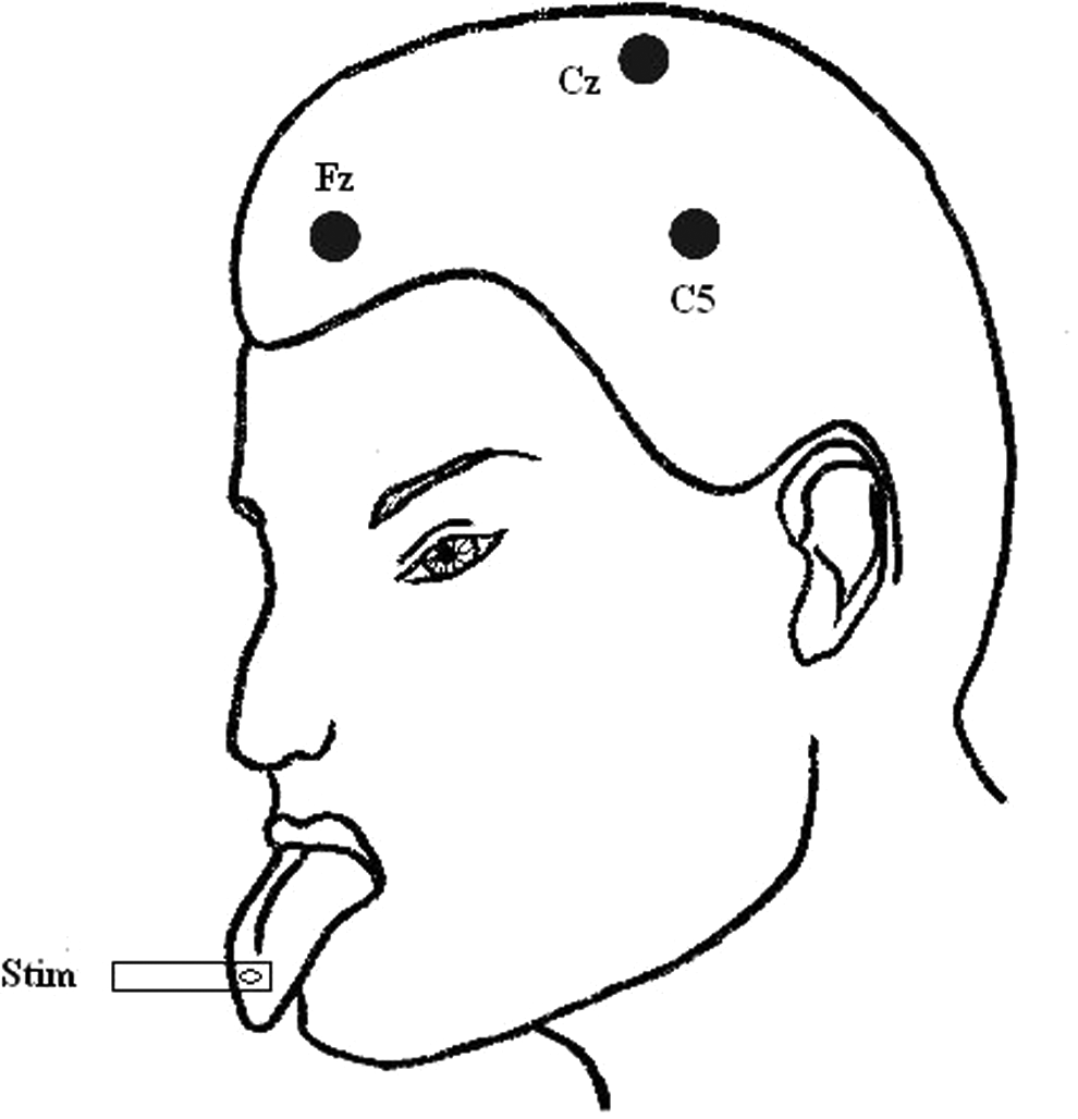
Modified electroencephalogram (EEG) electrodes were used for stimulation and they were located on the lateral side of the first two-thirds of the tongue. Active electrodes were situated on the contralateral side of the scalp, according to the International 10/20 system, at the middle position between C3 and T3 for the stimulation of right side of the tongue—C5 electrode. Both electrodes were referred to the frontal electrode, Fz. Electrode situated at the vertex, Cz, was used as the ground electrode.
At the beginning of each set of trials, the perceptive threshold for each participant was assessed. The intensity of stimulation during each set of trials was set at 3 times the perceptive threshold. It varied from 4.5 to 10 mA for patients and from 3.5 to 9 mA for healthy controls.
The cortical response was recorded from 4 surface disk electrodes situated at the surface of the scalp. Active electrodes were situated in the contralateral side of the scalp, according to the International 10/20 System, at the middle position between C3 and T3 for the stimulation of the right side of the tongue—C5 electrode—and at the middle position between C4 and T4 for the stimulation of the left side of the tongue—C6 electrode. Both electrodes were referred to the frontal electrode, Fz. Electrode sitauted at the vertex, Cz, was used as a ground electrode.
Responses obtained from electrical stimulation of the tongue were recorded with a Brain Products Vision Recorder (BrainProducts GmbH, Munich, Germany), and analysis of the recorded data was performed using a Brain Products Vision Analyzer (BrainProducts GmbH, Munich, Germany). Signals were filtered with bandpass from 0.1 to 1000 Hz. Sampling rate was 5000 Hz. For analysis, signals were divided into segments of 70 milliseconds duration (20 milliseconds before the stimulus and 50 milliseconds after) and averaged for each set of 300 trials. The grand average was computed from 2 averaged sets and used for analysis.
The obtained responses consisted of 3 main components (N1, P1, and N2) as shown in Figure 2. Latencies and peak-to-peak amplitude values (N1-P1 and P1-N2) were analyzed in order to detect the difference between healthy controls and patients with MS.
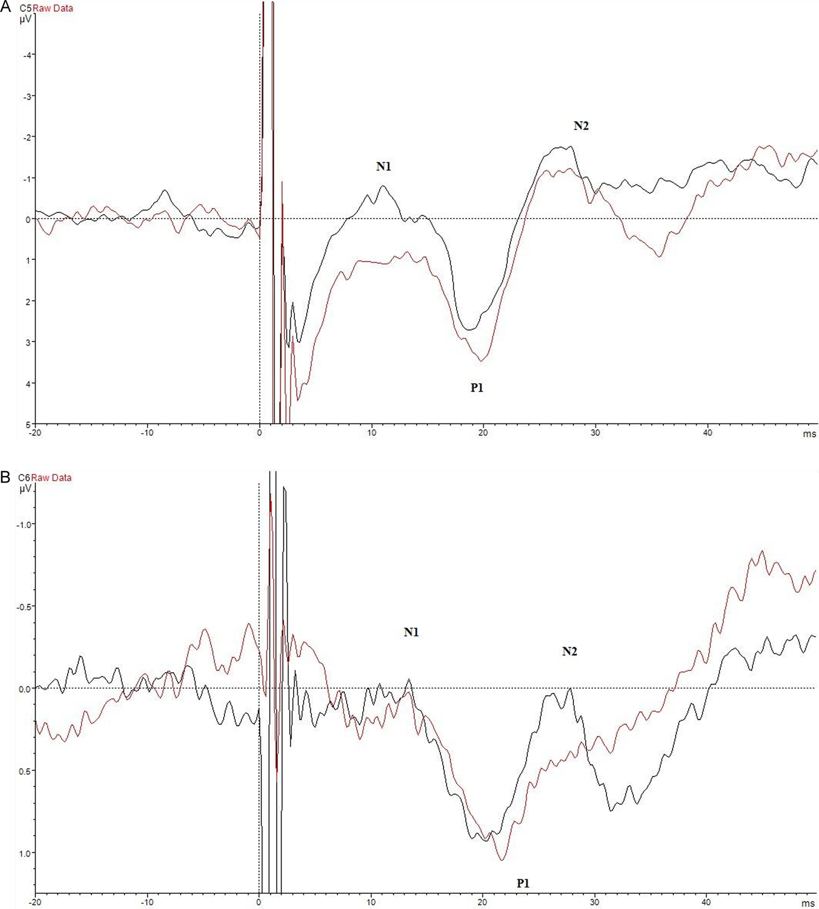
Superimposed tracings from 2 repeats of the tongue somatosensory evoked potentials (tSSEPs) in the healthy control. A, The tSSEPs from the stimulation of the right side of the tongue. B, The tSSEPs from the stimulation of the left side of the tongue.
Statistical analysis was performed using program SPSS statistics 17.0. (IBM Corp., NY, USA). Differences in the qualitative variables were analyzed by the chi-square (χ 2 ) test, while the differences in quantitative variables, in respect of distribution, were analyzed by the parametric t test or nonparametric Mann-Whitney test. P values less than .05 were considered statistically significant.
Results
There was no statistically significant difference between the groups regarding age (P = .670) and gender (P = 1). General characteristics of the patients are shown in Table 1. Of the 10 patients, (20%) 2 had clinically evident involvement of the brainstem, (50%) had brainstem lesions on the brain MRI, while 9 (90%) had prolonged latencies in tSSEP (Figure 3). Seven, (88%) of the 8 patients with no clinical evidence of brainstem pathology, had prolonged latencies/conduction block on tSSEP. One patient (patient 5) with no clinical evidence of brainstem dysfunction and normal tSSEP, had 1 brainstem lesion in brain MRI.
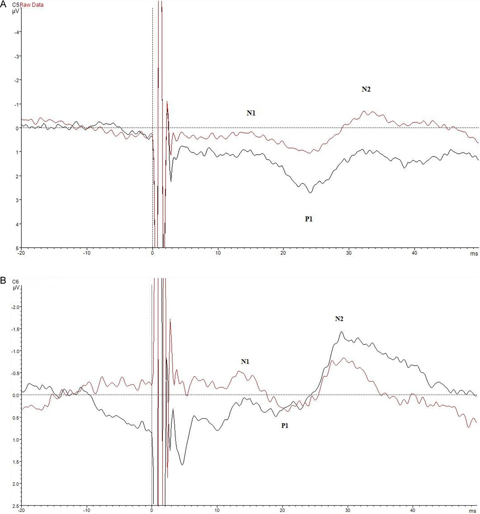
Superimposed tracings from 2 repeats of the tongue somatosensory evoked potentials (tSSEPs) in the patient with multiple sclerosis (MS). A, The tSSEP from the stimulation of the right side of the tongue showing prolonged latencies. B, The tSSEP from the stimulation of the left side of the tongue showing altered morphology of major potentials.
Characteristics of Studied Patients.
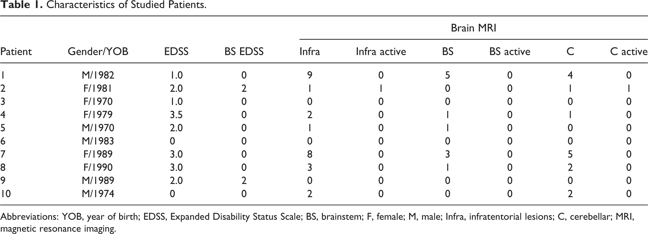
Abbreviations: YOB, year of birth; EDSS, Expanded Disability Status Scale; BS, brainstem; F, female; M, male; Infra, infratentorial lesions; C, cerebellar; MRI, magnetic resonance imaging.
When comparing MS patients with healthy controls, we found statistically significant prolongation of N1, P1, and N2 latencies for stimulation of the right side of the tongue. For left side stimulation, latencies for waves N1 and P1 were more than 2 seconds longer without reaching statistical significance. Wave N2 reached statistically significant prolongation (Table 2). There were no differences in peak-to-peak amplitudes on both the sides between patients and healthy controls.
Statistical Comparison Between MS and Healthy Controls.
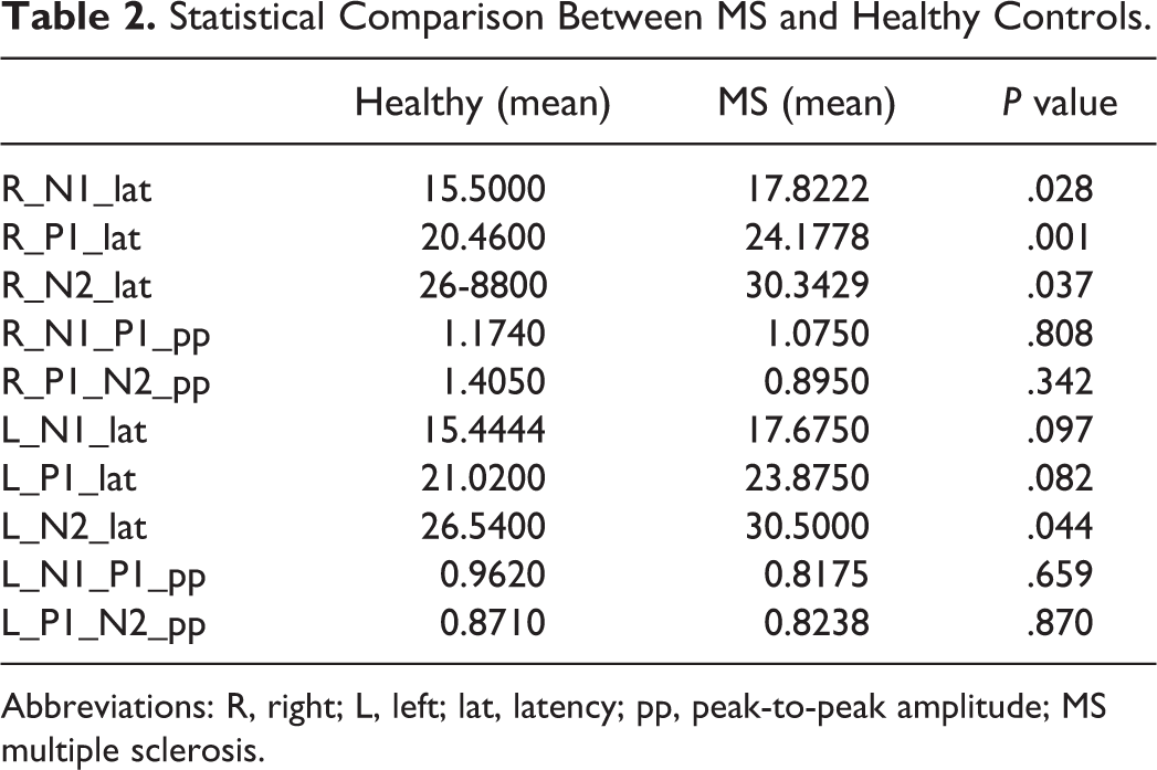
Abbreviations: R, right; L, left; lat, latency; pp, peak-to-peak amplitude; MS multiple sclerosis.
Discussion
In the present study, we have shown that patients with MS have significantly prolonged latencies of tSSEP compared to healthy controls. The tSSEP abnormalities seem to be present in a very high percentage, indicating that damage to the afferent trigeminal pathway is much more prevalent than previously thought. Differences in the results for the stimulation of the right and left side of the tongue can be explained with different lesion localization in the studied group and the relatively small numbers of patients included.
Symptoms attributed to trigeminal nerve (facial pain and non-painful facial sensory disturbance) are frequently encountered in patients with MS. The prevalence of these symptoms ranges between 3% and 7%. 9,10 Symptoms of afferent trigeminal pathway damage may or may not be related to brain MRI lesions in the brainstem, and vice versa. 10,11 Using high-resolution MRI at 3 T, the percentage of visible lesions rises to 23%, further delineating discrepancy between MRI involvement and trigeminal symptoms. 12 Although MRI is crucial in diagnosis of MS, several studies have shown that EP better correlates with disability of patients with MS than MRI. 13,14
Trigeminal somatosensory EPs have been used for the assessment of trigeminal (/) nerve pathology, and there are several described methods, most of which are bound to evoke scalp signals contaminated by myogenic activity. 15 Previous studies have shown impaired trigeminal function in patients with MS without clinical symptoms in the trigeminal area using trigeminal somatosensory EP. 7,16 Compared to the trigeminal somatosensory EP, tSSEP have excellent sensitivity for pathological processes involving the somatosensory afferents of the tongue; however, they do not seem to provide further information on the nature of the lesion. 8
The main limitation of this study is the small number of patients; however, despite this we have shown a clear difference in tSSEP latencies in patients with MS compared to healthy participants.
In conclusion, tSSEP seems to be an efficient method for evaluating the afferent trigeminal pathway in patients with early MS. Further confirmatory studies with a larger number of patients are needed.
Footnotes
Authors’ Contribution
The authors Tereza Gabelić and Magdalena Krbot Skorić contributed equally to the manuscript. Gabelić, Krbot Skorić, and Habek were involved in study concept and design. Gabelić, Krbot Skorić, Adamec, Mayer, and Habek acquired data. Gabelić, Krbot Skorić, Adamec, Mayer, and Habek analyzed and interpreted data. Gabelić, Krbot Skorić, and Habek drafted the manuscript. Gabelić, Krbot Skorić, Adamec, Mayer, and Habek critically revised the manuscript. Gabelić, Krbot Skorić, Adamec, Mayer, and Habek were involved in administrative, technical, and material support.
Declaration of Conflicting Interests
The authors declared no potential conflicts of interest with respect to the research, authorship, and/or publication of this article.
Funding
The authors received no financial support for the research, authorship, and/or publication of this article.