






Abstract
This study evaluates the recovery of vestibular nerve function after vestibular neuritis (VN) by vestibular-evoked myogenic potentials (VEMPs). Twenty-six patients with the diagnosis of VN were included. All patients underwent ocular VEMP (oVEMP) and cervical VEMP (cVEMP) recordings, at 6 days and 6 months from the onset of the symptoms. Of the 26 patients, 14 showed improvement on oVEMP at month 6 (group 1), and 12 showed no change or worsening on oVEMP at 6 months (group 2). At the same time, there was no change in the amplitudes of the cVEMP on either healthy or affected sides in both groups. Inability to perform the Fukuda test, and chronic white matter supratentorial lesions present on brain magnetic resonance imaging (MRI) were more frequent in patients with worse outcome on oVEMP (P = 0.044 and 0.045, respectively). Although involvement of the inferior branch of the vestibular nerve was not associated with oVEMP outcome, oVEMP latencies (N10 and P13) were associated with improvement or worsening in oVEMP amplitudes, showing that prolonged latencies correlate with 6-month improvement in oVEMP amplitudes (Pearson correlation −0.472, P = 0.041 and −0.580, P = 0.009, respectively). This study identified clinical, MRI and neurophysiological predictors of recovery in patients with superior VN, and offers additional insight into, and better understanding of, the role of VEMP in diagnosis and prognosis of patients with VN. Further studies are needed to validate this diagnostic procedure and to assess its clinical usefulness in VN management.
Introduction
VEMPs are a relatively new diagnostic procedure used in assessment of patients with VN 1 , which is characterized by the presence of vertigo, nausea, horizontal-rotatory nystagmus, and a positive head thrust test. 2 In the acute phase, VN is primarily a clinical diagnosis, and the triad of abnormal head thrust test, direction-fixed nystagmus, and absent skew deviation, differentiates it from stroke, with a specificity and sensitivity comparable to MRI in the first 24 hours. 3
Certain paraclinical investigations are necessary to exclude other possible causes of an acute vestibular syndrome, such as neuroimaging for suspected vascular causes. Other investigations confirm the diagnosis of VN, for instance, the traditionally used caloric test. However, there is accumulating evidence that VEMP could be more useful in assessing vestibular dysfunction, because of its ability to distinguish between lesions of superior and inferior branches of the vestibular nerve. 4
It is now well accepted that the saccule and inferior vestibular nerve represent peripheral origins of cVEMP recorded from the sternocleidomastoid muscle (SCM) in response to high-intensity air conducted tone bursts or mechanical stimulation. 5 Similarly, oVEMP to air conduction stimulation derives its peripheral origins predominately from the utricle and superior vestibular nerve. 5 The cVEMP and oVEMP can vary independent of one another and, in that way, provide topological information about the sites of impairment. Caloric testing is utilized to assess the ampullary branch of the superior vestibular nerve originating from lateral and superior semicircular canals, oVEMP test assesses the utricular branch of the superior vestibular nerve, while cVEMP test is used to evaluate function of the inferior vestibular nerve originating from the posterior semicircular canal and most of the saccule. 6 Studies have also shown that oVEMP produced in response to bone-conducted sound and air conduction stimulation, predominantly reflects utricular functions while cVEMP reflects saccular functions. 7
Caloric testing is known to be initially normal in inferior VN. 8 Having in mind that patients with inferior VN demonstrate normal head impulse test, and that conventional MRI has low sensitivity for stroke detection in posterior fossa within the first 24 hours, VEMP has a significant role in differentiating inferior VN from stroke. 3 The clinical course of VN is characterized by incomplete recovery and central compensation for a unilateral vestibular dysfunction. 9 Neurophysiologic assessment of vestibular nerve function recovery is usually evaluated by caloric testing, and there are few studies assessing vestibular recovery by VEMP.10–13 However, all these studies have used only cVEMP in follow-up of VN. As most patients included in these studies clinically had superior branch VN, which can be evaluated with oVEMP, they offer limited data. The present study evaluated recovery of vestibular nerve function after superior VN by oVEMP and cVEMP.
Patients and Methods
Patients met the following VN criteria: (1) vertigo attack lasting no more than 48 hours, (2) spontaneous horizontal-torsional nystagmus with the fast phase toward one side, (3) positive head thrust test to the other side, (4) absence of skew deviation (assessed by Maddox rod), and (5) a normal brain computed tomography scan. Patients were excluded if they had any other neurologic deficit, experienced hearing loss or tinnitus, had atrial fibrillation, non-regulated diabetes or arterial hypertension. Patients with nonregulated cerebrovascular risk factors were excluded, in order to exclude possible stroke in the posterior circulation.
The following clinical parameters were collected from all patients: (1) age, (2) gender, (3) side of the lesion, (4) grade of nystagmus: occurs only on one side (opposite to the affected side) grade 1; occurs on one side and straight ahead, grade 2; occurs on both sides and straight ahead, grade 3, (5) number of vomiting episodes in the past 24 hours, (6) nausea according to the visual-analog scale, (7) postural instability according to Fukuda test, 14 and (8) The European Evaluation of Vertigo scale. 15 All patients underwent brain MRI, during the first 3 months from initial presentation, to exclude any brainstem and cerebellar lesions. MRI was performed on 1.5 T with T1, T2, fluid-attenuated inversion recovery, diffusion-weighted imaging, and attenuated diffusion coefficient sequences. The study was approved by the Ethical Committee of the University Hospital Center Zagreb, and all patients signed an informed consent prior to their inclusion in the study.
All patients underwent VEMP recordings at 6 days and 6 months after their initial presentation of symptoms. Methods of recording and analysis of recorded data were designed according to previously described details. 16
During the experiment, participants sat in a comfortable chair. Patients were instructed to slightly move their head away from the back of the chair and push against an elastic band that was strapped over their forehead in order to activate SCM. Contraction of the muscle was maintained due to cooperation of patients in maintaining the same position during the test. Participants were also instructed to direct their gaze at the ceiling in order to activate ocular muscles (OMs). Evoked response from the SCM was recorded by an active surface electrode placed on the belly of the stimulated SCM, and, was referred to a surface electrode placed on the tendon of the same SCM. Evoked response from the OM was recorded from 2 surface electrodes situated 2 cm below the contralateral eye. The active electrode was situated closer to the eye and referred to the reference 1 cm below.
The stimuli were delivered by a pair of headphones in series of 50 trials to one ear at a time, and repeated twice for each ear, in order to provide reproducibility. Stimuli were acoustic clicks, 1 ms in duration at intensity of 130 dB sound pressure. Stimulation rate was 1 Hz.
Recordings were performed using a Brain Products Brain Vision Recorder (Brain Products GmbH, Germany), and the analysis of recorded data used a Brain Products Brain Vision Analyzer (Brain Products GmbH, Germany). Signals were filtered with a bandpass filter from 5 to 1000 Hz. For analytical purposes, the signals were divided into segments with duration of 120 ms (20 ms before stimulus and 100 ms after stimulus) and averaged for each set of 50 trials. Averaged responses from each set were used to calculate a grand average, and for further analysis.
Normalized baseline values were used as SCM amplitude data instead of the absolute amplitude value, because absolute amplitude of evoked response depends on the amplitude of the muscle activity (muscle contraction) and is not reliable. Normalized baseline amplitude value is calculated by dividing the absolute peak-to-peak amplitude (P13-N23) with the mean value of rectified muscle activity prior to stimulus.
As all patients had clinical involvement of the superior branch of the vestibular nerve, we divided them into 2 groups, according to oVEMP findings: group 1 consisted of patients who showed improvement in oVEMP at 6 months, demonstrated by increase in the value of N10-P13 peak-to-peak amplitude at month 6, and group 2 consisted of patients who showed no change or worsening of oVEMP at 6 months, demonstrated by no change or decrease in the value of N10-P13 peak-to-peak amplitude. Decreased amplitude of the N10-P13 on the oVEMP was defined as a decrease of 50 % compared to normative values for the laboratory, or decreased amplitude of 50% on the affected side compared to the healthy (non-affected side) side. If patients showed an increase in amplitude on oVEMP after 6 months, they were assigned to group 1, and if patient showed a decrease or no change in amplitude on oVEMP after 6 months, they were assigned to group 2.
Differences in distribution of qualitative variables were confirmed by Chi-square test, while the differences in quantitative variables, in respect to distribution, were analyzed by the parametric t test or non-parametric Mann-Whitney test. Pearson correlation was used to calculate the correlation between clinical, MRI, and VEMP parameters. P Values less than .05 were considered statistically significant. SPSS software, version 19, swas used to support our statistical analysis.
Results
Twenty-six patients were included in the study, from February 2011 to February 2012. Ages were between 27 and 77 years, and 14 were male, 12 were female.
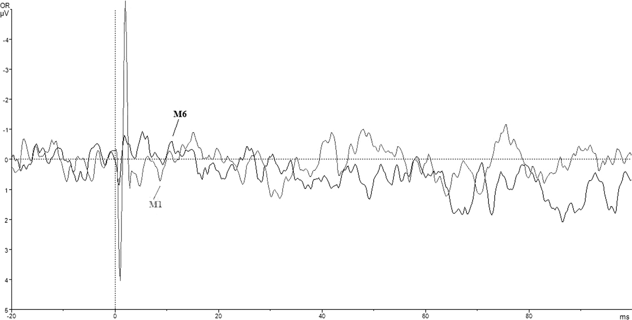
Of the 26 patients, 14 showed improvement in oVEMP at month 6 (group 1; Figures 1 and 2), and 12 showed no change or worsening in oVEMP at month 6 (group 2; Figures 3 and 4). Group 1 showed a statistically significant recovery of the affected vestibular nerve after 6 months (P = .003). In the same period, there was no change in the unaffected vestibular nerve, and the values of the affected side returned to normal values comparable to values on the unaffected side (Table 1). There were no changes in amplitudes of cVEMP on either healthy or affected side in group 1 (Table 2).

Tracings of oVEMP in one of the studied patients showing improvement at month 6. OR indicates right ocular; M0, initial VEMP; M6, VEMP at month 6; oVEMP, vestibular-evoked myogenic potentials.
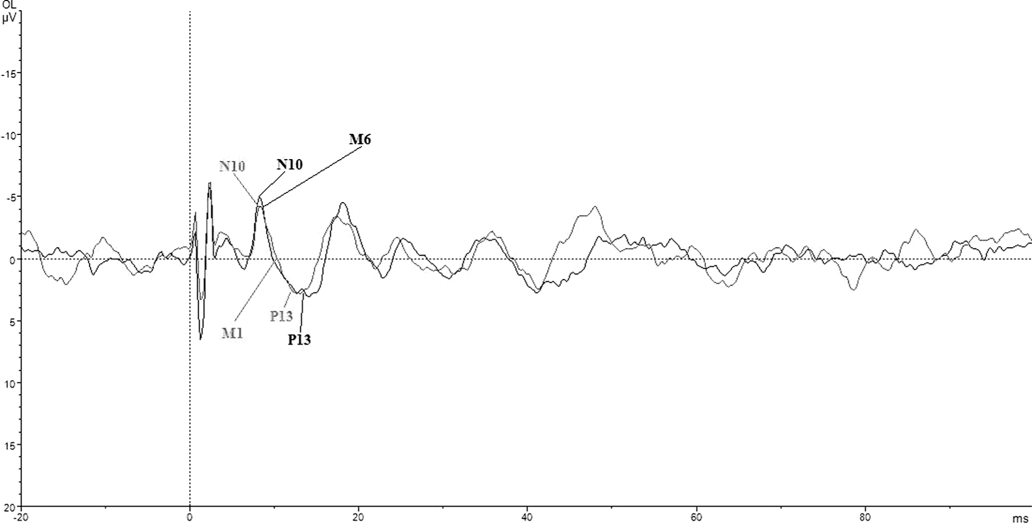
Tracings of ocular VEMP in the same patient as in Figure 1 but on the healthy side. OL indicates left ocular; M0, initial VEMP; M6, VEMP at month 6; VEMP, vestibular-evoked myogenic potentials.
Tracings of ocular VEMP in one of the studied patients showing no recovery at month 6. OR indicates right ocular; M0, initial VEMP; M6, VEMP at month 6; VEMP, vestibular-evoked myogenic potentials.

Tracings of ocular VEMP in the same patient as in Figure 3 but on the healthy side. OL indicates left ocular; M0, initial VEMP; M6, VEMP at month 6; VEMP, vestibular-evoked myogenic potentials.
Comparison Between Peak-to-Peak Amplitudes of oVEMP in 14 Patients in Group 1.
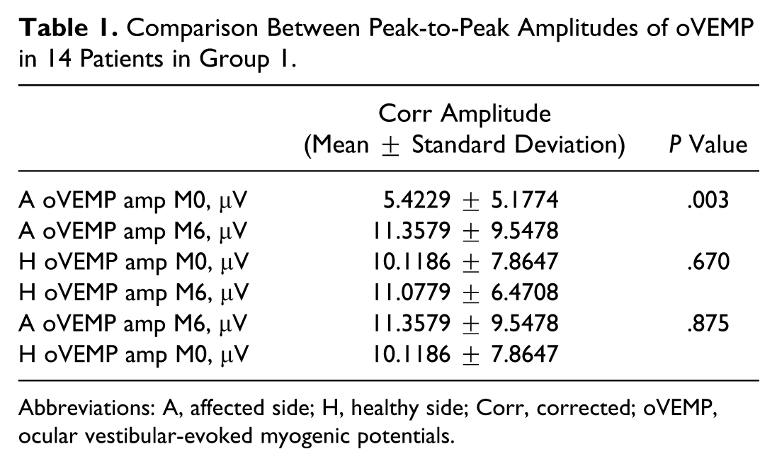
Abbreviations: A, affected side; H, healthy side; Corr, corrected; oVEMP, ocular vestibular-evoked myogenic potentials.
Comparison Between Normalized Amplitudes of cVEMP in 14 Patients in Group 1.
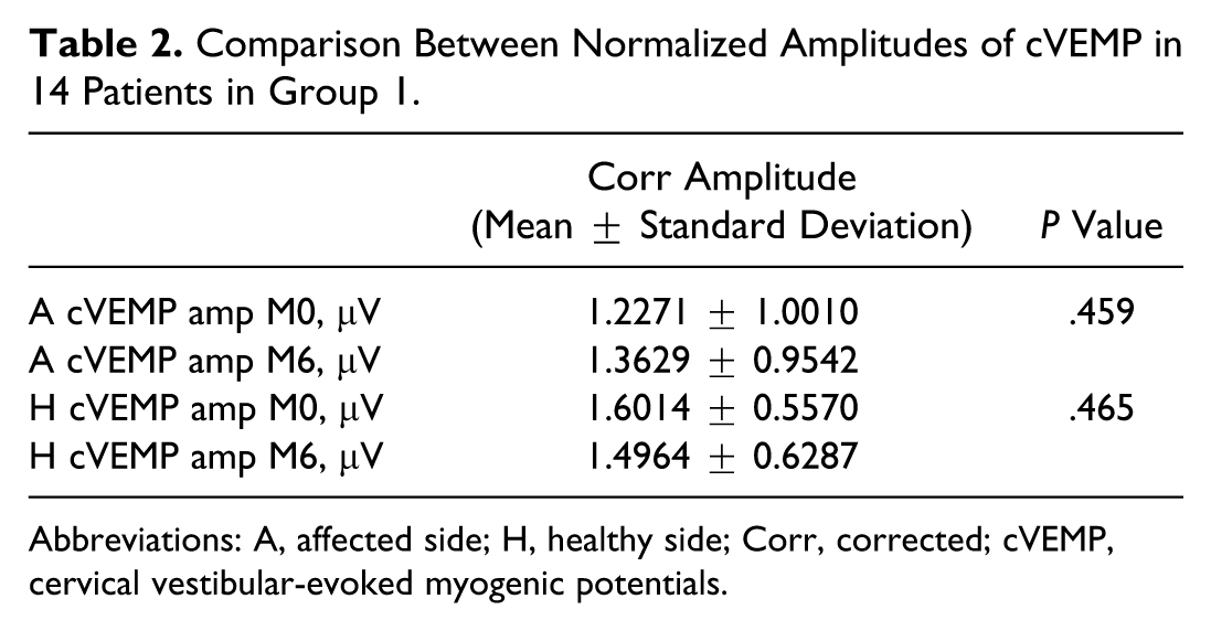
Abbreviations: A, affected side; H, healthy side; Corr, corrected; cVEMP, cervical vestibular-evoked myogenic potentials.
Group 2 showed worsening of the affected vestibular nerve after 6 months, leaning toward significance (P = 0.069). In the same period, there was no change in the unaffected vestibular nerve, and values of the affected side remained lower compared to values on the unaffected side, however, close to significance (P = .056; Table 3). At the same time, there was no change in amplitudes of cVEMP on either the healthy or the affected side in group 2 (Table 4).
Comparison Between Peak-to-Peak Amplitudes of oVEMP in 12 Patients in Group 2.
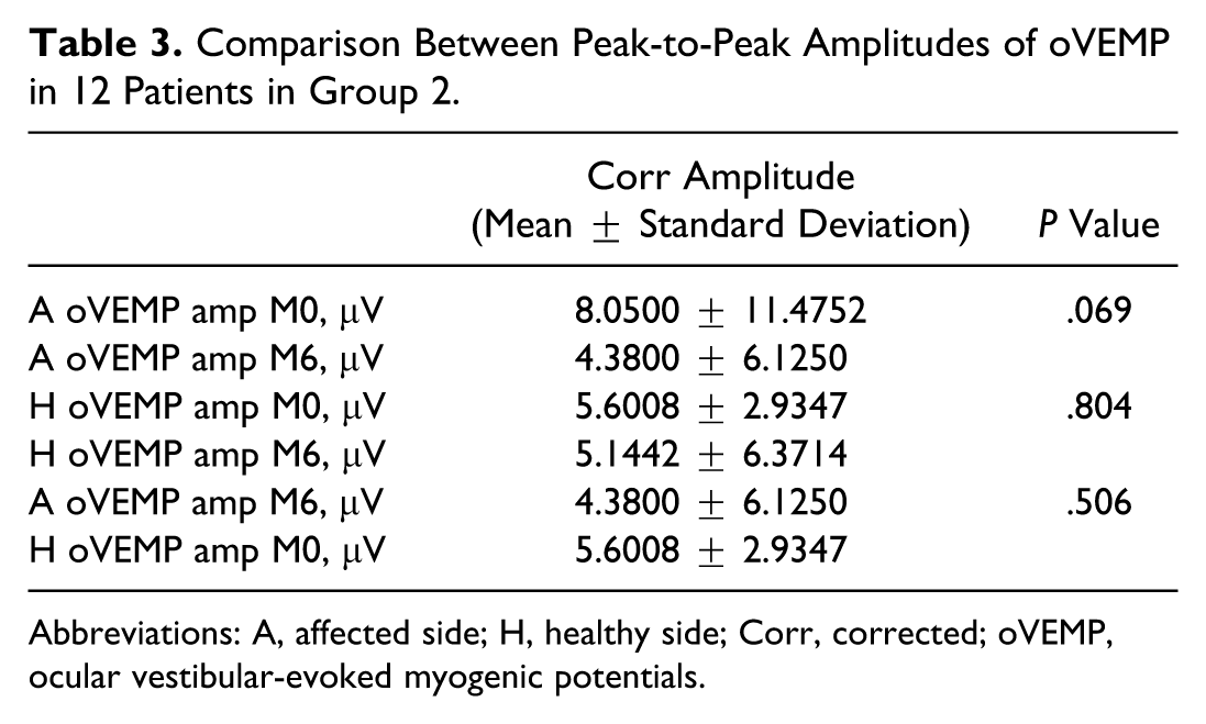
Abbreviations: A, affected side; H, healthy side; Corr, corrected; oVEMP, ocular vestibular-evoked myogenic potentials.
Comparison Between Normalized Amplitudes of cVEMP in 12 Patients in Group 2.
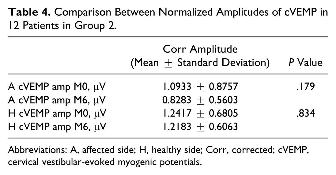
Abbreviations: A, affected side; H, healthy side; Corr, corrected; cVEMP, cervical vestibular-evoked myogenic potentials.
In 7 (26.9%) of 26 patients, initial oVEMP showed conduction block (absent potential) on the affected side, while the response was normal in all patients on the healthy side.
Differences in clinical and MRI findings between the groups are shown in Table 5. Inability to perform the Fukuda test, and chronic white matter supratentorial lesions present on brain MRI (Figure 5), were more frequent in patients with a worse outcome on oVEMP (P = .044 and .045, respectively).
Differences in Clinical and MRI Findings Between Groups (Group 1-14 Patients; Group 2-12 Patients).
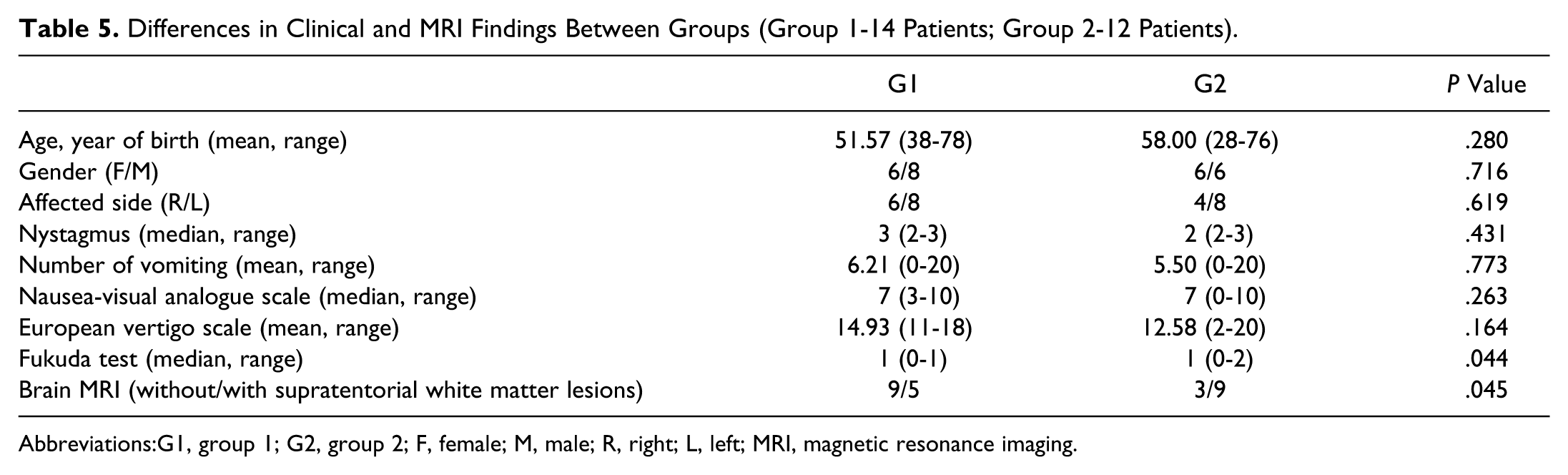
Abbreviations:G1, group 1; G2, group 2; F, female; M, male; R, right; L, left; MRI, magnetic resonance imaging.

An example of the brain magnetic resonance imaging, fluid-attenuated inversion recovery sequence, of one of the studied patients in group 2 (the group with worsening of ocular vestibular-evoked myogenic potentials amplitudes). Note the chronic hyperintensive white matter lesions.
Differences in neurophysiological findings between the groups are shown in Table 6. The inferior branch of the vestibular nerve was affected in 15 (57.7%) patients. Although involvement of the inferior branch of the vestibular nerve was not associated with oVEMP outcome, prolonged latencies of oVEMP were associated with better outcome. Pearson correlation confirmed that oVEMP latencies (N10 and P13) were associated with improvement or worsening in oVEMP amplitudes, showing that prolonged latencies are correlated with 6-month improvement in oVEMP amplitudes (Pearson correlation −0.472, P = .041 and −0.580, P = .009, respectively).
Differences in Neurophysiological Findings Between Groups (Group 1-14 Patients; Group 2-12 Patients).
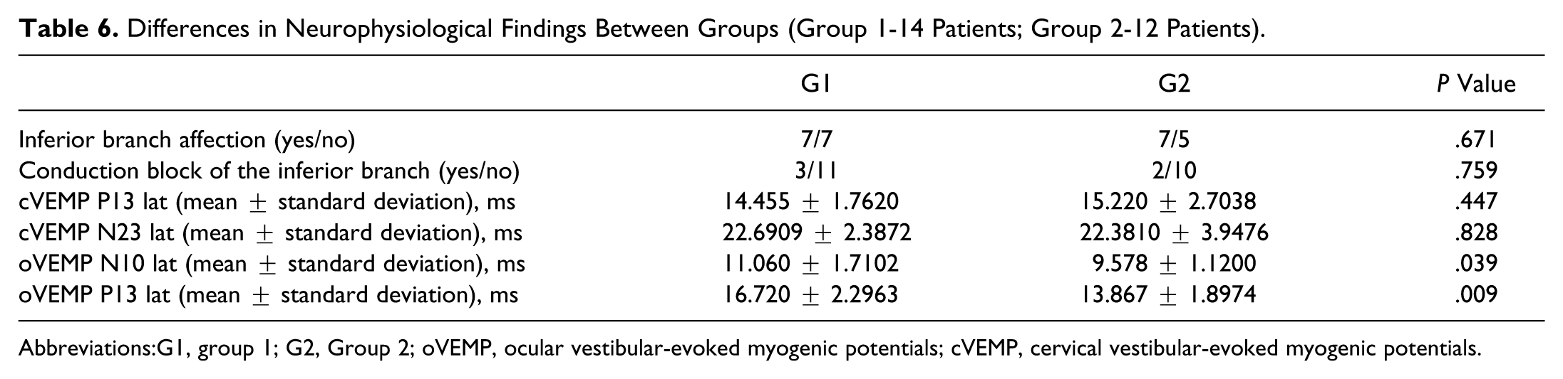
Abbreviations:G1, group 1; G2, Group 2; oVEMP, ocular vestibular-evoked myogenic potentials; cVEMP, cervical vestibular-evoked myogenic potentials.
Year of birth was negatively associated with involvement of inferior branch of the vestibular nerve and the MRI (Pearson correlation −0.414, P = .036 and −0.632, P = .001, respectively). These findings indicate that older patients have a lower chance of inferior nerve involvement and a higher chance of T2 hyperintensities in the supratentorial white matter. Female gender was associated with a higher number of vomiting episodes (Pearson correlation −0.424, P = .031). No other correlations between the studied parameters were found.
Discussion
The present study has demonstrated certain advantages in combining oVEMP and cVEMP in long-term monitoring of patients with VN. Inability to perform the Fukuda test, and chronic white matter supratentorial lesions present on brain MRI, were shown to be associated with worse outcome on oVEMP. Prolonged oVEMP latencies (N10 and P13) are in correlation with 6-month improvement in oVEMP amplitude.
It has been shown that both oVEMP and cVEMP response parameters demonstrate good test–retest reliability, and that simultaneous oVEMP and cVEMP may be a convenient screening tool for assessing crossed vestibuloocular reflex and ipsilateral sacculocollic reflex, which definitely shortens the diagnostic test time.17,18 Several studies have shown that VEMP has several advantages in the diagnosis and follow-up of patients with VN. A study by Shin et al demonstrated that oVEMP represents a crossed utriculoocular pathway, thus representing function of the superior branch of vestibular nerve, while cVEMP represents an ipsilateral vestibulocollic reflex pathway, which is mediated by the inferior branch of vestibular nerve. 4 The superior branch is therefore responsible for conducting afferents from the lateral and superior semicircular canals and utricular macula, while the inferior branch innervates the posterior semicircular canal and the saccular macula. 19 Besides the functional differences in the 2 branches of the vestibular nerve, there is a significant difference in the anatomy of their pathways. The superior branch travels through a longer and narrower tunnel in the temporal bone than the inferior branch, making it more susceptible to inflammation and subsequent edema-mediated injuries. 20
VN was traditionally thought of as a partial vestibular lesion, which affects only the superior branch. 21 However, there is an increasing number of studies that demonstrate the involvement of the inferior branch, in which VEMP plays an important diagnostic role. 22 In our study, 15 (57.7%) of the 26 patients had involvement of the inferior vestibular nerve, adding to recognition of inferior branch involvement in the pathophysiologic mechanism of VN.
Our results have shown recovery of vestibular function in 14 (53.8%) of the 26 patients after 6-month follow-up. Previous reports have demonstrated recovery of vestibular function in 30% to 50% of the patients with VN on cVEMP testing only.11,12 Murofushi et al reported recovery in 5 of 13 patients in a period of 6 months to 2 years. 11 In all, 4 of those 5 patients showed full recovery. Ochi et al reported recovery in 1 of 2 patients who showed abnormal VEMP initially in a period of 15 months. 12 In comparison with their results, our study has shown a greater percentage of recovery in a shorter period of time. The reason for this could be inpatient management. Murofushi and Ochi have not reported any specific treatment of patients in their study. All of our patients were treated with oral prednisone as described previously. 23
We have also identified several factors that are associated with the recovery of oVEMP. Inability to perform the Fukuda test was the only clinical finding associated with worse recovery of oVEMP. Also, chronic white matter lesions were more prevalent in patients who did not recover. This finding can be interpreted with lower capability of central compensation, which is important in the clinical recovery of patients with VN. 24 Vascular etiology of the VN can be involved in the pathogenesis of VN in patients with T2 hyperintensities on the MRI. 25
We found that prolonged latencies of oVEMP were associated with oVEMP recovery on follow-up. This finding can be interpreted in several ways. We have recently observed that prolonged latencies of both oVEMP and cVEMP are present in up to 80% of patients with multiple sclerosis, indicating that prolonged VEMP latencies are associated with a demyelinating process. 26 One can extrapolate that prolonged latencies in patients with VN indicate a lesion of myelin at the 8 cranial nerve root entry zone or the nerve itself. 27 Inferior branch involvement demonstrated by cVEMP was not associated with oVEMP recovery or worsening. Reasons for this can be found in the anatomical variations between the 2 branches. 20 Based on our findings, we conclude that although the inferior nerve is frequently affected in patients with superior VN, it does not contribute to the extent of the recovery, and is less prone to protracted dysfunction when compared to the superior nerve.
Possible limitations of this study could be the short follow-up period and the relatively small number of patients. In addition, we have not performed caloric testing. The reason for this is 3 fold. First, caloric testing does not differentiate between superior and inferior VN and is regularly negative in inferior VN, acutely. 8 Second, all of our patients were subjected to MRI, excluding stroke as a possible cause of acute vestibular syndrome. Third, the aim of our study was to evaluate the recovery of vestibular nerve function after VN by means of VEMP, in which caloric testing would bring no additional information. Nevertheless, to the best of our knowledge, this is the first study which used the combination of oVEMP and cVEMP in monitoring of patients with VN.
Conclusion
In conclusion, this study offers an additional insight into better understanding the role of VEMP in the diagnosis and prognosis of patients with VN. Further studies are needed to validate this diagnostic procedure, and to assess its clinical usefulness in VN management.
Footnotes
Author Contributions
Study concept and design: Adamec, Habek. Acquisition of data: Adamec, Krbot, Handžić Barušić, Bach, Gabelić, Habek. Analysis and interpretation of data: Adamec, Krbot, Handžić, Gabelić, Habek. Drafting of the manuscript: Adamec, Krbot, Habek. Critical revision of the manuscript for important intellectual content: Adamec, Krbot, Handžić, Barušić, Bach, Gabelić, Habek. Administrative, technical, and material support: Adamec, Krbot, Barušić, Bach, Gabelić, Habek.
Declaration of Conflicting Interests
The author(s) declared no potential conflicts of interest with respect to the research, authorship, and/or publication of this article.
Funding
The author(s) received no financial support for the research, authorship, and/or publication of this article.
