
Abstract
Background. Epilepsy is prevalent in the elderly, whose brain morphologies and skull electrical characteristics differ from those of younger adults. Here, using a multivariate definition of signal-to-noise ratio (SNR), we explored the detectability of epileptic spikes in scalp EEG measurements in elderly by forward simulations of hypersynchronous spikes generated at 78 cortical regions of interest (ROIs) in the presence of background noise. Methods. Simulated electric potentials were measured at 18, 35, and 70 standard 10–20 electrode positions using three reference methods: infinity reference (INF), common average reference (CAR), and average mastoid reference (M1M2). MRIs of six elderly subjects were used to construct finite element method (FEM) models with age-adjusted skull conductivities. Results. SNRs of epileptic spikes increased with increasing sizes of the brain electrical source areas, although medial and deep brain regions such as the hippocampus showed lower SNRs, consistent with clinical findings. The SNRs were greater in the 70-channel dataset than in the 18-channel and 35-channel datasets, especially for ROIs located closer to the head surface. In addition, the SNRs were lower for the CAR and M1M2 references than for the ideal INF reference. Moreover, we found comparable results in the standard FEM heads with age-adjusted skull conductivities. Conclusions. The results provide insights for evaluating scalp EEG data in elderly patients with suspected epilepsy, and suggest that age-adjusted skull conductivity is an important factor for forward models in elderly adults, and that the standard FEM head with age-adjusted skull conductivity can be used when MRIs are not available.
Keywords
Introduction
Epilepsy is the third most prevalent neurological disorder in elderly people aged greater than 65 years. 1 A recent study reported that 73.2% of elderly adults over 65 years old with epilepsy displayed interictal epileptiform discharges (IEDs). 2 Since the detection of IEDs suggests the presence of epileptic disorders, interictal EEG monitoring3–5 and automatic methods to detect seizure-related abnormal activity (synchronization) (Supplementary Introduction) provide useful information for the diagnosis of epilepsy. Electrical source imaging (ESI) can localize IEDs in the brain6,7 and provides useful information for the diagnosis and classification of epilepsy as well as the prognosis of epilepsy.5,8 ESI with high-density EEG recording with more than 64 channels provides accurate localization of IEDs based on postoperative clinical outcomes,9–11 while some studies have reported that ESI with standard EEG recording may also be useful for localizing IEDs.12–16
Accurate ESI requires accurate forward simulation of epileptic spikes in scalp EEGs from brain electrical signals against background noise. Accurate forward simulation depends on several factors, including accurate head modeling of the geometry and conductivity of head components (scalp, skull, cerebrospinal fluid [CSF], gray matter, and white matter) and the number of scalp electrodes.11,17–19 In this sense, the heads of elderly people may be different from those of young adults in terms of brain morphology and skull conductivity,11,20 while the optimization of skull conductivity is reported to be important for decreasing ESI estimation errors. 21 In addition, the locations and sizes of brain electrical sources may affect the detectability (ie, signal-to-noise ratio [SNR]) of epileptic spikes in scalp EEGs.22–26 Moreover, referencing methods may affect scalp EEG spike signals in actual EEG recordings. 27 The aim of the present study is to investigate the effects of the locations and sizes of brain electrical sources and of reference types on the detectability (SNRs) of epileptic spikes in scalp EEGs in elderly subjects. To this end, we computed forward simulations of scalp epileptic potentials in 18-, 35-, and 70-ch standard 10-20 locations under different reference methods from different locations and sizes of brain electrical source regions, and we analyzed SNRs of multivariate scalp epileptic potentials. For the forward simulation, finite element method (FEM) models were constructed from the MR images of individual elderly subjects, and skull conductivity was adjusted for the ages of the elderly subjects in this study. To analyze the impact of skull conductivity, SNRs were also analyzed using the standard head models with commonly used skull conductivity and those with age-adjusted skull conductivities.
Subjects and Methods
Participants
T1-weighted MR images acquired from six elderly subjects (74.7 ± 4.1 years, mean age ± standard error of the mean [SEM]; male, n = 3; female, n = 3) were used for the simulation study. The anonymized patients’ data (MR images of the heads) were obtained by opt-out according to Japanese ethical guidelines. The protocol in this study was carried out in accordance with the Declaration of Helsinki and was reviewed and approved by the Ethics Review Board for Human Research (permit no.: R2022065, approved on July 26, 2022).
General Experimental Procedures
First, individual FEM volume conductor models, each with an embedded cortical surface source model, were created based on individual MR images (Figure 1A) (Supplementary Methods-1). Spike generation in an irritative zone (IZ) or seizure onset zone (SOZ) was simulated as the synchronous activity of multiple dipoles in a cortical area with surface-normal currents (“macrocolumns”). The simulated IZs and/or SOZs were located in 78 regions of interest (ROIs) (Table 1). At each ROI, 11 different synchronous cortical patches consisting of macrocolumns (current dipole elements) were created. Since elderly people with recent-onset epilepsy mostly present with focal seizures,28,29 the ROIs in this study were considered one at a time. Potential distributions over the scalp generated by synchronous current dipole elements within each ROI were estimated by forward simulation using individual FEM models (Supplementary Methods-2). In this forward simulation, epileptic spikes in scalp EEGs with three different numbers (18, 35, and 70) of nested electrode channels (Figure 1B) were computed with three different reference types: infinity reference (INF), common average reference (CAR), and average mastoid reference (M1M2). Examples of scalp potentials are shown in Figure 1C. Finally, SNRs of epileptic spike distributions were computed by considering “noise” as asynchronous non-ROI brain activity.
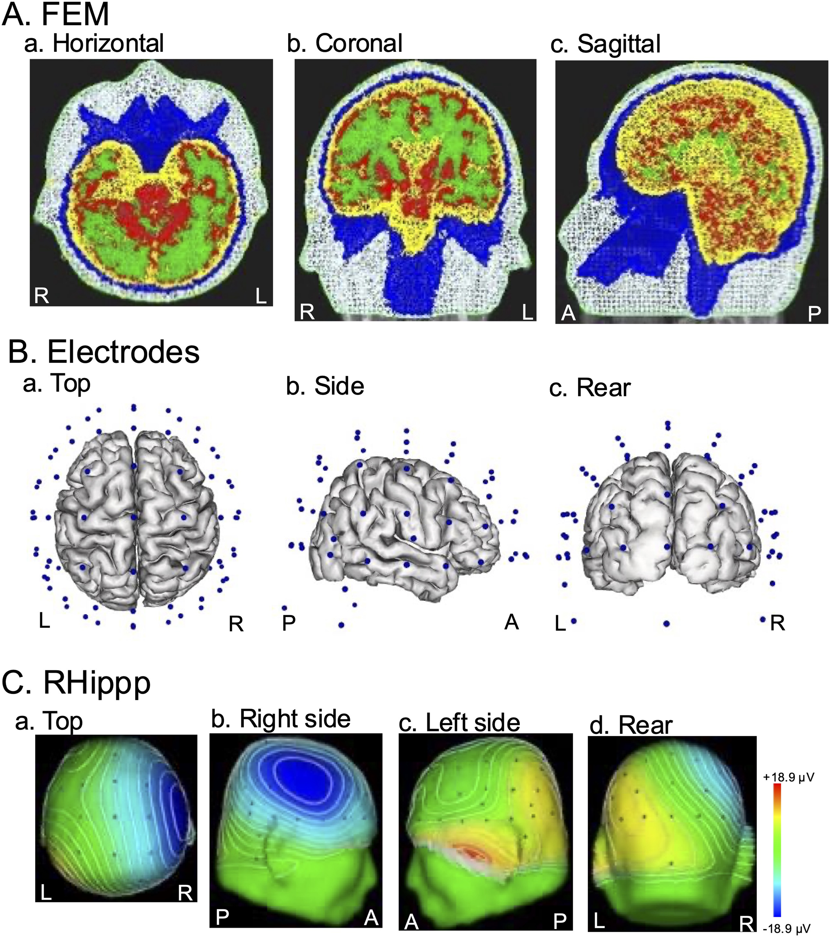
Schematic representation of the experimental methods. (A) An example of a 5-layer FEM head in one subject. The white, blue, yellow, red, and green areas indicate the scalp, bone (skull), cerebrospinal fluid (CSF), gray matter, and white matter, respectively. R, right; L, left; A, anterior; P, posterior. (B) Locations of the 70 electrodes in the same subject. The blue dots indicate the electrodes. (C) Potential distributions on the scalp after forward simulation of the brain electrical source area with an area expansion number of 10 without noises in the right posterior hippocampus (RHippp) of the same subject.
Anatomical Names and MIN Coordinates of the 78 ROIs.
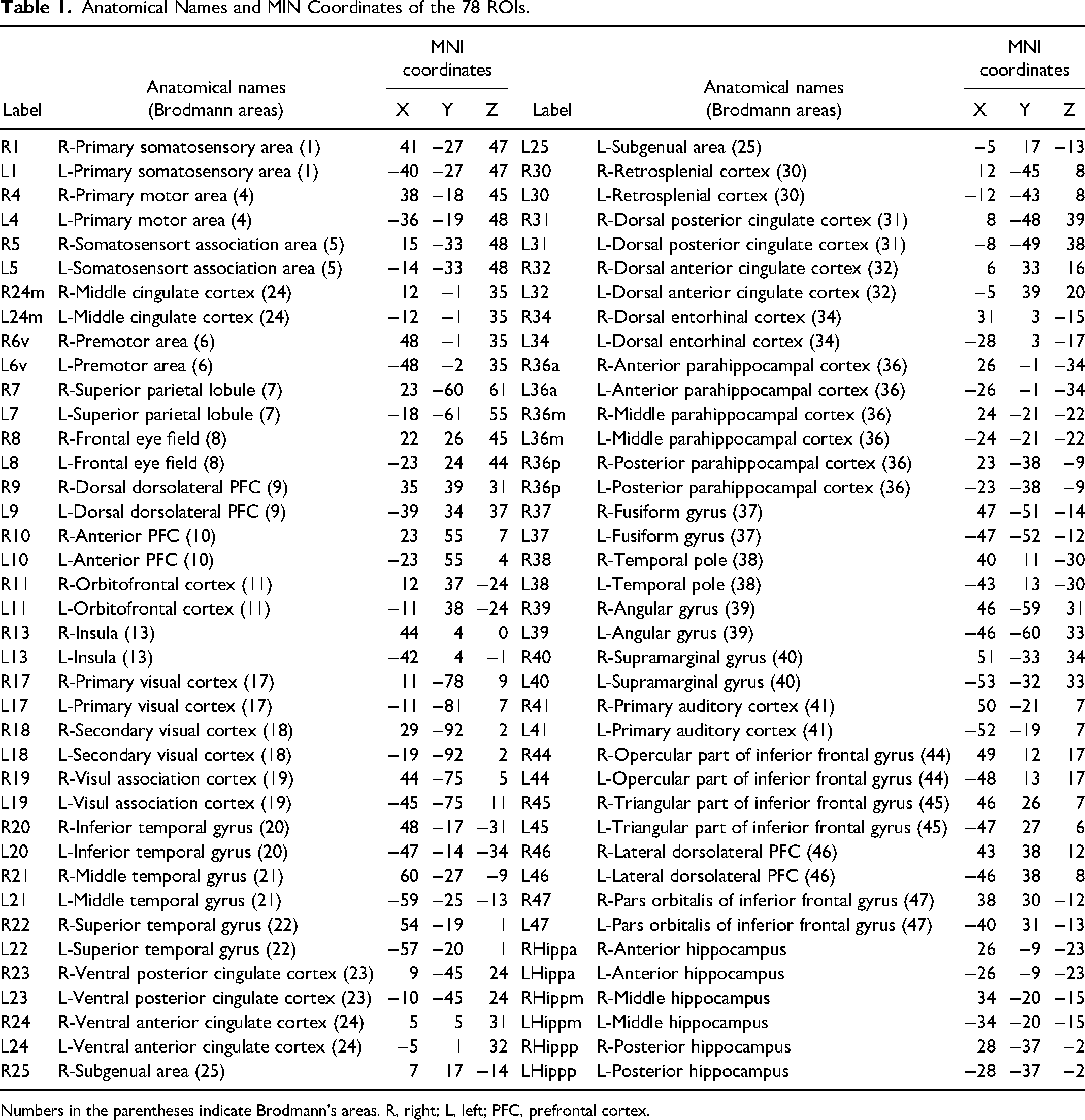
Numbers in the parentheses indicate Brodmann's areas. R, right; L, left; PFC, prefrontal cortex.
To analyze the effects of skull conductivity on SNRs, six artificial elderly heads were created using the standard head (ICBM 152 nonlinear asymmetric atlas; see below for details) with skull conductivity adjusted for individual subjects’ ages (Supplementary Methods-2), and the same analyses were performed using these standard head-derived datasets. In these analyses, the other components of the standard head (ie, scalp, CSF, gray matter, and white matter) had the same conductivity values as those used across the subjects as in the individual FEM head models. Thus, age-adjusted skull conductivity was the only difference between these artificial standard elderly head models. Furthermore, as a control for artificial standard elderly heads with age-adjusted skull conductivities, the standard head with commonly used skull conductivity (10 mS/m) (dubbed as “standard skull conductivity”) 30 was also used to compute SNRs.
Assessment of SNR as Spike Detectability in Scalp EEGs
In this paper, a signal of interest (SOI) was a synchronized group of macrocolumns, each simulated by current dipole, in a ROI patch with 11 different sizes (see former section and Supplementary Methods-2). To assess the detectability of epileptic spikes in scalp EEGs, SNRs of epileptic spikes were computed. The assumption in the former section indicates that all current dipoles in each ROI were summed and projected, via a forward matrix, to an electric potential signal vector in multi-channel sensor space. Furthermore, there are manifold possible realizations of background noise in source space: each of these projected to sensor space as a noise vector. SOI strength was measured in sensor space using a “Gram norm” (defined in Supplementary Methods-3) that equalizes channels and reverses inter-channel correlations under the “null assumption” that source space currents are independent and identically distributed. Importantly, the strength of each noise vector was measured (using the Gram norm) with respect to its projection onto the Gram-normalized signal vector. For example, if one realization of a Gram-normalized noise vector happens to be orthogonal to the Gram-normalized signal vector, then the noise strength is zero (on that realization). For multiple realizations, the strength of the SOI-relative background noise was estimated as the square root of average squared noise strengths, ie, root mean squared (RMS) noise. Finally, the SNR for each SOI is simply the signal strength divided by the estimated RMS noise (Supplementary Methods-3).
Statistical Analysis
All values were expressed as mean ± standard error of mean (SEM). To assess the impacts of ROI, extent of the brain electrical source areas, and number of electrodes on SNRs under INF reference, SNR data were examined with repeated-measures 3-way analysis of variance (ANOVA) with factors of ROI, extent of the brain electrical source areas, and number of electrodes. To analyze the effects of reference types (INF, CAR, and M1M2), SNR data were analyzed by repeated measures 2-way ANOVA with factors of ROI and reference type. In these ANOVA statistical tests, sphericity was assessed, and the degrees of freedom were corrected by the Greenhouse‒Geisser method where appropriate. Post hoc multiple comparisons among the groups were performed by Bonferroni and Tukey tests. Statistical significance was set at p < 0.05. All statistical analyses were performed using GraphPad Prism version 9.5.1 (GraphPad Software, San Diego, CA) and SPSS Statistics (v. 28, SPSS Inc., Chicago).
Results
Effects of ROIs, Extent of Brain Electrical Source Areas, and Electrode Numbers
To analyze the effects of ROI, expansion number of the brain electrical source areas, and number of electrodes on SNRs under INF reference, repeated measures 3-way ANOVAs with factors of ROI, number of electrodes, and expansion number of the brain source area were performed separately for the right and left hemispheres. In the right hemisphere, there were significant main effects of ROI, expansion number of brain electrical source areas, and electrode number, while all possible interactions among the three factors, including interaction between ROI and expansion number of brain electrical source areas, interaction between ROI and electrode number, interaction between expansion number of brain electrical source areas and electrode number, and interaction among ROI, expansion number of brain electrical source areas, and electrode number, were also significant (Supplementary Table S2). In the left hemisphere, comparable results were observed in the repeated measures 3-way ANOVA (Supplementary Table S2).
In both hemispheres, the main effects of ROI and interactions between ROI and other factors were significant. Since SNRs were most prominent at area expansion number of 10, a subsidiary repeated measures 1-way ANOVA on SNRs among the ROIs was performed under the following conditions (area expansion number of 10, 70-ch recording, and INF reference) in each hemisphere. The results in the right hemisphere indicated that there was a significant main effect of ROI [F (3.746, 18.73) = 16.91, p < 0.0001]. Figure 2A shows post hoc comparisons of SNRs among the different ROIs (Figure 2A, Supplementary Table S3). SNRs in the right posterior hippocampus (RHippp) were smaller than those in the other ROIs (p < 0.05, 0.01, 0.0001, Tukey tests). Furthermore, SNRs were also smaller in other ROIs in the deep and medial brain regions, including the hippocampus (RHippa, RHippm), parahippocampal cortex (R36a, R36 m, R36p), retrosplenial cortex (R30), posterior cingulate cortex (R23), medial occipital cortex (primary visual cortex) (R17), insula (R13), orbitofrontal cortex (R11), and subgenual cingulate cortex (R25) (Supplementary Table S3). In the left hemisphere, comparable results were observed: there was a significant main effect of ROI [F(3.885, 19.43) = 28.31, p < 0.0001] (Figure 2B, Supplementary Table S4).
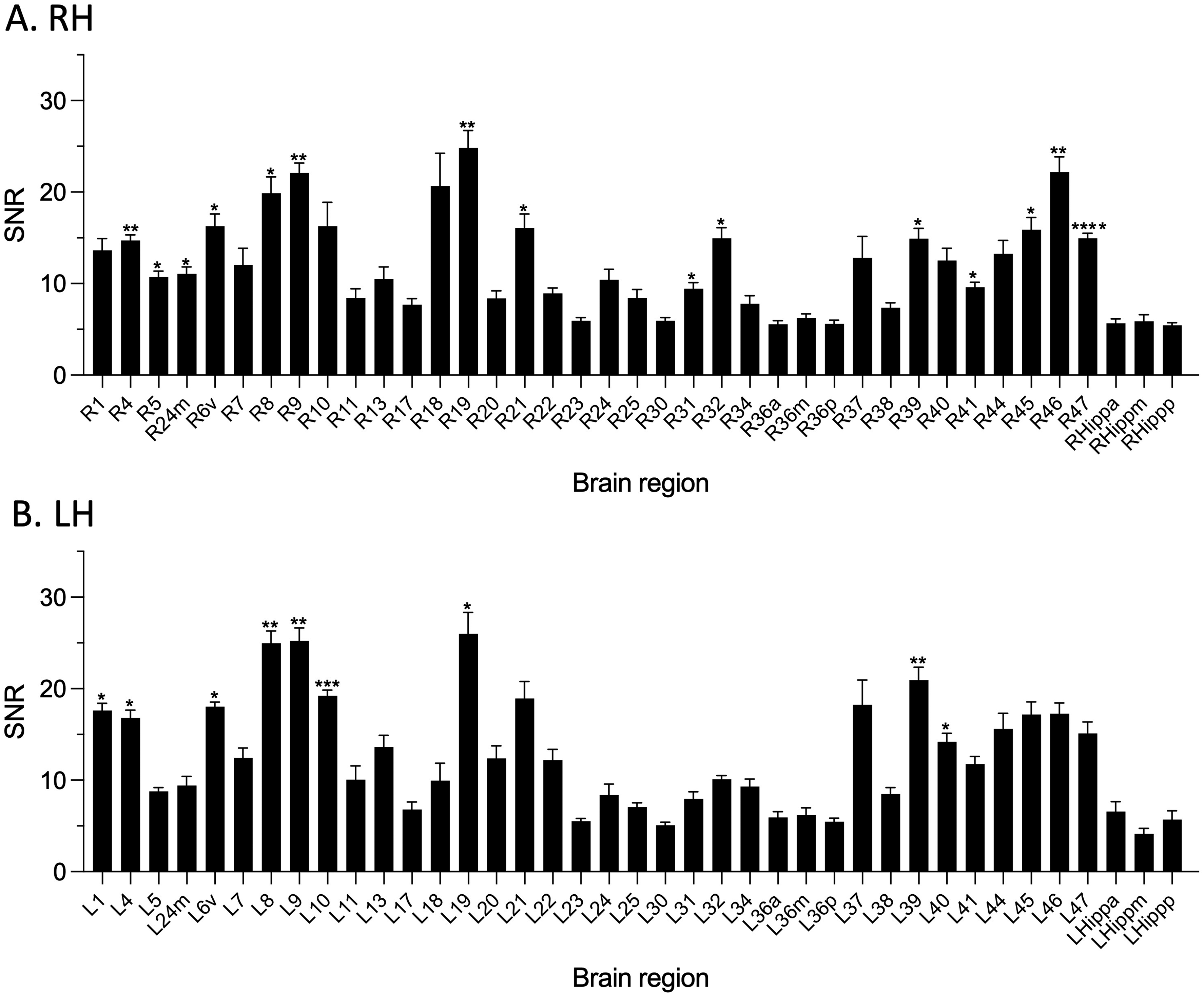
Post hoc comparison of SNRs among the different ROIs in the right hemisphere (RH) (A) and left hemisphere (LH) (B) after repeated measures 1-way ANOVA under the following conditions [area expansion number of 10, 70-ch recording, and infinite (INF) reference]. Asterisks indicate significant differences from the posterior hippocampus: *, p < 0.05; **, p < 0.01; ***, p < 0.001; ****, p < 0.0001 (Tukey test).
In both hemispheres, the main effect of expansion number of the brain electrical source areas was significant under INF reference. Post hoc tests for the main effect of the area expansion number indicated that SNRs were greater in the wider areas (larger expansion numbers) than in the smaller areas (smaller expansion numbers) in all possible comparisons among the expansion numbers [p < 0.001, Bonferroni test in both hemispheres]. These findings indicate that SNRs increased as the expansion number increased, as shown in Figure 3. Furthermore, rising rates of SNR were lower in the deep and/or regions (Supplementary Results).
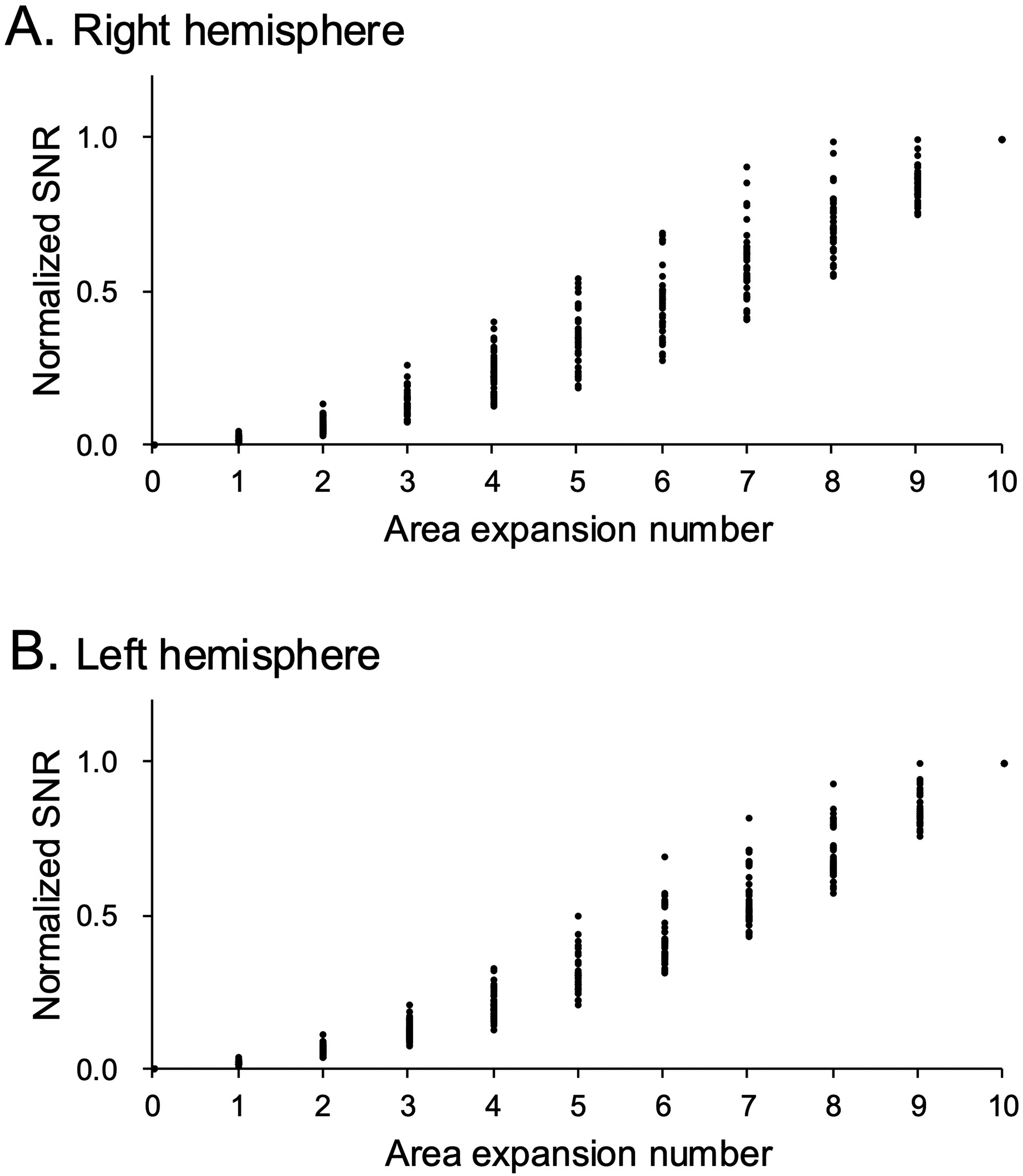
Relationships between normalized SNRs in the 70-channel recordings under infinite (INF) reference and extents of the brain electrical source areas in all ROIs in the right (A) and left (B) hemispheres. In each ROI in each subject, the SNRs were normalized by the average SNR at expansion number 10 for the six subjects. The average normalized SNR, averaged over the six subjects, was then plotted for each expansion number in each ROI.
In both hemispheres, the main effect of electrode number was significant. Post hoc tests of the main effect of electrode number indicated that SNRs were greater in the 70-channel dataset than in the 35-channel [p < 0.0001, Bonferroni test in both hemispheres] and 18-channel [p < 0.0001, Bonferroni test in both hemispheres] datasets. Furthermore, there were significant interactions between the number of electrodes and the remaining two factors (ROI and expansion number of brain electrical source areas) (Supplementary Table S2), suggesting that this superiority of the 70-channel recording was dependent on ROIs and expansion numbers of the brain electrical source areas. To examine the effects of electrode number and expansion number of the brain electrical source areas on SNRs in specific ROIs, subsidiary 2-way ANOVAs on SNRs under INF reference with factors of electrode number and expansion number of the brain electrical source areas were performed in the right superior parietal lobule (RtSPL) (R7) and right posterior hippocampus (RHippp). In the RtSPL located in a shallow region of the brain (Figure 4A), there were significant main effects of expansion number [F(1.232, 6.162) = 36.60, p = 0.0007] and electrode number [F(1.035, 5.175) = 104.8, p < 0.0001] and a significant interaction between expansion number and electrode number [F(1.742, 8.711) = 32.19, p = 0.0001]. Post hoc comparisons indicated that SNRs were significantly greater for 70 channels than for 18 channels and 35 channels and greater for 35 channels than for 18 channels across all expansion numbers. The improvement in SNRs with increasing electrode number was more evident for larger area expansion numbers. However, in the RHippp, which is located in a deep and medial brain region (Figure 4B), the impact of electrode number was less evident. The same analysis by repeated measures 2-way ANOVA indicated that there were significant main effects of expansion number [F(1.148, 5.740) = 253.4, p < 0.0001] and electrode number [F(1.239, 6.193) = 36.18, p = 0.0007] and a significant interaction between expansion number and electrode number [F(2.221, 11.11) = 19.91, p = 0.0002]. Similarly, post hoc comparisons indicated that SNRs were greater for 70 channels than for 35 channels and 18 channels for most expansion numbers except for expansion number 7.
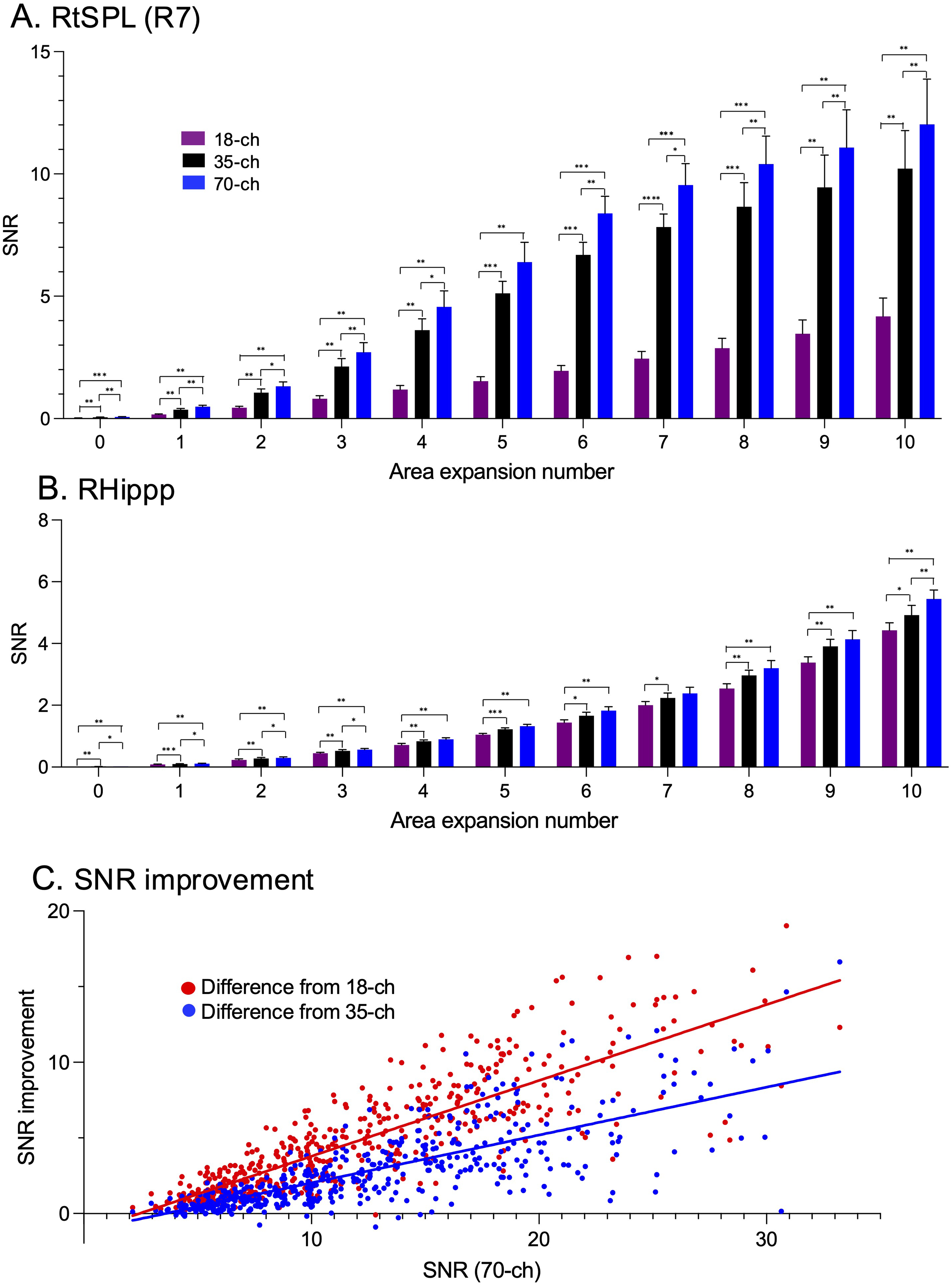
Relationships between SNRs under infinite (INF) reference and the number of electrodes. A, B: Relationships between SNRs and area expansion numbers in the right superior parietal lobule (RtSPL) (R7) (A) and right posterior hippocampus (RHippp) (B). *, p < 0.05; **, p < 0.01; ***, p < 0.001; ****, p < 0.0001 (Tukey test). C: Relationships between SNR improvements in the 70-channel recordings from the 18-channel (red dots) and 35-channel (blue dots) recordings and SNRs in the 70-channel recordings.
A visual comparison of the data for R7, located in a shallow brain region, and RHippp, located in a deep brain region, suggests that the impact of electrode number (ie, SNR improvements from the 18- and 35-channel datasets in the 70-channel datasets) is more evident at the ROIs located in shallow brain regions with larger SNRs. To examine this idea, we analyzed the relationships between SNRs in 70-channel recordings and the SNR improvement from 18-channel and 35-channel recordings under INF reference. Figure 4C shows the relationships between SNRs in the 78 ROIs (area expansion number 10, INF reference and 70-channel) and the SNR improvements from the 18- and 35-channel datasets (area expansion number 10 and INF reference). Linear regression analyses of the data indicated that SNRs in the 70-channel recordings were significantly and positively correlated with the improvements in SNRs from 18-channel recordings [F(1, 466) = 1171, p < 0.0001] and 35-channel recordings [F(1, 466) = 581.1, p < 0.0001]. Furthermore, the slopes of the regression lines were significantly greater for the improvements from the 18-channel recordings than those from the 35-channel recordings [F(1, 932) = 88.35, p < 0.0001]. These findings indicate that SNR improvements by larger electrode numbers are significantly effective, but its impact on detecting signals is limited in ROIs with low SNRs, such as deep and medial brain regions.
Effects of Reference Types
To analyze the effects of reference types, we analyzed each of the 9 datasets with different simulation conditions in each hemisphere [39 ROIs×3 expansion numbers of brain electrical source areas (expansion number = 0, 5, and 10)×3 reference types (INF, CAR, and M1M2)] by repeated measures 2-way ANOVA with factors of ROI and reference type. In the 18-channel dataset with an area expansion number of 0 in the right hemisphere, there were significant main effects of ROI [F(4.225, 21.12) = 8.368, p = 0.0003] and reference type [F(1.427, 7.134) = 81.40, p < 0.0001] and a significant interaction between ROI and reference type [F(4.658, 23.29) = 3.076, p = 0.0306] (Table 2A). Post hoc tests of the main effect of reference type indicated that SNRs were greater in the INF than in the CAR (p = 0.0008, Bonferroni test) and M1M2 (p = 0.0003, Bonferroni test). In the other 18-channel datasets with area expansion numbers of 5 and 10, the same results were observed (Table 2A). In the 35-channel datasets, including those with an expansion number of 5, as well as the 70-channel datasets, including those with an expansion number of 10, the same results were observed: there were significant main effects of ROI and reference type, and significant attenuation of SNRs was observed in the M1M2 and/or CAR (Table 2A).
Statistical Results (p-Values) of Repeated Measures 2-way ANOVA on the Data Derived from the Individual FEM Heads in the Right (A) and Left (B) Hemispheres.
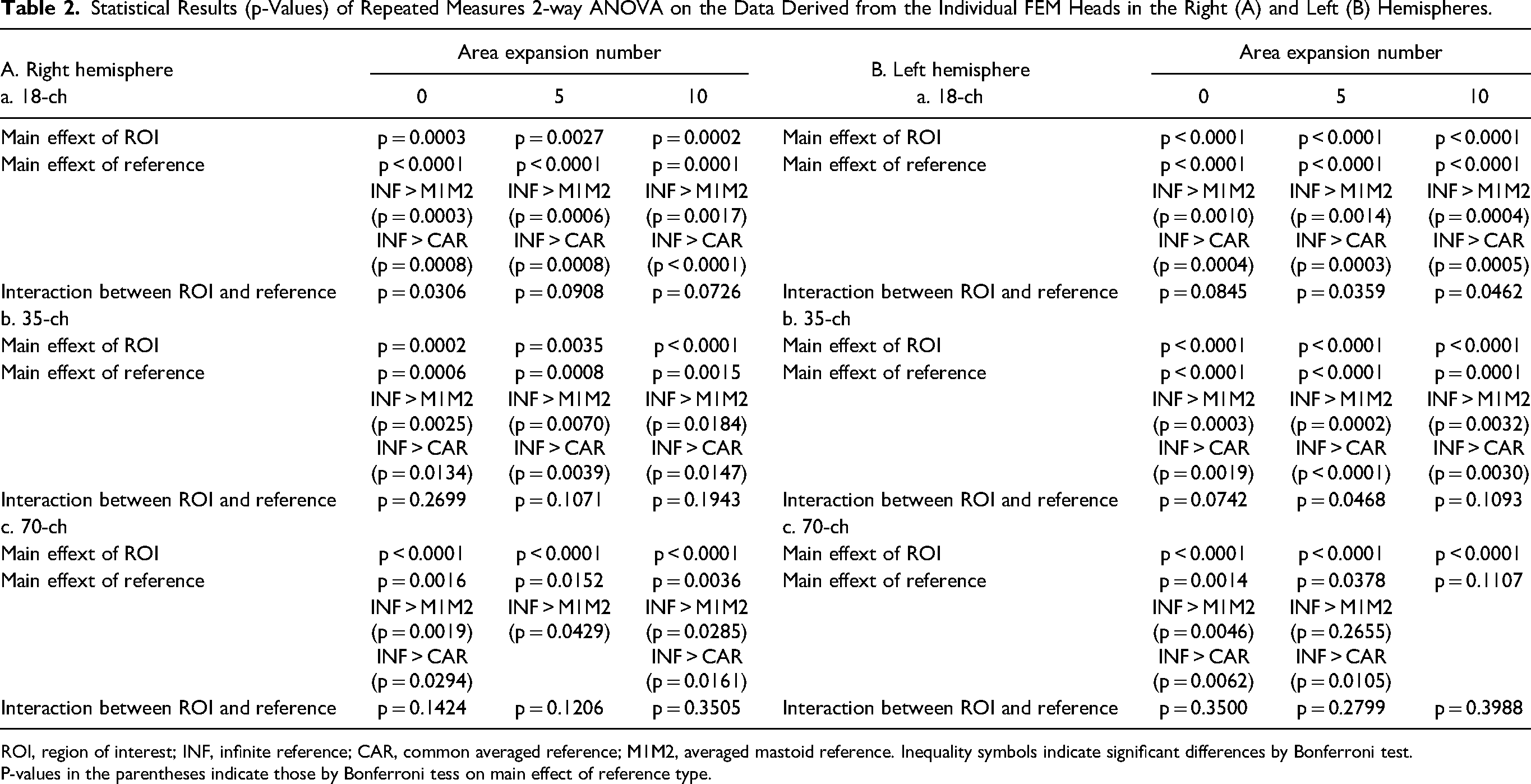
ROI, region of interest; INF, infinite reference; CAR, common averaged reference; M1M2, averaged mastoid reference. Inequality symbols indicate significant differences by Bonferroni test.
P-values in the parentheses indicate those by Bonferroni tess on main effect of reference type.
The same trend was observed in the left hemisphere (Table 2B). In all datasets except those in the 70-ch recordings with an expansion number of 10, there was a significant main effect of reference type. In these datasets, SNRs were greater for the INF than for the M1M2 and CAR. Taken together, these results indicated that SNRs were attenuated in the M1M2 and/or CAR compared to the INF.
Effects of the Skull Conductivity
The above results indicated that ROIs, area expansion numbers, electrode numbers and reference types significantly affected SNRs. Skull conductivity may also affect SNRs. To examine this idea, we analyzed linear correlations (linear regression analysis) of SNRs in 70-channel recordings under INF reference between the individual FEM heads and artificial standard elderly heads with skull conductivity adjusted for individual ages (Figure 5A). The results indicated that SNRs were significantly correlated between the two head models in the right [F(1, 2572) = 8820, p < 0.0001, R2 = 0.7442] (Figure 5Aa) and left [F(1, 2572) = 12024, p < 0.0001: R2 = 0.8238] (Figure 5Ab) hemispheres. This finding suggests that differences in skull conductivity may contribute to variation in SNRs in the elderly. However, the above correlations could be ascribed to factors other than skull conductivity such as morphological factors of the heads, since the standard head has many morphological characteristics similar to those in the individual FEM heads. We further analyzed correlations of SNRs between the individual FEM heads and the standard head with the standard skull conductivity instead of artificial standard elderly heads with age-adjusted skull conductivities (Figure 5B). The results indicated that degrees of goodness of fit (R2) to the regression lines were relatively lower in Figure 5B than in Figure 5A, although SNRs were significantly correlated between the individual FEM heads and standard head with the standard skull conductivity in the right [F(1, 2572) = 1441, p < 0.0001, R2 = 0.3592] (Figure 5Ba) and left [F(1, 2572) = 1157, p < 0.0001: R2 = 0.3103] (Figure 5Bb) hemispheres. These findings suggest that SNRs in the standard head with the standard skull conductivity, compared to those in artificial standard elderly heads with age-adjusted skull conductivities, deviated more from those in individual FEM heads. Figure 5C shows a comparison of the absolute deviation of SNRs from those of the individual FEM heads between the standard head with the standard skull conductivity and artificial standard elderly heads with age-adjusted skull conductivities. The results indicated that the SNR deviations were significantly smaller for the artificial standard elderly heads with age-adjusted skull conductivities than for the standard head with the standard skull conductivity in the right (p < 0.0001, paired t-test: Figure 5Ca) and left (p < 0.0001, paired t-test: Figure 5Cb) hemispheres. These findings indicated that skull conductivity significantly affects SNRs in forward simulation in elderly adults.
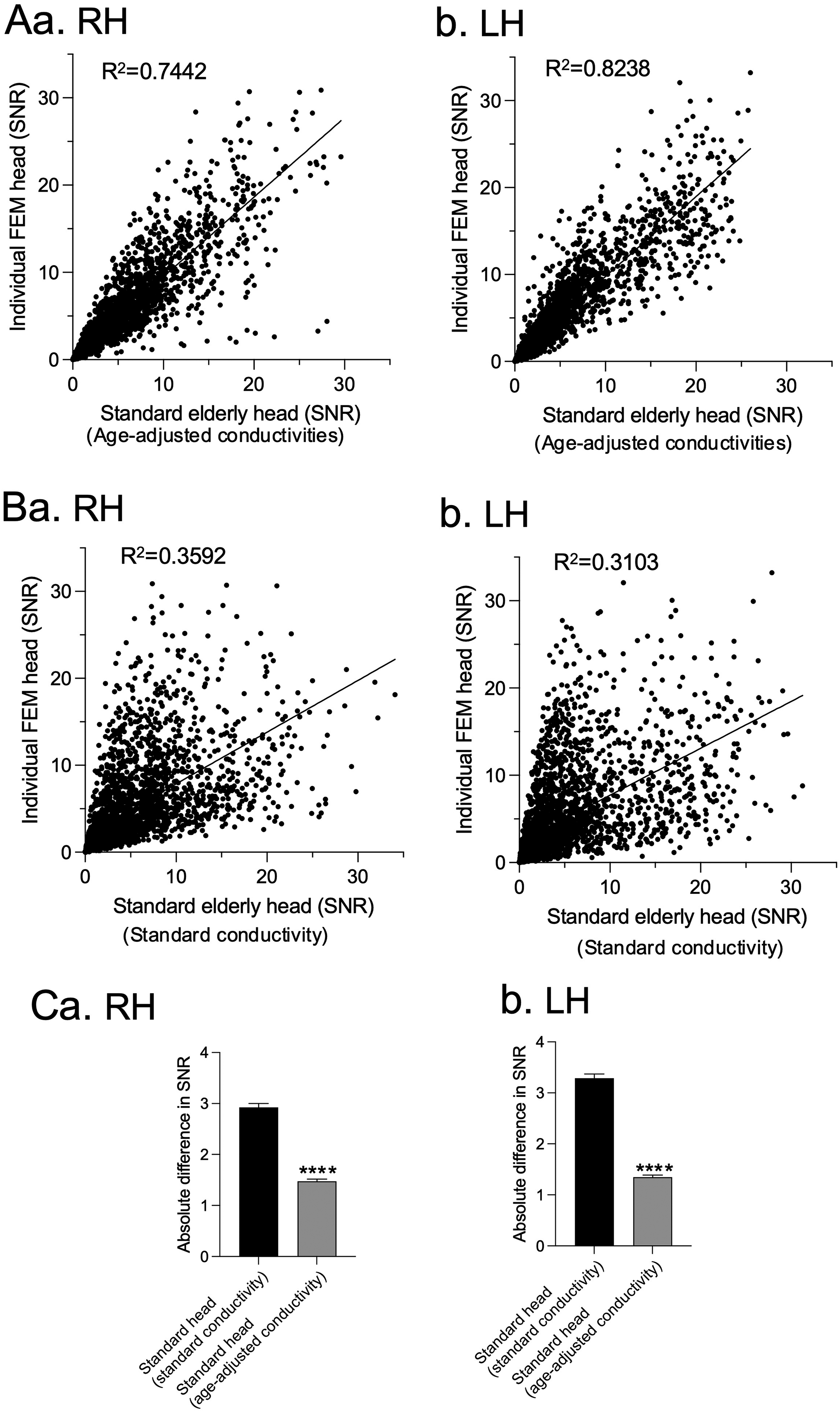
Effects of skull conductivity on SNRs in aged adults. A: Relationships between SNRs in the individual FEM heads and those in the artificial standard heads with skull conductivity adjusted for individual ages in the right hemisphere (RH) (a) and left hemisphere (LH) (b). B: Relationships between SNRs in the individual FEM heads and those in the standard head with the standard skull conductivity in the right hemisphere (RH) (a) and left hemisphere (LH) (b). C: Comparison of SNR deviations from those in the individual FEM heads between standard heads with standard skull conductivity and artificial standard heads with age-adjusted skull conductivities in the right hemisphere (RH) (a) and left hemisphere (LH) (b). ****, p < 0.0001.
Furthermore, we performed the same statistical comparison of SNRs under INF reference derived from artificial standard elderly heads, as shown in the initial section, and found comparative results. The analyses by repeated measures 3-way ANOVAs with factors of ROI, number of electrodes, and expansion number of brain electrical source area indicated that in both hemispheres, there were significant main effects of ROI, expansion number of brain source area, and number of electrodes, while all possible interactions among the three factors were also significant (Supplementary Table S7). These findings indicated that age-related changes in skull conductivity significantly affect SNRs and that at least individual differences in skull conductivity were involved in the results in the aged adults.
Discussion
Epilepsy in elderly individuals is a significant and increasingly prevalent problem (see Introduction), and EEG recording is fundamental for the diagnosis of epilepsy. In the present study, we simulated the SNRs of epileptic spikes in scalp EEGs using realistic FEM models based on MR images of individual elderly subjects with skull conductivity adjusted for their age. To the best of our knowledge, this is the first study focused on elderly subjects.
Effects of Sizes of the Brain Electrical Source Areas
In the present study, SNRs of scalp epileptic spikes progressively increased as the extent of the brain electrical source area increased across all 78 brain electrical source areas, although the slopes of the progression were lower in the medial and deep brain regions. It has been proposed that for detecting epileptic signals at the scalp through EEG, it is necessary for an area of the cortex ranging from 6 to 10 cm2 to be active.31,32 Tao et al clinically examined the extent of the active cortical area by performing simultaneous recording of scalp EEGs and electrocorticograms (ECoGs) in the medial temporal lobe and reported that cortical areas greater than 10 cm2 were required to be active for cortical epileptic signals to be detected in scalp EEGs.22,33 However, the use of silastic sheets for ECoG recording and holes in the skull significantly affect current spread from brain electrical sources,34,35 suggesting the need for simulation studies. A simulation study using the MNI standard head model reported that the signal-to-background ratio (SBR), comparable to SNRs in the present study, increased as the extent of brain electric source areas in the left temporo-parieto-occipital region increased and that cortical epileptic activity was detectable in scalp EEGs when cortical epileptic source areas greater than 6–8 cm2 were active. 23 These findings, along with the present findings, are consistent with physiological findings that focal epileptic activity with synchronization proceeds by the recruitment of neighboring neuronal networks, 36 eventually becoming detectable in scalp EEGs when a sufficient extent of the area becomes synchronous.22,33
Effects of Locations of the Electrical Source Areas
In the present study, SNRs were smaller in the ROIs in the deep and medial brain regions, including the hippocampus, parahippocampal cortex, retrosplenial cortex, posterior cingulate cortex, medial occipital cortex (primary visual cortex), insula, orbitofrontal cortex, anterior cingulate cortex, and subgenual cingulate cortex. These findings are consistent with those of human clinical studies involving simultaneous recording of ECoGs and/or stereotactic electroencephalograms (SEEGs) as well as scalp EEGs, which reported that epileptic spikes were visible in ECoGs and/or SEEGs but often invisible in scalp EEGs in the medial temporal cortex, including the hippocampus, insula, medial frontal cortex, and orbitofrontal cortex.25,37–39
Effects of Recording Methods
The number of electrodes significantly affected SNRs under INF reference. SNRs usually increased as the number of electrode channels increased: SNRs were often greater in the 70-channel dataset than in the 18- and 35-channel datasets. However, the impact of the number of electrodes was more evident at ROIs located shallowly in the brain with wider area extents, while the impact was less evident in the ROIs in the deep and medial brain regions such as the hippocampus. A previous clinical study reported that the average amplitude of epileptic spikes in scalp EEG originating from the mesial temporal lobe was very small (7.1 µV) and was below background noise levels. 40 Thus, the low impact of the number of electrodes on SNRs in the deep and medial brain regions might be ascribed to the low amplitudes of the spikes originating from those brain regions. This idea is consistent with the finding that if the active area was small, even in ROIs located in the shallow part of the brain (eg, Figure 4A), the improvement degree of SNRs by the larger number of electrodes was small.
Previous studies reported that, compared with INF reference, CAR and M1M2 modulated scalp EEG potentials since CAR and M1M2 themselves become active through volume conduction from brain electrical source areas. 41 In the present study, we investigated how reference types affect SNRs in scalp EEGs. The results indicated that SNRs were smaller in CAR and M1M2 than in INF for all electrode numbers except for the 70-channel recordings with an area expansion number of 10 in the left hemisphere. A previous simulation study using a 3-layer boundary element method (BEM) model of the MNI head reported that potential attenuation due to referencing CAR and M1M2 was relatively universal across different electrode numbers. 42 These previous results were similar to those obtained using the individual FEM heads of elderly adults in this study. These findings suggest the need to develop appropriate referencing methods with less attenuation effects in actual recordings from elderly adults.
Effects of Skull Conductivities
The present results indicated that SNRs were significantly correlated between the individual FEM heads and artificial standard elderly heads with skull conductivities adjusted for individual ages and indicated that even for artificial standard elderly heads, comparable results were observed via 3-way ANOVA with factors of ROI, area expansion number, and electrode number. Furthermore, the deviation of SNRs from those in the individual FEM heads was smaller for the artificial standard elderly heads with age-adjusted skull conductivities than for the standard head with the standard skull conductivity. These findings suggest that skull conductivity significantly affects SNRs in elderly adults and, consequently, that adjusting skull conductivity is important in forward simulation in elderly adults; and they are consistent with previous studies indicating that skull conductivity is an important factor for forward and/or inverse simulations in other age groups ranging from neonates to middle-aged adults.30,43–45 The findings further suggest the artificial standard elderly heads with age-adjusted skull conductivities can be used in lieu of individual FEM heads when MR images are not available.
Supplemental Material
sj-docx-1-eeg-10.1177_15500594251323625 - Supplemental material for Detectability in Scalp EEGs of Epileptic Spikes Emitted from Brain Electrical Sources of Different Sizes and Locations: A Simulation Study Using Realistic Head Models of Elderly Adults
Supplemental material, sj-docx-1-eeg-10.1177_15500594251323625 for Detectability in Scalp EEGs of Epileptic Spikes Emitted from Brain Electrical Sources of Different Sizes and Locations: A Simulation Study Using Realistic Head Models of Elderly Adults by Makoto Takenaka, Mark E. Pflieger, Tomokatsu Hori, Yudai Iwama, Jumpei Matsumoto, Tsuyoshi Setogawa, Atsushi Shirasawa, Hiroshi Nishimaru and Hisao Nishijo in Clinical EEG and Neuroscience
Supplemental Material
sj-xlsx-2-eeg-10.1177_15500594251323625 - Supplemental material for Detectability in Scalp EEGs of Epileptic Spikes Emitted from Brain Electrical Sources of Different Sizes and Locations: A Simulation Study Using Realistic Head Models of Elderly Adults
Supplemental material, sj-xlsx-2-eeg-10.1177_15500594251323625 for Detectability in Scalp EEGs of Epileptic Spikes Emitted from Brain Electrical Sources of Different Sizes and Locations: A Simulation Study Using Realistic Head Models of Elderly Adults by Makoto Takenaka, Mark E. Pflieger, Tomokatsu Hori, Yudai Iwama, Jumpei Matsumoto, Tsuyoshi Setogawa, Atsushi Shirasawa, Hiroshi Nishimaru and Hisao Nishijo in Clinical EEG and Neuroscience
Supplemental Material
sj-xlsx-3-eeg-10.1177_15500594251323625 - Supplemental material for Detectability in Scalp EEGs of Epileptic Spikes Emitted from Brain Electrical Sources of Different Sizes and Locations: A Simulation Study Using Realistic Head Models of Elderly Adults
Supplemental material, sj-xlsx-3-eeg-10.1177_15500594251323625 for Detectability in Scalp EEGs of Epileptic Spikes Emitted from Brain Electrical Sources of Different Sizes and Locations: A Simulation Study Using Realistic Head Models of Elderly Adults by Makoto Takenaka, Mark E. Pflieger, Tomokatsu Hori, Yudai Iwama, Jumpei Matsumoto, Tsuyoshi Setogawa, Atsushi Shirasawa, Hiroshi Nishimaru and Hisao Nishijo in Clinical EEG and Neuroscience
Supplemental Material
sj-xlsx-4-eeg-10.1177_15500594251323625 - Supplemental material for Detectability in Scalp EEGs of Epileptic Spikes Emitted from Brain Electrical Sources of Different Sizes and Locations: A Simulation Study Using Realistic Head Models of Elderly Adults
Supplemental material, sj-xlsx-4-eeg-10.1177_15500594251323625 for Detectability in Scalp EEGs of Epileptic Spikes Emitted from Brain Electrical Sources of Different Sizes and Locations: A Simulation Study Using Realistic Head Models of Elderly Adults by Makoto Takenaka, Mark E. Pflieger, Tomokatsu Hori, Yudai Iwama, Jumpei Matsumoto, Tsuyoshi Setogawa, Atsushi Shirasawa, Hiroshi Nishimaru and Hisao Nishijo in Clinical EEG and Neuroscience
Supplemental Material
sj-xlsx-5-eeg-10.1177_15500594251323625 - Supplemental material for Detectability in Scalp EEGs of Epileptic Spikes Emitted from Brain Electrical Sources of Different Sizes and Locations: A Simulation Study Using Realistic Head Models of Elderly Adults
Supplemental material, sj-xlsx-5-eeg-10.1177_15500594251323625 for Detectability in Scalp EEGs of Epileptic Spikes Emitted from Brain Electrical Sources of Different Sizes and Locations: A Simulation Study Using Realistic Head Models of Elderly Adults by Makoto Takenaka, Mark E. Pflieger, Tomokatsu Hori, Yudai Iwama, Jumpei Matsumoto, Tsuyoshi Setogawa, Atsushi Shirasawa, Hiroshi Nishimaru and Hisao Nishijo in Clinical EEG and Neuroscience
Supplemental Material
sj-xlsx-6-eeg-10.1177_15500594251323625 - Supplemental material for Detectability in Scalp EEGs of Epileptic Spikes Emitted from Brain Electrical Sources of Different Sizes and Locations: A Simulation Study Using Realistic Head Models of Elderly Adults
Supplemental material, sj-xlsx-6-eeg-10.1177_15500594251323625 for Detectability in Scalp EEGs of Epileptic Spikes Emitted from Brain Electrical Sources of Different Sizes and Locations: A Simulation Study Using Realistic Head Models of Elderly Adults by Makoto Takenaka, Mark E. Pflieger, Tomokatsu Hori, Yudai Iwama, Jumpei Matsumoto, Tsuyoshi Setogawa, Atsushi Shirasawa, Hiroshi Nishimaru and Hisao Nishijo in Clinical EEG and Neuroscience
Supplemental Material
sj-xlsx-7-eeg-10.1177_15500594251323625 - Supplemental material for Detectability in Scalp EEGs of Epileptic Spikes Emitted from Brain Electrical Sources of Different Sizes and Locations: A Simulation Study Using Realistic Head Models of Elderly Adults
Supplemental material, sj-xlsx-7-eeg-10.1177_15500594251323625 for Detectability in Scalp EEGs of Epileptic Spikes Emitted from Brain Electrical Sources of Different Sizes and Locations: A Simulation Study Using Realistic Head Models of Elderly Adults by Makoto Takenaka, Mark E. Pflieger, Tomokatsu Hori, Yudai Iwama, Jumpei Matsumoto, Tsuyoshi Setogawa, Atsushi Shirasawa, Hiroshi Nishimaru and Hisao Nishijo in Clinical EEG and Neuroscience
Supplemental Material
sj-xlsx-8-eeg-10.1177_15500594251323625 - Supplemental material for Detectability in Scalp EEGs of Epileptic Spikes Emitted from Brain Electrical Sources of Different Sizes and Locations: A Simulation Study Using Realistic Head Models of Elderly Adults
Supplemental material, sj-xlsx-8-eeg-10.1177_15500594251323625 for Detectability in Scalp EEGs of Epileptic Spikes Emitted from Brain Electrical Sources of Different Sizes and Locations: A Simulation Study Using Realistic Head Models of Elderly Adults by Makoto Takenaka, Mark E. Pflieger, Tomokatsu Hori, Yudai Iwama, Jumpei Matsumoto, Tsuyoshi Setogawa, Atsushi Shirasawa, Hiroshi Nishimaru and Hisao Nishijo in Clinical EEG and Neuroscience
Supplemental Material
sj-docx-9-eeg-10.1177_15500594251323625 - Supplemental material for Detectability in Scalp EEGs of Epileptic Spikes Emitted from Brain Electrical Sources of Different Sizes and Locations: A Simulation Study Using Realistic Head Models of Elderly Adults
Supplemental material, sj-docx-9-eeg-10.1177_15500594251323625 for Detectability in Scalp EEGs of Epileptic Spikes Emitted from Brain Electrical Sources of Different Sizes and Locations: A Simulation Study Using Realistic Head Models of Elderly Adults by Makoto Takenaka, Mark E. Pflieger, Tomokatsu Hori, Yudai Iwama, Jumpei Matsumoto, Tsuyoshi Setogawa, Atsushi Shirasawa, Hiroshi Nishimaru and Hisao Nishijo in Clinical EEG and Neuroscience
Supplemental Material
sj-docx-10-eeg-10.1177_15500594251323625 - Supplemental material for Detectability in Scalp EEGs of Epileptic Spikes Emitted from Brain Electrical Sources of Different Sizes and Locations: A Simulation Study Using Realistic Head Models of Elderly Adults
Supplemental material, sj-docx-10-eeg-10.1177_15500594251323625 for Detectability in Scalp EEGs of Epileptic Spikes Emitted from Brain Electrical Sources of Different Sizes and Locations: A Simulation Study Using Realistic Head Models of Elderly Adults by Makoto Takenaka, Mark E. Pflieger, Tomokatsu Hori, Yudai Iwama, Jumpei Matsumoto, Tsuyoshi Setogawa, Atsushi Shirasawa, Hiroshi Nishimaru and Hisao Nishijo in Clinical EEG and Neuroscience
Supplemental Material
sj-docx-11-eeg-10.1177_15500594251323625 - Supplemental material for Detectability in Scalp EEGs of Epileptic Spikes Emitted from Brain Electrical Sources of Different Sizes and Locations: A Simulation Study Using Realistic Head Models of Elderly Adults
Supplemental material, sj-docx-11-eeg-10.1177_15500594251323625 for Detectability in Scalp EEGs of Epileptic Spikes Emitted from Brain Electrical Sources of Different Sizes and Locations: A Simulation Study Using Realistic Head Models of Elderly Adults by Makoto Takenaka, Mark E. Pflieger, Tomokatsu Hori, Yudai Iwama, Jumpei Matsumoto, Tsuyoshi Setogawa, Atsushi Shirasawa, Hiroshi Nishimaru and Hisao Nishijo in Clinical EEG and Neuroscience
Footnotes
Acknowledgments
This study was supported by University of Toyama and University of East Asia.
Data Availability
Since the data in the manuscript and Supplemental Materials include patient data, they are available upon reasonable request.
Declaration of Conflicting Interests
The authors declared the following potential conflicts of interest with respect to the research, authorship, and/or publication of this article: MEP was, and AS is affiliated with companies that develop and market the software used in this study. The other authors declare that the research was conducted in the absence of any commercial or financial relationships that could be construed as a potential conflict of interest.
Funding
The authors disclosed receipt of the following financial support for the research, authorship, and/or publication of this article: This work was supported by the University of East Asia, University of Toyama,
Ethical Approval
The anonymized patients’ data (MR images of the heads) were obtained by opt-out according to Japanese ethical guidelines. The protocol in this study was carried out in accordance with the Declaration of Helsinki and was reviewed and approved by the Ethics Review Board for Human Research at the University of Toyama (permit no.: R2022065, approved on July 26, 2022).
Supplemental Material
Supplemental material for this article is available online.
References
Supplementary Material
Please find the following supplemental material available below.
For Open Access articles published under a Creative Commons License, all supplemental material carries the same license as the article it is associated with.
For non-Open Access articles published, all supplemental material carries a non-exclusive license, and permission requests for re-use of supplemental material or any part of supplemental material shall be sent directly to the copyright owner as specified in the copyright notice associated with the article.