






Abstract
Objectives
Childhood Occipital Visual Epilepsy (COVE) is a self-limited epileptic syndrome that typically begins in late childhood or adolescence characterized by brief visual seizures. The recent 2022 International League Against Epilepsy (ILAE) classification distinguishes COVE from photosensitive occipital lobe epilepsy (POLE), emphasizing the absence of photic-induced seizures in COVE. In this study, we aimed to describe the clinical and electrophysiological features of patients with COVE diagnosed according to the new ILAE criteria.
Methods
This retrospective cohort study analyzed 30 patients diagnosed with COVE at a tertiary epilepsy center between 1988 and 2023. Patients were selected based on ILAE 2022 criteria, and all cases with intermittent photic stimulation (IPS)-induced seizures were excluded.
Results
Most patients (93%) presented with elementary visual hallucinations, such as colorful lights. Orofacial seizures occurred in 7%, and 37% had nocturnal seizures. EEG abnormalities were primarily occipital and resolved in 85% of cases over time. Generalized spike-wave discharges (GSWDs) were rare (5%), and only one patient developed juvenile myoclonic epilepsy during follow-up. At final follow-up, 77% of patients achieved seizure freedom, and 47% discontinued medication.
Conclusion
COVE is an epileptic syndrome associated with a favorable prognosis. By excluding photosensitivity in light of the newly proposed diagnostic criteria from the ILAE, future research should focus on a more homogenous group of COVE patients to enhance understanding of this syndrome. Accurate classification using updated ILAE criteria allows for clearer clinical delineation and more reliable outcome predictions.
Keywords
Introduction
Childhood occipital visual epilepsy (COVE), formerly known as late-onset benign occipital epilepsy or Gastaut syndrome, is an epileptic syndrome that typically manifests in later childhood or adolescence. 1 In most cases, the syndrome is self-limited and responds well to anti-seizure medications. Affected individuals usually exhibit normal neuropsychological development and cognitive function. COVE is characterized by brief seizures during wakefulness, often manifesting as visual phenomena such as multicolored circles. Complex visual hallucinations and ictal blindness may also occur, sometimes accompanied by motor symptoms and impaired awareness. 2 Seizures generally resolve by puberty, with most patients maintaining normal developmental outcomes. 3
Since the initial description, COVE has been the subject of controversy regarding precipitating factors such as photosensitivity. Gastaut, who defined this syndrome in 1982, 4 included patients exhibiting photosensitivity during intermittent photic stimulation (IPS). 5 However, in the following years, in 1996, Guerrini et al described photosensitive occipital lobe (POLE) epilepsy, characterized by photic-induced occipital seizures and occipital EEG paroxysms. 6
In the most recent report by the International League Against Epilepsy (ILAE) on the classification of childhood-onset epileptic syndromes, COVE and POLE are categorized under the self-limited focal epilepsies of childhood (SeLFE). 1 They constitute a subgroup in which seizures may persist beyond puberty. The clinical course differs between these two syndromes: while photosensitivity is not a defining feature of COVE, photic-induced seizures and occipital paroxysms triggered by IPS are mandatory diagnostic criteria for POLE.
Distinguishing between COVE and POLE remains challenging due to inconsistencies in the literature, particularly in studies focusing on COVE that also include individuals with IPS sensitivity. In a recent cohort study analyzing POLE patients based on the new ILAE diagnostic criteria, they identified a subgroup classified as ‘probable POLE’. This subgroup comprised patients with occipital seizures who exhibited either photosensitivity during IPS or seizures triggered by clinical photic-stimulating factor. 7 They suggest that the features of this subgroup were similar to those of the group with the definite POLE. Notably, the clinical features of this “probable POLE” subgroup closely resembled those of patients meeting the criteria for definite POLE.
In this study, we aim to describe the characteristics of a more homogenous group of patients with COVE by excluding the cases with occipital paroxysms triggered by IPS and visually induced occipital lobe seizures, in light of the recent definition of ILAE. By means of this, we aim to contribute a more refined understanding of these syndromes.
Materials and Methods
Study Design and Participants
This study was designed as a retrospective analysis. Patients diagnosed with COVE according to the ILAE 2022 criteria 1 who were followed up at our tertiary Epilepsy Clinic for at least two years were included in the study.
Clinical and Demographic Data
Data were collected from the patients’ medical record, including age, sex, history of febrile seizures (FS), family history of epilepsy, family history of FS, family history of migraine, age of seizure onset, history of headache, intellectual status, and cranial MRI findings, seizure manifestations, duration, and frequency of seizures, circadian distribution, antiseizure medications (ASM) and clinical outcomes.
EEG Data
EEG recordings were performed while patients were awake for 30 min and/or prolonged lasting up to two hours, which included both sleep and wake periods. Scalp electrodes were placed according to the 10-20 international system. The time constant was 0.3 s, and the high-frequency filter was standardized at 70 Hz. IPS with flash frequencies ranging from 1 to 60 Hz was applied in all cases, followed by four minutes of hyperventilation (HV), and eye-opening and eye-closure reactivities were noted in all recordings. The video-EEG recordings were obtained using Galileo Sirius BB E980 3040 600.
The background activity was documented during EEG recordings. Focal discharges were categorized as unilateral and bilateral and further localized as frontal (Fp1-2, Fz, F3-4), centrotemporal (C3-T3, C4-T4), central (Cz), and posterior (temporo-parieto-occipital; T5-O1, P3-O1, T6-02, P4-O2). Additionally, generalized spike-wave discharges (GSWD) were also analyzed.
Patients with at least two sleep EEG recordings were further evaluated for changes in the EEG findings during follow-up. A more than two-fold increase in the frequency of focal discharges during sleep was defined as the sleep enhancement of EEG abnormalities.
Between October 1988 and January 2023, a total of 30 patients met the inclusion criteria. The patients’ medical records and EEG findings were analyzed retrospectively.
Results
Clinical and Demographic Findings
The study included 30 patients over a 35-year period. The demographic and clinical characteristics of the patients are summarized in Table 1. Cranial imaging revealed arachnoid cysts in two patients, while the results were normal in the remaining cases.
Demographical and Clinical Characteristics of Patients.
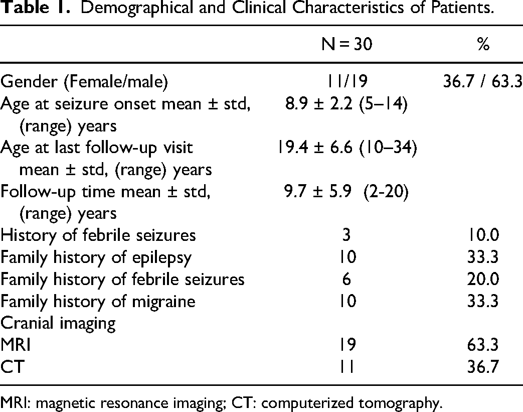
MRI: magnetic resonance imaging; CT: computerized tomography.
Regarding the neuropsychological status, one of these patients had a borderline IQ (IQ:86) and another one had been diagnosed with attention deficit hyperactivity disorder (ADHD). According to the parent's reports and school achievements, three patients had minor learning difficulties, one of whom also exhibited behavioral problems. The frequency of these findings was comparable to the prevalence of such neuropsychological profiles in the general population. 8
Seizure Characteristics
The seizure characteristics of patients are summerized in Table 2. Elementary visual hallucinations were —most often described as colorful, circular, or linear lights—were reported in 28 patients (93%). One patient experienced diplopia, and another reported blurred vision. Six patients (20%) described micropsia-macropsia while one experienced metamorphopsia.
Seizure Characteristics.
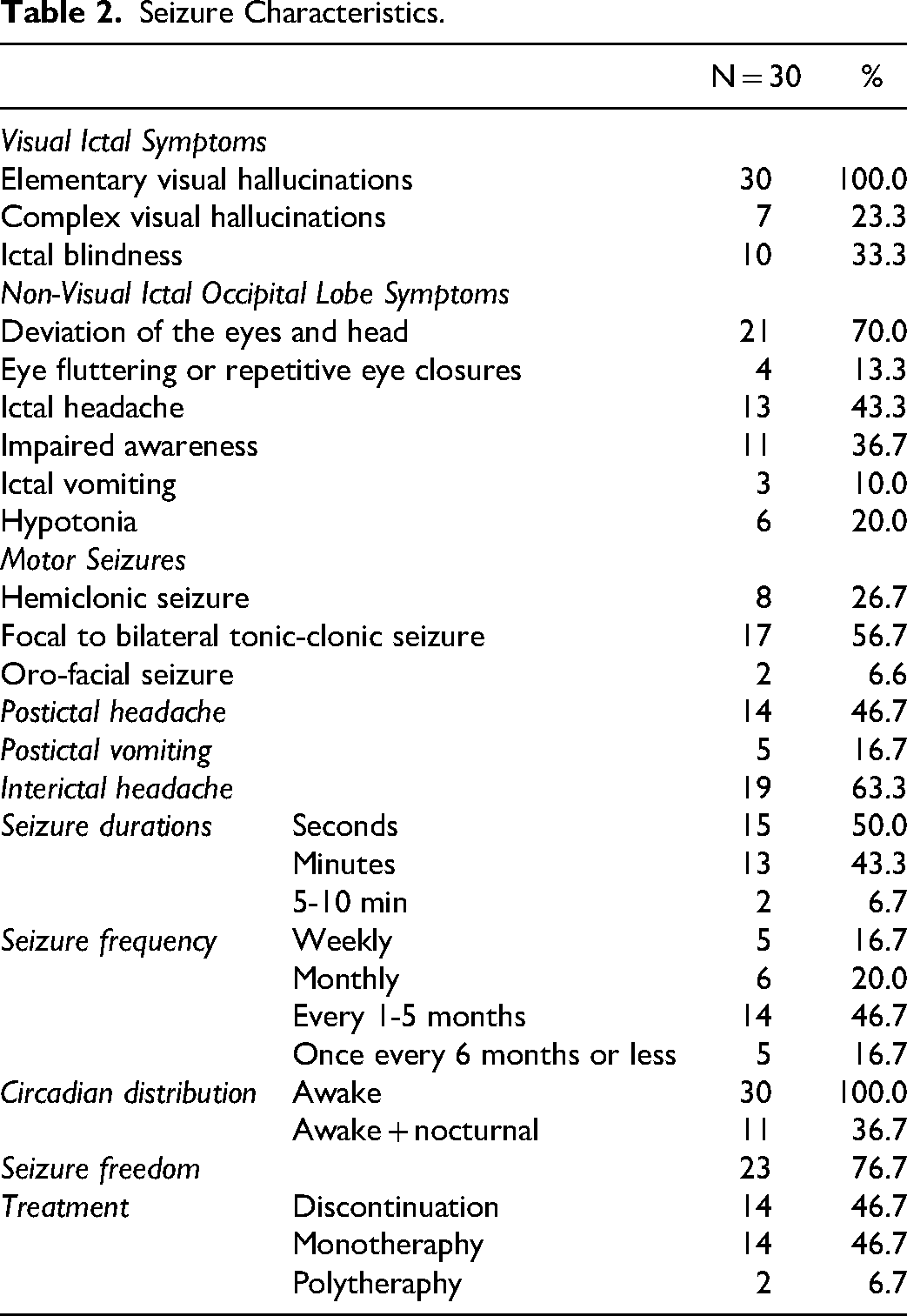
Oro-facial seizures were reported in two patients (7%). One patient experienced an orofacial seizure at age 10, with sleep EEG showing right centrotemporal discharges. The second patient had an orofacial seizure following her initial visual seizure, and a sleep EEG later revealed left centroparietal discharges.
According to parental reports, seizures lasted for a few seconds in 50% of cases and a few minutes in 43%. In two patients (7%) seizures lasted 5-10 min. Seizure frequencies were reported as weekly at 16.7% of cases, and monthly at 20%, every 1-5 months in 47%, and once every 6 months or less in 17%.
All patients were experienced seizures while awake. However, 11 patients (37%) had nocturnal seizures as well. Among them, one had an orofacial seizure, one had elementary visual hallucinations, and the rest experienced focal to bilateral tonic-clonic seizures. Focal to bilateral tonic-clonic seizures occurred during wakefulness in 8 patients (27%), during sleep in 6 (20%), and during both sleep and wakefulness in 3 (10%).
EEG Findings
A total of 148 EEGs were performed, with each patient undergoing between 1 and 10 recordings (mean 5.0 ± 2.1). Background activity was normal in all cases. Interictal EEG findings from the first abnormal EEGs were presented in Table 3.
Interictal EEG Findings on First Abnormal EEGs.
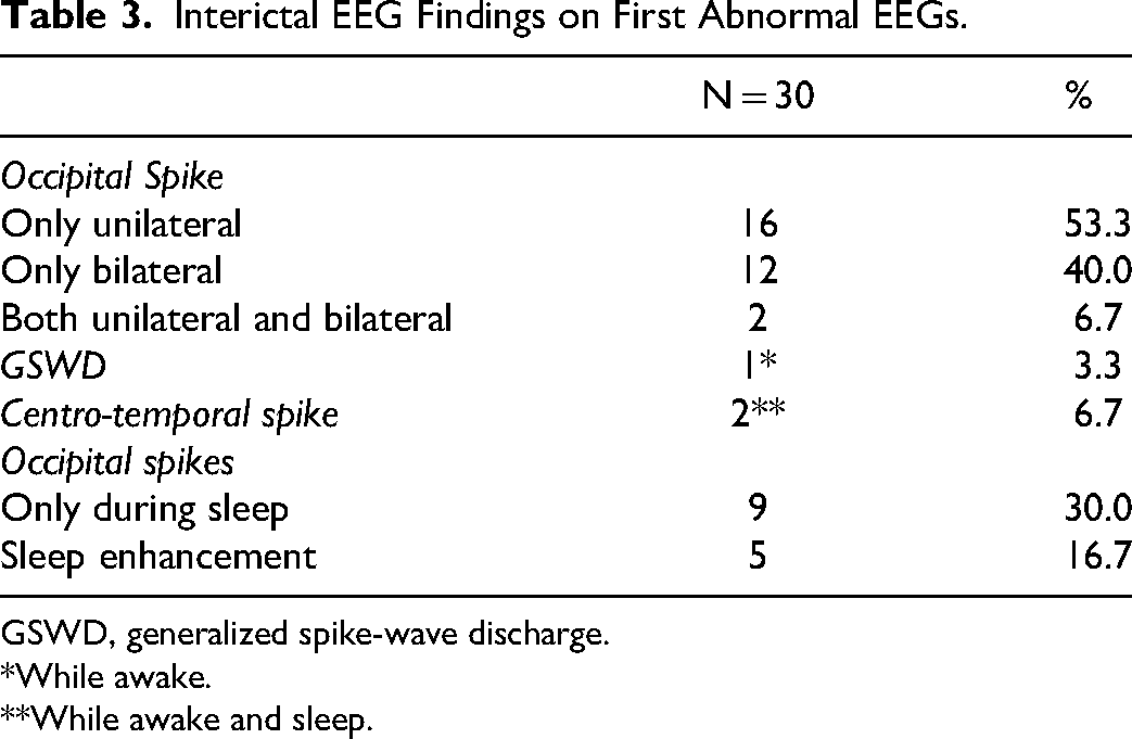
GSWD, generalized spike-wave discharge.
*While awake.
**While awake and sleep.
Twenty patients had at least two sleep EEGs. In 9 of them (45%), occipital paroxysms were observed only during sleep. Sleep enhancement of EEG abnormalities was identified in five patients (25%). An increase in the number of focal discharge during sleep was noted in one patient (5%). In the follow-up EEGs, the lateralization of focal discharges remained unchanged; however, a new foci appeared in three patients (15%) (one bi-frontal, one centrotemporal, one centroparietal - this patient had oro-facial seizures). Additionally, GSWD were observed in one patient (5%) who was later diagnosed with juvenile myoclonic epilepsy (JME).
EEG abnormalities resolved in 17 of 20 patients (85%) after a mean of 5.7 years from seizure onset. In the remaining 3 patients, EEG abnormalities persisted in the final sleep EEG. Among these, occipital spikes were observed exclusively during sleep in 2 patients. Of the 17 patients with normalized final EEGs, seizures continued in 4 patients. Conversely, of the 3 patients with persistent EEG abnormalities, only one had ongoing seizures.
Treatment and Course of Illness
At the final follow-up, seizure freedom was achieved in 23 patients (77%) after a mean of 4.7 years (range:2 months to 19 years). The mean duration of seizure freedom was 6.2 years (range: 1-13 years). However, EEG abnormalities persisted in five of 23 patients. Treatment was discontinued in 14 patients (47%) after a mean seizure-free period of 3.9 years (range:1-6 years), and these patients were followed up without ASM for a mean of 3.3 years (range:1 month to 8 years). At the final follow-up, 14 patients (47%) were on monotherapy, with 11 receiving carbamazepine (CBZ) and 3 receiving valproate (VPA). Two patients (%7%) were on polytherapy (lamotrigine (LTG)+VPA, VPA + CBZ).
The most frequently used ASMs were CBZ (n = 21) and VPA (n = 13). Other ASMs included vigabatrin (n = 2), levetiracetam (n = 1), LTG (n = 1), ethosuximide (n = 1), phenobarbital (n = 1), and barbexaclone (n = 1).
Seven patients (23%) continued to experience seizures after a mean follow-up of 8.2 years (range: 1.9-19.1 years). Among them, two experienced weekly seizures, one monthly, two every 1-5 months, and two every 6 months or less. In two patients, the seizures persisted for more than 18 years. Visual seizures remained in 5 of the 7 patients. One patient experienced both visual seizures and eye deviation while another had both visual and myoclonic seizures starting at age 20. EEG at that age revealed GSWD during eye closure and IPS. He was treated with valproic acid and responded well, experiencing only one myoclonic seizure per year, only when he was tired. This patient was also diagnosed with JME.
Discussion
First described in 1982, COVE was defined as a benign epilepsy of childhood with distinctive visual seizures. Gastaut included patients with photosensitivity in his descriptive study of COVE. 4 However, in 1996, Guerrini et al introduced idiopathic photosensitive occipital lobe (IPOLE) epilepsy as a separate entitiy. 6 Despite this, photosensitive patients were often considered as a subset of COVE, rather than a distinct syndrome of POLE. 9 Caraballo et al also suggested that POLE is not an independent epilepsy syndrome, but a variant of COVE with particularly high photosensitivity2,10 . On the other hand, the ILAE Task Force classified photic-induced visual seizures as a part of reflex epilepsy syndrome named “idiopathic photosensitive occipital lobe epilepsy”11,12 . More recently, newly proposed ILAE diagnostic criteria defined photic-induced focal sensory visual seizures and occipital paroxysms facilitated by eye-closure and IPS are the key diagnostic features of POLE. However, photosensitivity was not included in the diagnostic criteria of COVE 1 . In a recent cohort analyzing POLE patients, a subgroup exhibiting photosensitivity during IPS or photic-triggered seizures was identified. The clinical features of this ‘probable POLE’ subgroup were found to closely resemble those of patients with definite POLE. 10 In this study we only included patients with COVE according to the new diagnostic criteria proposed by ILAE; in order to better define its clinical and electrophysiological characteristics.
During the course of illness, SeLFEs may coexist with each other or overlap with other genetic generalized epilepsies (GGEs)7,9,13,14 . Although 13% of patients with SeLFE have been reported to experience different types of SeLFE at the same or at different times, this overlap is relatively rare in COVE (2%) compared to the other self-limited epilepsies of childhood 15 . In our cohort, 7% of patients had orofacial seizures, a finding consistent with Fortini et al's report of 6%. 16 . Moreover, while absence seizures have been described in COVE patients in previous studies,1,15,16 no history of absence seizures was found in our study group. However, one patient was diagnosed with JME during follow-up. The coexistence of different types of epilepsies appears to be more frequent in photosensitive epilepsies and other GGEs7,17,18 . For example, Cerrahoğlu Şirin et al reported that just over one-third patients of with POLE had additional diagnosis of GGE 7 . The lower rate of overlap between SeLFEs and JME may be attributed to the absence of the IPS-dependent nature of COVE.
Frontal, centrotemporal, and generalized EEG abnormalities have been previously reported in COVE, 1 including centrotemporal spikes in interictal EEGs. 12 Extra-occipital paroxysms were reported in 33.3% of the patients by Wakamoto et al, 19 and centrotemporal spikes were reported in 24% by Caraballo et al notably higher than in our cohort. 2 GSWDs were reported at rates of 17% by Wakamoto et al, 19 27% by Caraballo et al2% and 35% by Verrotti et al 3 ; all higher compared to our cohort. We believe this difference is largely due to the inclusion of IPS-sensitive individuals in those studies, suggesting potential overlap with GGE. Notabely, IPS sensitivity was observed in 15%2% to 41% 3 of the patients in these studies. Cerrahoğlu Şirin et al, in their analysis of POLE using the ILAE criteria, found that 34% of POLE patients exhibited interictal GSWDs—a much higher rate than in our COVE cohort.
We hypothesize that GSWDs are less prevalent in focal epilepsy syndromes compared to the frequency of focal abnormalities in GGEs. For instance; in a study performed involving 220 patients diagnosed with SeLFE syndromes, GSWDs were reported in 19% of them 14 . Among those diagnosed with COVE in this population, the GSWD rate was 19%, which is higher than that observed in our cohort. In another study that evaluating generalized paroxysms in focal epilepsies; 11% of participants were found to have GSWDs on EEG. 20 The study speculated that interictal GSWDs were associated with female gender and parental consanguinity, suggesting a resemblance to photosensitivity. This hypothesis may partially explain the low GSWD rate in our cohort, as patients with photosensitivity were excluded according to the new diagnostic criteria. Conversely, several studies have documented the presence of focal EEG features in various types of GGEs, with prevalence rates as high as 65% in some studies.20–24
The coexistence of occipital lobe epilepsy (OLE) and migraine may create difficulties in diagnosis. Migraine auras may resemble auras in OLE, but they are shorter in duration, usually have more gradual onset and are not followed by other features of seizures.9,25
COVE has a generally favorable prognosis with appropriate ASM treatment, consistent with the self-limited nature of the disease. The most commonly used ASMs are CBZ or VPA monotherapies. Seizure remission occurs in approximately half of the patients 2 to 4 years after onset. Studies report 76%to 82%of the patients achieve seizure freedom with ASM monotherapy.2,3 In our cohort, the seizure freedom rate was 77%, similar to the 80% reported by Verrotti et al 3 In contrast, prognosis in POLE varies: some patients achieve remission, while others continue to experience photic-induced seizures. 1 Although not statistically significant, recurrence appears more likely in POLE patients with coexisting GGE. 7
In conclusion, following the recent publication of the ILAE, there is a growing need to study the proposed syndromes in greater detail. For many years, it has been debated whether POLE is a variant of COVE or a distinct epileptic syndrome. With the latest ILAE classification, COVE and POLE are both classified under SeLFEs. While these two syndromes share certain clinical features, such as seizure characteristics, occipital spikes on EEG, and the persistence of seizures beyond puberty, their prognoses differ significantly. COVE is associated with a higher rate of seizure remission and lower coexistence with genetic GGEs. Additionally, interictal GSWD are observed less frequently in COVE compared to POLE. Therefore, a clear distinction between these syndromes is essential. Future studies on COVE should exclude photosensitivity in accordance with the recent ILAE criteria to better delineate its clinical and electrophysiological characteristics.
Footnotes

Ethical Approval
The study protocol was developed in accordance with the principles set out in the Declaration of Helsinki. Ethical approval was obtained from the Institutional Ethical Committee (approval number: 825959).
Consent to Participate
This study is not under consideration by any other journal at the same time, and it has not been accepted for publication elsewhere in any language
Funding
The authors received no financial support for the research, authorship, and/or publication of this article.
Declaration of Conflicting Interests
The authors declared no potential conflicts of interest with respect to the research, authorship, and/or publication of this article.
Data Availability Statement
The datasets analysed during the current study are available from the corresponding author on reasonable request.