
Abstract
Background. Sentinel lymph node biopsy (SLNB) is a standard staging procedure in breast cancer and skin melanoma patients. Radioactive colloid (RC) and blue dye are the routinely used markers for staining. The new dye used in this procedure, indocyanine green (ICG), seems to have true potential in near-infrared–guided SLNB. The aim of this study was to analyze 1-year morbidity after SLNB using RC and ICG or RC and ICG conjugated to human serum albumin (ICG:HSA) in breast cancer and skin melanoma patients. Methods. Forty-nine patients diagnosed with breast cancer and 10 patients with skin melanoma underwent SLNB using ICG with RC and ICG:HSA with RC. A total of 47 SLNB patients without the need for additional lymphadenectomy were evaluated approximately 1 year (11-13 months) for the presence of tattoo, extremity swelling, nerve dysfunction/pain, range of motion, and stiffness. Results. From the 47 patients examined, long-term morbidity was present in 3 (6.4%). In 1 patient, tattoo persisted for 11 months. Mild lymphedema was seen in 1 patient, and 1 patient exhibited minor functional deficit. Conclusions. Using ICG or ICG:HSA seems to be safe, and long-term morbidity in SLNB patients is low. However, skin discoloration may appear as it does after the use of blue dye, and an increased number of harvested nodes might be associated with an increased number of iatrogenic lymphedema.
Introduction
The current staging standard for breast cancer and skin melanoma is sentinel lymph node biopsy (SLNB). The 2 markers used routinely in SLNB are radioactive colloid (RC) and blue dye (BD). Numerous studies have reported postoperative complications related to the use of these dyes.1-5 Among these complications are allergic reactions, persistent tattoo, and lymphedema. Recently, the use of indocyanine green (ICG) as a sentinel lymph node (SLN) marker has been described in an increasing number of publications.6-12
One important issue in the use of ICG in SLNB is the necessity to remove a larger number of lymph nodes than are resected with RC or BD. According to Hojo et al, 13 an average of 1.9 to 2.0 SLNs are resected when using BD or RC, while ICG stained an average of 3.8 to 4.1 SLNs, depending on the type of lymphatic drainage. Tagaya et al 14 observed an average of 2.3 SLNs using BD, while fluorescence navigation with ICG indicated an average of 5.5 SLNs. The greater number of resected lymph nodes can lead to increased cases of lymphedema even with the relatively minimally invasive nature of SLNB.
The purpose of our study was to analyze morbidity after SLNB using ICG or ICG:HSA (indocyanine green combined with human serum albumin) in melanoma and breast cancer patients.
Methods
Sentinel lymph node biopsy was performed using ICG and ICG:HSA with RC on 49 patients diagnosed with breast cancer between September 2010 and November 2010 and 10 patients diagnosed with skin melanoma between September 2010 and December 2010, both groups at Greater Poland Cancer Center, Poznan. Detailed information about patients, preparation of ICG and ICG:HSA, and SLNB procedure is described in our previous articles.15,16 Fifty patients who underwent SLNB without the need for additional lymphadenectomy were asked to visit our Cancer Centre for 1-year follow-up (between 11 and 13 months after operation). A total of 47 patients were examined. Limb circumference, of both the operated and nonoperated extremity, was measured preoperatively. The same procedure was performed during the follow-up visit. We measured mid-calf and mid-thigh, as well as mid-arm and mid-forearm. Lymphedema was defined as mild, moderate, or severe as follows: Mild lymphedema consisted of 2 to 3 cm increase of calf or biceps circumference with minimal or no symptoms; moderate lymphedema included 3 to 4 cm increase in circumference or moderate symptoms; and severe lymphedema included ≥4 cm increase in circumference or disabling symptoms.
At the time of the follow-up visit, 11 to 13 months postoperatively, skin discoloration was also evaluated and the patients were asked about persistence of skin discoloration and the length of time until it resolved after SLNB. Changes in range of motion as well as nerve dysfunction/pain and stiffness were also evaluated at this visit via physical examination and questioning the patients for such complaints.
Long-term complications were calculated as present at least 11 months after operation.
Written and oral consent was obtained from all study participants and the protocol of the study was approved by the local ethics committee.
Results
Long-term complications in the group of 47 patients that underwent SLNB using not only RC but also ICG or ICG:HSA were seen in 3 patients (6.4%).
Lymphedema was not observed in any of the patients who had undergone SLNB for breast cancer. Among the patients treated for skin melanoma, a female patient who underwent SLNB at the right inguinal with resection of 5 SLNs (among these 3 RC + ICG positive and 2 only ICG positive) had right lower limb edema. The observed lymphedema was mild (2.5 cm increase in circumference compared to preoperative measurement).
One breast cancer patient had tattoo persistence of 11 months. She had been operated at another hospital because of suspicion of benign changes (tumorectomy), but the histopathology report (available 3 weeks later) had confirmed invasive ductal carcinoma T1c. This patient was qualified for surgery to increase the resection margin at the tumor focus and undergo SLNB at the Greater Poland Cancer Center in Poznan, which was performed 28 days after the original surgery. The marker was injected at 4 sites around areola, and the patient underwent classical radiotherapy 6 weeks after the second surgery with the addition of antiestrogen treatment. Green discoloration was observed at 6 months postoperation (Figure 1), and disappeared in the 12th month. Other patients confirmed that green discoloration was not present at all (eg, in the group after mastectomy) or lasted for 2 to 3 weeks after the operation.
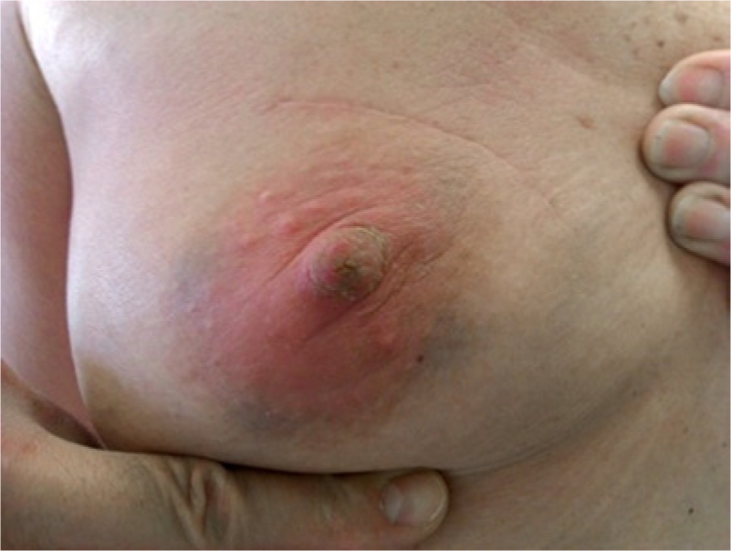
Green discoloration observed at 6 months postoperation.
None of the patients had nerve dysfunction/pain. One patient, after breast conserving treatment of the upper outer quadrant and SLNB, reported minor functional deficit where the post-SLNB wound site caused some problems with arm mobility, namely problems with arm elevation. No additional lymph nodes stained only by ICG were harvested during SLNB.
It is worth mentioning that none of the patients had any kind of allergic reaction during or after operation.
Discussion
Indocyanine green is visible in near-infrared light and allows for visualization of lymphatic pathways in addition to the SLN itself. Because of the presence of iodine particles, which stabilize ICG, this dye is contraindicated in patients with an allergy to iodine or seafood (which tends to contain large quantities of iodine). It is worth noting, however, that the risk of adverse reactions to the ICG injection is small (approximately 0.003% at doses exceeding 0.5 mg/kg). 17 The technique can therefore be used safely in those patients who have slight or mild iodine allergies in whom radiographic intraoperative cholangiography is contraindicated.18,19 The amount of iodine included in ICG as a stabilizing agent is extremely small (<0.1 mg/mL) in comparison with the amount of iodine in radiocontrast agents (50-300 mg/mL). ICG-related eye toxicity is dose dependent20,21 and the risk is negligible in fluorescent angiography, considering the small amount of ICG (2.5 mg) injected.
Tattoo persistence after BD use is one of the reasons that some surgeons do not use BD in SLNB. As reported by Guiliano, 22 the use of BD can lead to a tattoo persisting even up to 1 year, and postoperative radiotherapy and breast edema may prolong this side effect. In the case of our one patient with persistent tattoo, breast edema after her first surgery and additional postoperative radiotherapy may have contributed to the length of tattoo persistence. Silberstein et al 23 report persistence of skin discoloration for even 9 months postoperatively after BD use in skin melanoma.
In addition to prolonged discoloration, 22 side effects of BD use include allergic reaction (up to 2%) with skin rash—with anaphylactic reaction20,24; blue-colored urine for up to 48 hours 24 ; interference with intraoperative oximetry monitoring25,26; and cutaneous necrosis. 27 In our group of patients, no one exhibited any of the above side effects except for skin discoloration.
Another concern is the observed greater number of SLNs resected when using ICG. On one hand, we have the publication of Tagaya et al 14 —who list an average of 5.5 SLNs stained with ICG compared with an average of 2.3 SLNs stained with BD—and on the other hand, we have the publication of Huttemann et al 28 —who, in 18 patients, detected an average of 1.27 SLNs using RC but detected an additional 2 SLNs using near-infrared fluorescence with ICG:HSA, which increased the average number of resected SLNs to 1.38. In the case of ICG alone, the number of resected SLN did not differ from the number resected using RC.14,28 In addition to an increase in the number of resected lymph nodes, the cases of lymphedema also increase, which was demonstrated in randomized trials as SLNs versus lymphadenectomy groups.1-5
Our study demonstrated that, in the case of 10 melanoma patients, a total of 4 additional lymph nodes were resected in 3 of these patients. In our 49 patients with breast cancer, a total of 13 additional lymph nodes were resected from 11 patients. A total of 17 lymph nodes staining only for ICG or ICG:HSA were resected from our patients. The average number of resected lymph nodes in the breast cancer patients was 2.43 for ICG compared with 2.07 for RC, and 2.14 for ICG:HSA compared with 2.00 for RC. An average of 1.9 lymph nodes were resected by ICG or ICG:HSA in the melanoma patients, compared with 1.6 resected after detection by RC. The larger number of resected lymph nodes was not associated with an increase in the number of patients with lymphedema. Only 1 patient, with right thigh melanoma, who had 5 lymph nodes resected (3 lymph nodes stained by RC and 2 additional lymph nodes stained by ICG) was found to have mild lymphedema of the right lower limb. None of the other patients in this study were found to have lymphedema. Functional deficit was not associated with increased number of ICG or ICG:HSA harvested nodes.
Keeping in mind that morbidity reduction is a goal of SLNB and new tracers with decreased long term side effects are being sought, ICG may be seen as a safe alternative to BD. The long-term complications over 6 months after operation using ICG or ICG:HSA are low and comparable to other studies that used RC or BD. Nevertheless, for the first time we report the persistence of a tattoo for 10 months, and lymphedema of the leg where a greater number of lymph nodes were resected.
Footnotes
Acknowledgements
We are indebted to Dr Margarita Lianeri for her analysis and editorial assistance.
Declaration of Conflicting Interests
The author(s) declared no potential conflicts of interest with respect to the research, authorship, and/or publication of this article.
Funding
The author(s) received no financial support for the research, authorship, and/or publication of this article.