






Abstract
Background. Clinical effectiveness and safety of biological and synthetic adhesives in digestive closures have been evaluated. Their use is becoming more prevalent, as rigidity and inflexibility are its more remarkable weaknesses. However, little is known about their role in gastric and anastomotic closures. Moreover, usefulness of novel flexible types of synthetic adhesives as n-butyl-cyanoacrylate has not been assessed yet. Materials and Methods. One centimeter long gastrotomy was performed in 24 male Wistar rats, which were divided depending on the type of closure method employed: manual USP 5/0 silk interrupted suture versus sutureless closure with Histoacryl Flexible (n-butyl-cyanoacrylate with softener) or Histoacryl Double Component (n-butyl-cyanoacrylate with softener and hardener). Microscopic evaluation of the suture viability and integrity was performed, and adhesion formation during the cicatrization process were assessed. During an 8-week follow-up clinical and histopathological aspects as well as hematologic and inflammatory biomarkers were studied. Results. No differences among groups where found in any of the clinical, analytical, or histopathological issues assessed except for a higher incidence rate of adhesions in the Histoacryl Double Component group when compared with hand-sewn suture group (P = .04). Our results support experimental studies in large mammals (pigs) for further study of sutureless hollow viscera closure.
Introduction
The most critical aspect for the surgical outcome is the correct technical execution, especially when suturing anastomoses. Many local and systemic factors have been pointed out as crucial in determining suture’s success. Moreover, a number of technical details such as correct hemostasis and vascularization, border apposition or tension-free closure, among others, are described.1,2 Despite the fact that systemic and local factors depend on patient’s tissue characteristics and cannot be modified during the surgical procedure, technical aspects may be crucial.
Gastric sutures and anastomosis are commonly performed not only during resective procedures but also after peptic ulcer complications repairing or even endoscopic-access closure after transgastric natural orifice trans-endoscopic surgery (NOTES). Although infrequent, gastric suture dehiscence is a life-threatening complication. It occurs in about 4.2% of patients after gastrectomy 3 and in 4% of patients after primary perforated ulcer suture. 4 Moreover, novel approaches such as transgastric NOTES are occasionally associated with postoperative abdominal contamination that may contribute notably to increase the procedure-related morbimortality.5,6
Many different compounds have been used as alternative options to classic manual or mechanical sutures. 7 They can be subdivided into biological (fibrin, thrombin, collagen, tissular transglutaminase, polysaccharides) or synthetic (acryl, cyanoacrylate, polyethylene glycol, and urethane-based substances). An ideal alternative suture procedure using adhesives should be easy to apply, quickly solidify, create mild intra-abdominal inflammatory response in order to avoid adherences, as also minimal toxicity. Landegren et al 8 observed a transient cytotoxic effect in the first days of in vitro cell cultures, which rapidly decreased in the first 2 weeks explaining the promising results when using cyanoacrylates for nerve repair. Despite some other critical comments,9,10 cyanoacrylate tissue adhesives characteristics can facilitate an optimal gastric wound closure. They can be uniformly and quickly applied and their physical properties allow the approach of the wound edges, isolating the peritoneal cavity from the gastrointestinal lumen without impairing vascularization.
Cyanoacrylate adhesives (CAs) have been previously employed in vascular,11,12 nerve, 13 and more recently, gastrointestinal anastomosis.13-16 However, cyanoacrylate has been almost exclusively used in experimental colonic anastomosis showing promising results in terms of safety and feasibility in both small and large animals.14-16 Furthermore, the clinical experience with this adhesive in the gastroesophageal area is limited to gastrojejunal anastomosis 17 and postoperative leak or cutaneous fistulae.18-20 Nonetheless, because of the lack of clinical and experimental reports, the role of CA in gastric closure remains unclear.
Our hypothesis was if synthetic adhesives can achieve a permanent junction between hollow visceral structures, inducing by their solely application in gastric closure a correct wound healing, thus avoiding leakage. Plus, our aim was to evaluate the technical feasibility and safety of suture-less gastric closure using 2 new different types of n-butyl-cyanoacrylate flexible adhesives as a single supportive element after longitudinal gastrotomy comparing with traditional nonabsorbable manual suture. As primary endpoint we evaluated the presence of anastomotic leak and histological integrity during cicatrization. Clinical and analytic issues as well as the presence of adherences and kidney or liver toxicity were also assessed.
Materials and Methods
Materials
Two new CAs were tested. Histoacryl Flexible (HF) and Histoacryl Double Component (H2K) (both developed by B. Braun), which are n-butyl-cyanoacrylate–based adhesives that includes absorbable “softeners,” which increases flexibility and elasticity, while H2K includes both “softener” and absorbable “hardener” components to be applied with a gasless spray system. HF composition includes 75 weight% 2-cyanoacrylic acid n-butyl ester, 24.85 weight% triacetin, 0.14 weight% myristic acid, and D&C violet No. 2 as a colorant. HF is sterile applied with a 1-mL polypropylene syringe. H2K is applied on the tissue with a gasless double syringe spray. One containment—the “softener”—includes 60 weight% 2-cyanoacrylic acid n-butyl ester and 40 weight% triacetin, while the other container includes the hardener along with 5 weight% histidin pH 3,2 and 150 ppm FD&C green No. 3. The mixture of both components results in a rapid polymerization and the creation of very flexible layers (50 μL of CA HF and approximately 100 μL of CA H2K were applied).
Animals
Thirty-nine adult Wistar male rats (250-350 g; Harlan S.A. Barcelona, Spain) housed and fed under standard laboratory conditions were used in the present study. All animals received standard care, with free access to food and water. Local (Catalonian Directive D214/1997) and European Union Guidelines for Ethical Care of Experimental Animals (EC Directive 86/609/EEC for animal experiments) were strictly observed. Local Ethics Committee for Animal Experimentation permission was also obtained. Animals were divided into 3 experimental groups (n = 8 each) depending on what kind of closure material was employed. Following the Local Ethics Committee request, 6 animals were used for pilot study in order to assess the feasibility and safety of the adhesive closure (bursting pressure test, defined in cm H2O units as the pressure at the point when water leakage was macroscopically evident). Pressure test was carried out in 2 rats for each of the 3 methods immediately after the anastomosis via water infusion by direct 24Fr catheter gastric puncture after clamping esophageal and duodenal lumen. This is, cyanoacrylates H2K, HF, and single-interrupted nonabsorbable sutures (IS), respectively, showing no significant differences, although higher pressures were obtained in glue groups (12.5, 18, and 24 cm H2O).
Surgical Procedure
The animals received liquid diet for 8 hours prior to surgery and during the first 24 postoperative hours. No premedication was administrated and general inhaled anesthesia was employed (oxygen 1 L/min plus 2% of Sevofluorane). A midline laparotomy was performed under standard aseptic conditions, and after a 1-cm long gastrotomy 3 different types of closures were performed: manual interrupted 5/0 silk suture, HF closure, and H2K closure.
Fifty microliters of HF and approximately 100 μL of H2K were applied: H2K containing 2 substances mixed in situ by a diffuser during its application, and HF single component applied directly.
Just before CA use, 3 equidistant fixating sutures were placed to facilitate the apposition of the wound edges during 5 seconds. All of them were removed after the adhesive was applied (Figure 1). Blood samples were obtained via direct vena cava puncture after the procedure. The abdominal wall was closed with a 2/0 silk interrupted suture.

Picture showing the 3 different types of closures that were performed: Histoacryl Flexible (A), manual interrupted 5/0 silk suture (B), and Histoacryl Double Component (C and D).
Follow-up
After surgical intervention, all animals were kept in individual cages with free access to a meloxicam oral solution (1 mg/kg). Liquid diet was administrated during the first postoperative day. Posteriorly, subcutaneous buprenorfin (0.05-0.1 mg/kg) analgesia was added if mild/severe pain was observed or suffering was detected according to Ethics Committee recommendations.
In order to detect indirect anastomotic leak signs, peripheral blood samples were obtained from saphenous vein 2 and 4 weeks after surgery, including leukocyte count, basic biochemistry with glucose, sodium and potassium plasma levels, and C-reactive protein as inflammatory biomarker. Eight weeks after the first procedure all animals underwent an iterative laparotomy. Liver and kidney tissue samples as well as the surrounding gastric area involved in the suture were resected for histopathological analysis. Incidence, extension, and consistency of adhesions, if present, were evaluated as well as other macroscopic findings (intra-abdominal liquid or collections, suture dehiscence of hemorrhagic foci). In addition, blood samples from vena cava were obtained and animals were finally euthanized by exsanguination. Every animal was daily evaluated in order to record clinical (weight, secretions) or behavioral (position, vocalizations, movement, aggressiveness) changes during postoperative period.
Blood Samples
Central and peripheral venous samples (1 mL) were collected in standard laboratory tube with ethylenediamine tetra-acetic acid: 0.4 mL to perform a standard hemogram (leukocytes, platelets, and hematocrit), the rest was centrifuged (300g × 10 minutes) and collected in and preserved until their analysis. Standard hemogram and biochemical plasma measurements (glucose, sodium, potassium, and C-reactive protein) were performed. The amount of blood was determined following recommendations of the “Guidelines for Survival Bleeding of Mice and Rats” described elsewhere.
Histopathological Examination
Resected perianastomotic parts of stomach, plus liver and kidney samples were fixed in a 10% formaldehyde solution and embedded in paraffin for hematoxylin eosin and Masson’s trichromatic stain. Microscopic evaluation of the suture viability and integrity was performed. Moreover, numerical assessment of acute and chronic inflammation (0-3), granulation tissue (0-3), presence of foreign body inclusions (0-2), and liver (1-3) or kidney damage (1-4) according to Ehrlich-Hunt classification was taken. 21
Statistical Analysis
Numerical variables are expressed as medians (25th-75th percentile). The nonparametric test (Kruskal-Wallis and Mann-Whitney U) was employed and a 95% confidence level was considered significant. Statistical analysis was carried out using the Statgraphics Plus 5.1 package (Statpoint Technologies, Inc, Warrenton, VA, USA) for Microsoft Windows.
Results
Clinical and Macroscopic Findings
No animals were sacrificed during the procedure or follow-up because of suffering, associated behavioral changes, or direct complications. Every postoperative period was uneventful and no significant differences were found between groups when animal weights were weekly assessed (P = nonsignificant; Kruskal-Wallis test; Table 1). No dehiscence or leaks were observed in any of the experimental groups. Neither intra-abdominal abscesses nor macroscopic inflammatory changes were identified. When adhesions were evaluated, no differences were found between the 2 cyanoacrylate groups or between IS and CA groups. However, the presence and extension of adhesions was significantly higher in H2K closures when compared with IS closures (P = .04; Mann-Whitney U test; Table 2 and Figure 2).
Animals’ Weights (g) During the Postoperative Period. a
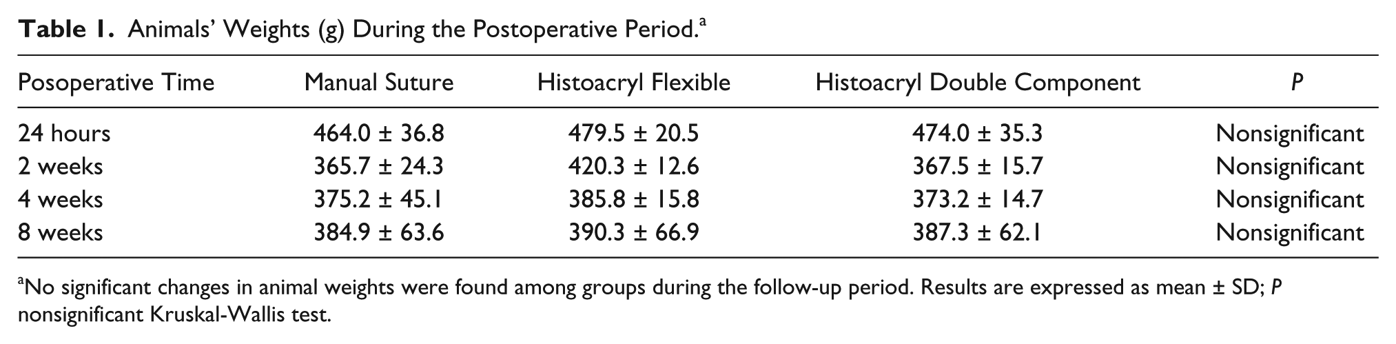
No significant changes in animal weights were found among groups during the follow-up period. Results are expressed as mean ± SD; P nonsignificant Kruskal-Wallis test.
Incidence and Extent of Adhesions. a

When adhesions were evaluated, no differences were found between the 2 cyanoacrylate groups or between manual and Histoacryl Flexible suture groups. However, the presence and extension of adhesions were significantly higher in those sutures performed with Histoacryl Double Component when compared with manual suture (P = .04; Mann-Whitney U test).
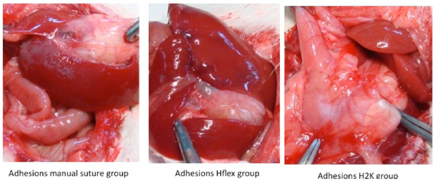
Comparison between observed postoperative adhesions in each experimental group: Manual suture group (A), Histoacryl Flexible closure group (B), and Histoacryl Double Component closure group (C).
Laboratory Tests
No differences among experimental groups were found in any of the laboratory tests performed (white blood count, ionogram, and C-reactive protein) (P = nonsignificant; Kruskal-Wallis test; Table 3).
Laboratory Tests.

No differences among experimental groups were found in any of the laboratory tests performed. Results are expressed as mean ± SD; P = nonsignificant (ns; Kruskal-Wallis test). All values are in the standard range of this rat breed.
Microscopic Findings
After histological examination, no anastomotic leaks or disruptions were identified. Furthermore, no differences in the appearance and location of acute or chronic inflammation signs, foreign body inclusions, granulation tissue, and liver and kidney damage where found among the 3 experimental groups (P = nonsignificant; Kruskal-Wallis test; Table 4, Figure 3), which is in good agreement with the in vitro biocompatibility tests formerly executed according to ISO 10993 for HF and H2K.

After histological examination, no anastomotic leaks or disruptions were identified. Moreover, no differences in the appearance and location of acute or chronic inflammation signs, gigantocellular reaction, granulation tissue, liver and kidney damage where found among the 3 experimental groups. Variables are expressed as median and 25th-75th percentile; P = nonsignificant (ns; Kruskal-Wallis test).
Numerical assessment of acute and chronic inflammation (0-3), granulation tissue (0-3), presence of giant cellular reaction (0-2), and liver (1-3) or kidney damage (1-4) was performed according to Ehrlich-Hunt classification.
Kidney damage: 1 glomerular congestion; 2 glomerular and vascular congestion; 3 glomerular congestion and vascular congestion plus dilatation; 4 glomerular congestion, vascular congestion, and dilatation plus hemorrhage.
Liver damage: 1 vascular dilatation; 2 vascular dilatation and congestion; 3 vascular dilatation and congestion plus hemorrhage.
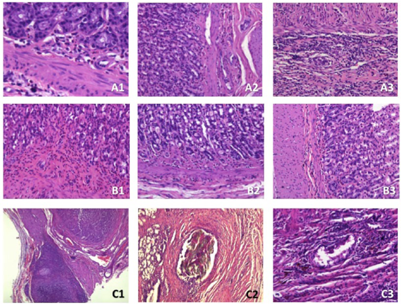
Comparative picture showing microscopic findings: gastric wall acute inflammation signs in manual suture (A1; 40×), Histoacryl Flexible (A2; 20×), and Histoacryl Double Component (A3; 20×) groups; gastric mucosal and submucosal chronic inflammation signs in manual suture (B1; 20×), Histoacryl Flexible (B2; 20×) and Histoacryl Double Component (B3; 20×) groups; and transgastric giant cellular reaction in manual suture (C1; 10×), Histoacryl Flexible (C2; 20×) and Histoacryl Double Component (C3; 20×) groups.
Discussion
Our results show that Histoacryl technical modifications can be safely used to close gastric visceral wounds with no significant local or systemic side effects in agreement with the previously performed biocompatibility tests (data not shown). Despite the fact that there are several studies that evaluate different types of CA in digestive anastomosis in comparison to sutures, the role of those adhesives in gastric sutures remains poorly understood. Moreover, to the best of our knowledge, this is the first time that a sutureless gastric closure with Histoacryl Flexible has been evaluated. Also, it is the first time that 2 different modifications of n-butyl-cyanoacrylate (HF and H2K) and 2 different application systems have been evaluated.
Previous experiences have studied gastrointestinal anastomosis with Histoacryl in rats. When compared with standard manual sutures, no differences in leakage and adhesions incidence or histopathological changes were identified. However, a shorter operative time was observed when the anastomosis was glued. 17 On the other hand, the rigidity of the previously tested glues may lead to small fissures that can evolve to intestinal leaks. Manual USP 5/0 silk interrupted suture was used to clearly discriminate the reaction to foreign body generated by strange material in the scar tissue, allowing its comparison with the cyanoacrylate group.
In our study, we used only superficial CA to join the 2 edges of the gastric wall. In any case, the healing process was not in contact with strange elements. Furthermore, no meshes were used and, therefore, no inert materials (or microabscesses) could alter the fibroblasts recruitment. In this sense, no differences on the histological features between CA groups were identified. Our findings suggest that the cyanoacrylate does not remain between the facing edges of the gastric walls tissue but above them, unlike what happens in the aforementioned mesh fixation. The collected data show similar outcomes between the 2 types of Histoacryl and the standard suture, except for the higher incidence of adhesions of H2K when compared with silk interrupted suture. Minor adhesions were observed in the HF group. In general, adhesions are due to the affinity of proteins like fibrinogen to the polymer surface. This is consistent with the fact that the larger spraying area with a slightly thicker film developed stronger adhesions. For that reason, we are suspicious that the healing process could be altered by the cyanoacrylate layer thickness; so in this study a very shallow film was applied. The appropriate technique is therefore a precise application of the glue only to the wound edges without protrusion. The observed differences between the 2 types of CA could be explained because the spray device covered a greater surface area than necessary.
Although our study was not focused on bursting pressure after closure, higher pressures were obtained when adhesives were applied. This is contrary to prior findings, pointing out that glued colonic anastomosis were not as resistant as hand-sewn anastomosis.16,22
Most of the previously published studies assessing the role of cyanoacrylate in gastrointestinal sutures are circumscribed to local or intraperitoneal issues. We have investigated the possible changes in several hematological and biochemical parameters during the follow-up after the surgical procedure. Our results indicate that there are no differences, suggesting the use of both flexible and double-component Histoacryl does not generate substantial changes in homeostasis. Thus, it could be assumed that both CA create a similar inflammatory response, just like traditional suture material does.
One of the possible applications of CA could be the closure of gastrointestinal perforations, 23 specially for peptic ulcer perforations. n-Butyl-cyanoacrylate has been previously successfully employed to close gastric perforations in a rat model. 24 When compared with standard suture, topical cyanoacrylate achieved similar results in terms of safety (anastomotic leakage) with better histopathological profile but with significantly increased adhesion formation. In contrast to our study, this n-butyl cyanoacrylate was employed as a reinforcement and sealer rather than as a substitute for suture material, because gastric perforations where closed using clips. Furthermore, to the date there is no evidence about the real efficacy of n-butyl-cyanoacrylate neither in a sepsis scenario nor under extreme acidic pH conditions. Then, it is our intention that it cannot be recommended yet to use this kind of glue to treat such conditions.
Although our results show that CA gastric closure does not seem to affect the normal wound healing patterns in terms of integrity or gastric wall cicatrization when compared with traditional suture, this is consistent with previous reports showing no differences when compared with other technical options. 23 Nevertheless, although it seems that Histoacryl modifications do not generate aberrant inflammatory patterns on the healing of the gastric wall, it has been pointed out previously that n-butyl-cyanoacrylate can affect the plasticity of vagal afferents impairing the nerve remodeling. 25 This should be taken into account specially when previous conditions altering gastric motility (eg, diabetes mellitus) are present.
In addition, the use of n-butyl-cyanoacrylate to perform gastric closures, gastrointestinal anastomosis, or anastomosis can be useful especially in clinical situations that require a safe and quick procedure such as high-risk patients. Moreover, it seems reasonable to assume that it can also be applied as an optimal reinforcement in high-risk gastrointestinal anastomosis such as those performed in oncological or bariatric surgery.
The vast majority of the gastric sutures or anastomosis are performed using hand-sewn or mechanical staplers. In contrast to these reliable options, glued anastomoses are quicker and easier to perform. They can avoid all the factors depending on an individual surgeon’s technique present in hand-sewn anastomosis and cause less tissue damage than mechanical anastomosis.
Considering all of this, our positive results drive us to propose experimental studies in large mammals (eg, pigs) for further study of sutureless hollow viscera closure.
Footnotes
Acknowledgements
The authors thank the collaboration of B. Braun Surgical S.A. in supporting this work.
Author Contributions
Study concept and design: Joan F. Julián Ibáñez
Acquisition of data: Jordi Tarascó Palomares, Jordi Navinés López, Erich K. Odermatt, Miguel A. Pacha González, Natàlia Guardia Torner
Analysis and interpretation: Jordi Tarascó Palomares, Jordi Navinés López, José M. Balibrea del Castillo, Luis F. del Castillo Riestra
Study supervision: Pau Turon Dols, Jaime Fernández-Llamazares Rodríguez
Authors’ Note
The present work has been developed under agreement between B. Braun Surgical S.A. and The Catalonian Public Health Institute (ICS), Barcelona, Spain.
Declaration of Conflicting Interests
The author(s) declared no potential conflicts of interest with respect to the research, authorship, and/or publication of this article.
Funding
The author(s) received no financial support for the research, authorship, and/or publication of this article.