
Abstract

To the Editor:
After we have described our previous method,1,2 a novel system capable of quantitatively assessing facial muscle movements was developed and is here presented. It automatically describes a set of morphological measurements and uses depth cameras together with advanced computer vision techniques, to perform detailed 3-dimensional (3D) characterization (Figure 1).
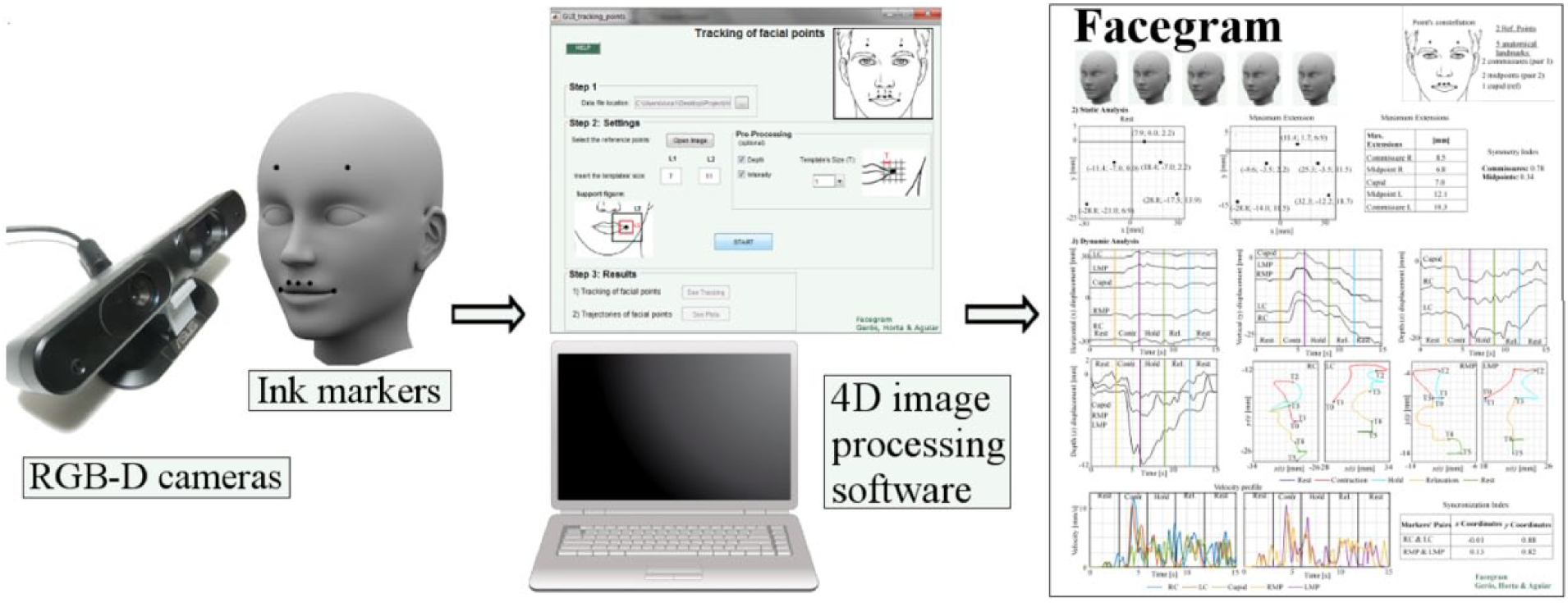
The Facegram (3D) concept.
The system is simple and of great clinical value. In a clinical setup, the clinician places ink dots/markers on the face of the subject identifying anatomical landmarks whose movement should be characterized. The face is recorded by the camera while performing prespecified movements. The analysis results are organized as an innovative and standard medical tool named FACEGRAM-3D. Given the complexity of facial musculature, it is not feasible to collapse the spatiotemporal analysis into a single plot. Instead, it aggregates a set of plots and measures (Supplemental Digital Content–1 [SDC1] available in the online version of the article).
The report is organized in 3 information blocks: subject’s data, static analysis, and dynamic analysis. Patient’s information, such as name/age/sex/date are presented at the top, as well as a representative frame at each phase of the movement (with its duration in seconds, Figure 2). The quantitative analysis is divided into 2 different categories: static and dynamic.
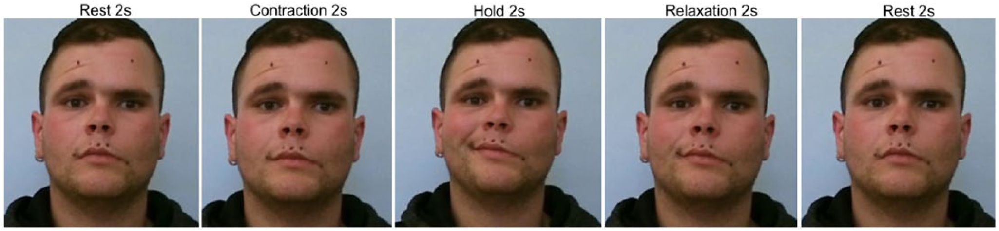
All trajectories represent markers’ movement during 5 phases of movement: Rest, Contraction (Contr.), Hold, Relaxation (Rel.), and Rest.
The components regarding static analysis are the 3D positions of each anatomical point at rest and at maximum extension, a table with maximum extensions for each marker and a symmetry index. This metric, between 0 and 1, is intended to provide information about the relative position of points at rest for each associated pair of anatomical landmarks (in the case of smile: R/L commissures and L/R midpoints).
The dynamic analysis section starts with plots of the x, y, and z coordinates for each landmark as a function of time, and parametric trajectories of each associated pair of markers in the xy-, xz-, and yz-plane. The velocities of each landmark as a function of time is also presented. Finally, a synchronization index is calculated for each pair of markers and each direction (horizontal/vertical) separately. This coefficient provides information about the correlation between the trajectories in a markers pair. If synchrony exists, the index is approximately 1 (Figure 3).
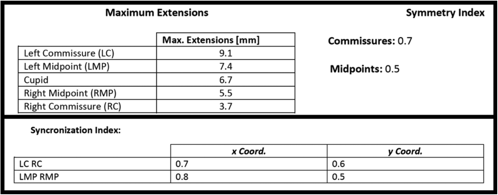
Symmetry index (static analysis) and synchronization index (dynamic analysis).
In this patient (Supplemental Digital Content–2 [SDC2], right facial paralysis), asymmetry at rest and maximum extension was detected especially among the commissures, and a cupid deviation to the left. The amplitude of excursion was greater at the left side. Symmetry index was higher for the commissures. The parametric curves for the left side were described by smooth lines but for the right side they had abrupt changes. The synchronization indexes showed higher correlation/synchronization for horizontal movements in the midpoints.
Postoperatively, 10 months after gracilis muscle transplantation and nerve coaptation to the masseteric branch (Supplemental Digital Content–3 [SDC3]), asymmetry at rest and at maximum extension was not as evident and symmetry indexes were also higher. Regarding vertical and horizontal displacement, the movement appeared to be synchronized with less fluctuation in depth displacement, and the anatomical pair’s trajectories were smoother and had similar velocity profiles. Synchronization index values were higher for the vertical displacement.
Further studies are needed to validate the potential of this technology.
Footnotes
Author Contributions
Study concept and design: Ricardo Horta, Ana Gerós, Paulo Aguiar.
Acquisition of data: Ricardo Horta, Ricardo Nascimento, Ana Gerós.
Analysis and interpretation: Ricardo Horta, Ricardo Nascimento, Ana Gerós, Paulo Aguiar, Alvaro Silva, José Amarante
Study supervision: Ricardo Horta
Declaration of Conflicting Interests
The author(s) declared no potential conflicts of interest with respect to the research, authorship, and/or publication of this article.
Funding
The author(s) received no financial support for the research, authorship, and/or publication of this article.
References
Supplementary Material
Please find the following supplemental material available below.
For Open Access articles published under a Creative Commons License, all supplemental material carries the same license as the article it is associated with.
For non-Open Access articles published, all supplemental material carries a non-exclusive license, and permission requests for re-use of supplemental material or any part of supplemental material shall be sent directly to the copyright owner as specified in the copyright notice associated with the article.