
Abstract
Dural suturing in transsphenoidal surgery requires well-honed technical skills. We have developed a semiautomatic dural suturing device and confirmed its effectiveness by comparing it with the conventional method. This device significantly shortens the suturing time compared with the conventional method. The dural suturing time in transsphenoidal surgery could be decreased significantly by use of this novel device.
Need
In transcranial surgery, watertight direct dural suturing is widely performed to prevent cerebrospinal fluid leak because it is the simplest and most practical and reliable method.1-5 However, a considerable amount of operating time is spent on suturing the dura when performing transsphenoidal surgery (TSS). According to Nishioka et al, 5 dural suturing increases the operating time by 30 minutes. Therefore, there is a need for surgical instruments that allow easier, secure, and more rapid dural suturing. We have developed a semiautomatic suturing device with a novel mechanism that can be used to stitch the dura.
Technical Solution
The ideal dural suturing device must satisfy the following criteria: (1) the device should be able to pass the suture threads through the dura in 2 directions, that is, from the nasal cavity side to the pituitary side and vice versa; (2) the suture thread should be able to be passed through multiple portions of the dura without needing to be drawn outside of the body; and (3) the outside diameter of the nasal insertion component is 3 mm or less, which is the same as for the surgical tools generally used in TSS. Our prototype suturing device is shown in Figure 1A. The device measures 340 × 100 × 11 mm. The nasal insertion component has an outer diameter of 3 mm and a length of 230 mm. Novel suturing device. (A) Appearance of the suturing device. (B) Schematic cross-sectional view of the portion at the distal end.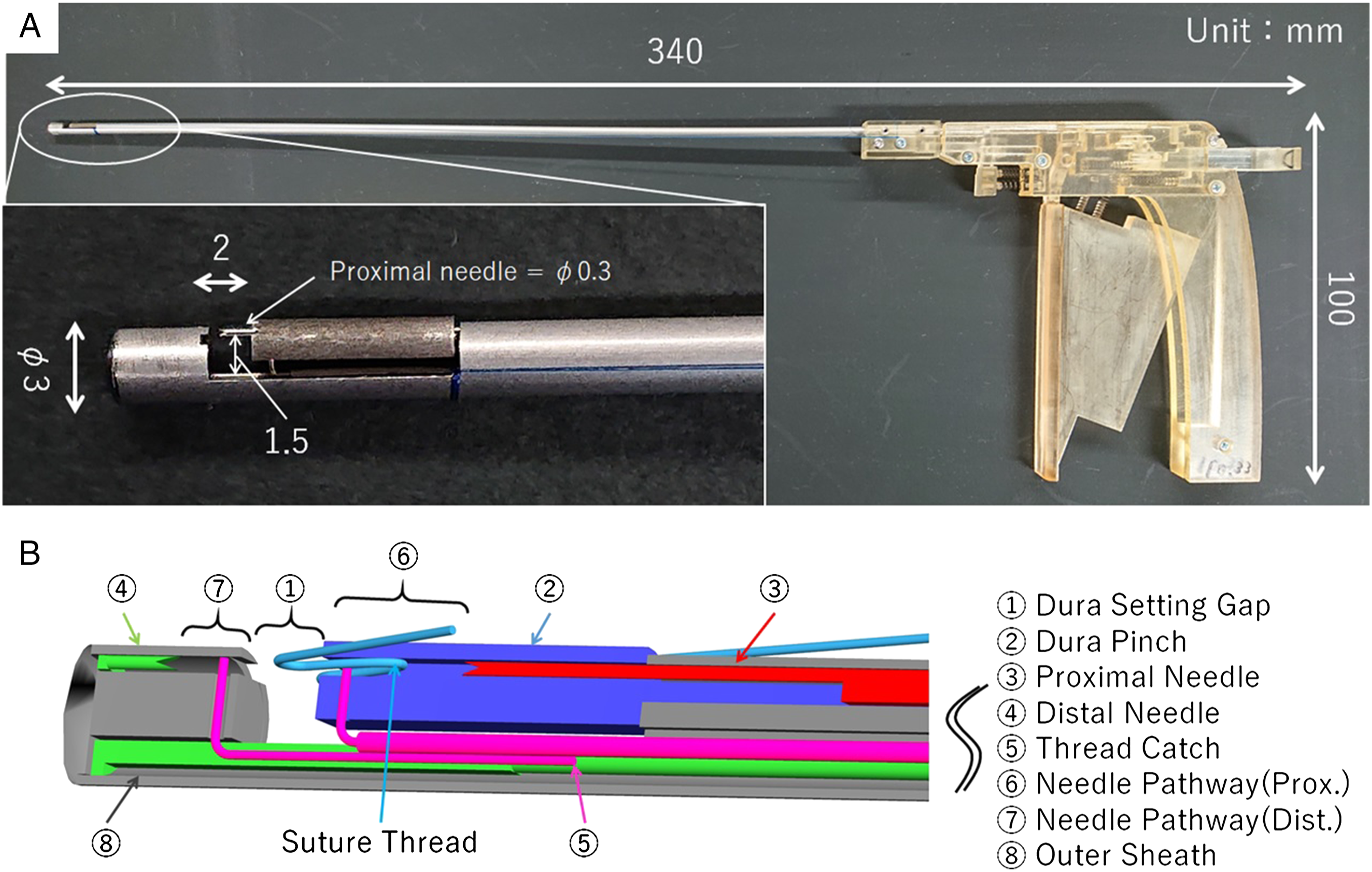
Figures 1B and 2 show a cross-sectional view of the main components of the tip and the suturing mechanism. When suturing in any direction, the objective is achieved by passing the thread through the dura with a needle on one side, and the thread catch on the other side grasps the suture. This design and mechanism meet the above 3 criteria. Suturing procedure. Suturing (A) from the nasal cavity side to the pituitary side and (B) from the pituitary side to the nasal cavity side.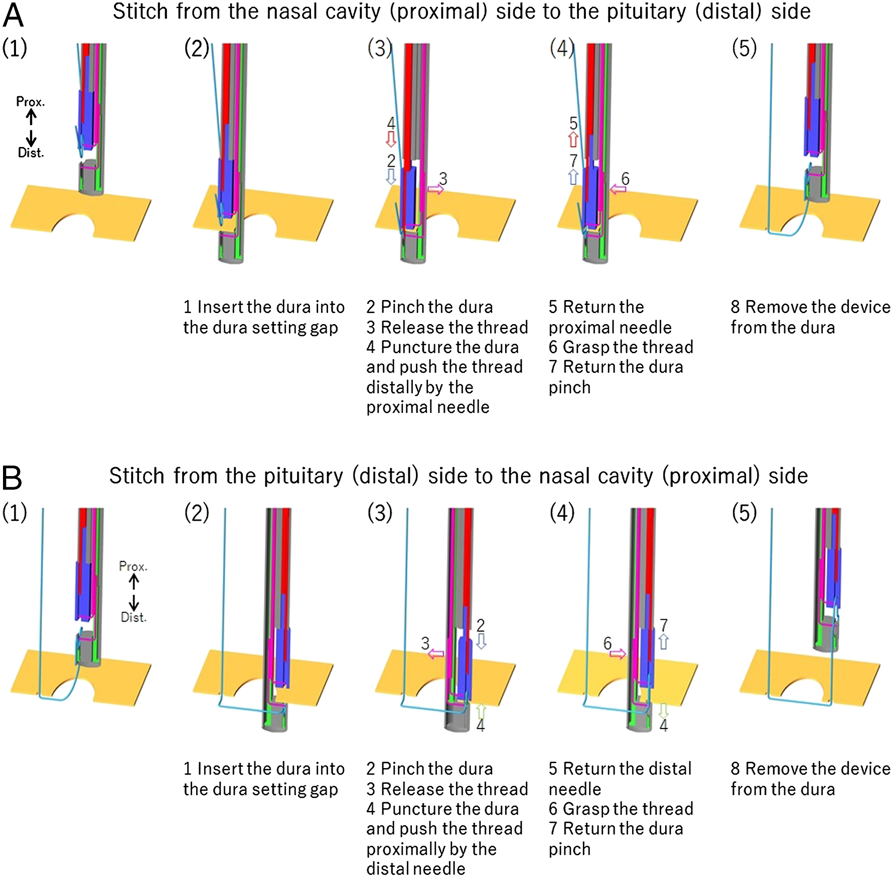
Proof of Concept
Two artificial dura mater preparations were placed on a goniometer stage with a gap of 5 mm. A silicon tube (outer diameter, 13 mm; length, 100 mm) was fixed vertically on the artificial dura mater. As shown in Figure 3A, the suture thread was passed through 2 points of the dura to connect the edges of the artificial dura mater. All sutures were carried out while observing the endoscopic image (Figure 3B). Experimental system. (A) Suture method, (B) complete experimental system, and (C) suturing using the suture instrument.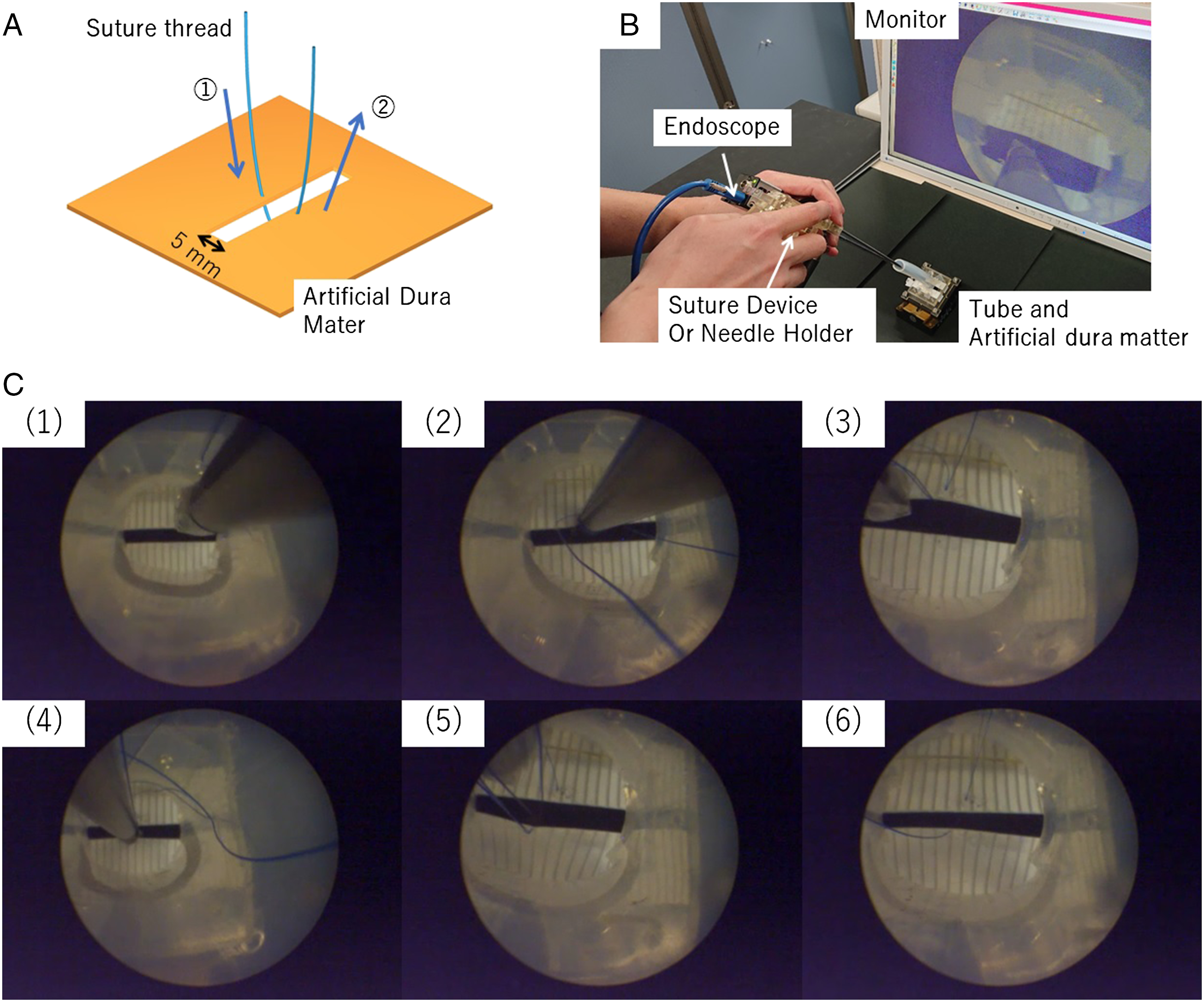
An engineer (subject 1) who was not a medical practitioner but was skilled in the use of the suturing device manipulated the developed suture device while a surgeon with more than 10 years of experience performing TSS on a daily basis (subject 2) and a young surgeon who had never previously sutured the dura during TSS (subject 3) used a conventional needle holder. The 3 subjects each performed 5 suturing trials. The suturing time and numbers of device reinsertions and needle drops were compared between the 3 subjects.
Results
Figure 3C shows the suturing procedure when using the prototype device. The mean suturing time (with standard deviation) was 49.8 ± 3.6 seconds in subject 1, 86.2 ± 39.8 seconds in subject 2, and 267.0 ± 145.0 seconds in subject 3. There was a significant difference in suturing time between subjects 1 and 2 (P = .037) and between subjects 1 and 3 (P = .027). The results tended to be less variable when suturing with the novel device than when suturing with the needle holder.
The device was taken out of the tube 1.0 ± 1.3 times by subject 2 and 24.6 ± 4.2 times by subject 3. The needle was dropped into the tube .0 ± .0 times by subject 2 and 24.6 ± 4.2 times by subject 3. Use of the novel suture device (by subject 1) did not require the needle to be changed and there was no risk of dropping the needle. The suturing time required by subjects 2 and 3 was longer because of the time required for device reinsertion and retrieving the dropped needle.
Next Steps
A comparative crossover study in which a larger number of operators perform suturing using this novel device and a conventional needle holder is needed in the future.
Conclusion
We have developed a semiautomatic suturing device to assist dural suturing in TSS. Surgeons with limited experience in performing TSS can use this device as effectively as experienced surgeons with little variation in suturing time.
Footnotes
Acknowledgments
The authors are grateful to Dr T Kawamata for clinical advice and allowing us the opportunity to observe a TSS being performed.
Author Contributions
Study conception and design: Shohei Suzuki, Etsuko Kobayashi, Ken Masamune, Yoshihiro Muragaki
Acquisition of data: Shohei Suzuki, Kenichi Hododuka, Kosaku Amano
Analysis and interpretation of data: Shohei Suzuki, Etsuko Kobayashi, Ken Masamune, Yoshihiro Muragaki
Study supervision: Ken Masamune, Yoshihiro Muragaki
Declaration of Conflicting Interests
The authors declared the following potential conflicts of interest with respect to the research, authorship, and/or publication of this article: Shohei Suzuki was a doctoral student at Tokyo Women’s Medical University from April 2015–March 2019 and is now an employee of Kaneka Corporation, which funded all research expenses, including the cost of manufacturing the suturing device and the experimental systems, and receives a salary from Kaneka Corporation. Kaneka Corporation owns the following 3 patent applications related to this research: PCT/JP2019/000063, JP2018/186972 (pending), and PCT/JP2019/033432. Etsuko Kobayashi, Kenichi Hododuka, Kosaku Amano, Ken Masamune, and Yoshihiro Muragaki have no conflicts of interest or financial ties to disclose.
Funding
The authors disclosed receipt of the following financial support for the research, authorship, and/or publication of this article: All research expenses related to this study, including the manufacturing cost of the suturing devices and experimental systems, were met by Kaneka Corporation, Tokyo, Japan.