






Abstract
Background/Need
Laparoscopic abdominal surgery requires navigating unique technical challenges with precision, dexterity, and a thorough understanding of anatomy. There is a need for higher-fidelity training models to assist in improving trainee competence. This manuscript introduces a novel modular abdominal simulation environment (MASE) with the ability to insufflate under standard parameters to accommodate laparoscopic and robotic surgery training and assessment.
Methodology and Device Description
CT scans of a deidentified patient pelvis and spine are processed, reconstructed, and modified into 3D printable files, then printed using a high-fidelity resin printer. Silicone skin is developed to cover the MASE and mechanically fixed to create an air-tight seal. Insufflation capability is tested by measuring the pre- and post-insufflation height of the model, as well as internal pressure.
Preliminary Results
MASE meets the following criteria: anatomical accuracy, scale-to-life, and re-usability. Its ability to be insufflated via a Veress needle at Palmer’s point recreates a pneumoperitoneum (increasing in height by 108%), allowing for effective port placement and clear visualization with a laparoscope. The platform successfully supports fundamentals of laparoscopic surgery (FLS) tasks including intracorporeal knot tying and peg transfer both with laparoscopic tools and robotic system.
Current Status
Current work includes a more efficient locking mechanism, incorporation of the retroperitoneal space, and addition of synthetic/explant organs for high-fidelity abdominal simulation. MASE combines high anatomical fidelity, realistic tissue simulation, and procedural versatility with reproducibility. Future testing includes stiffness characterization of the silicone skin and validation for surgical resident training.
Introduction
Abdominal surgery requires a high degree of technical skill and precision, and surgical simulation models have shown to improve the technical skills of trainees.1-3 A recent meta-analysis showed improvement in real operating performance after training with simulation. 4
Training models can be qualified in terms of fidelity and accessibility. Low-fidelity trainers offer simple, inexpensive, and accessible platforms of tissue-like structures to practice basic skills such as suturing or knot-tying but lack the realism needed. More advanced silicone and synthetic simulators provide improved anatomical accuracy but do not fully replicate the tactile feedback of live tissue. 5
Higher fidelity models include animal and cadaveric models, which have been used for training complex procedures and entire operations. However, ethical concerns, cost, and logistical challenges limit their use. 5 Additionally, cadavers have limited availability and are not as reusable as synthetic or virtual models. 6
Despite the multitude of current training models, no single platform currently combines patient-derived anatomical geometry, insufflation, re-usable modularity, and low-cost reproducibility. In this manuscript, we propose to bridge said gaps with the development of a modular abdominal simulation environment (MASE) for minimally invasive surgery simulation, compatible for both laparoscopic and robotic scenarios.
Methods
CT scans of deidentified patients (IRB: Pro00062197) are reconstructed into a 3D surface using medical imaging software (3DSlicer, Boston, MA, USA). The segmented volume is then exported as a stereolithography file and edited using 3D modeling software (Blender, Eindhoven, Netherlands) to remove segmentation noise. The post-processed pelvis is imported into computer-aided design (CAD) software (Fusion360, San Francisco, CA, USA) to add inserts into the pelvis. The final pelvis model is printed in four segments on a 3D printer (ELEGOO Saturn 3, Shenzhen, China) using resin (SirayaTech Fast, San Gabriel, CA, USA) and assembled (Figure 1A). The general dimension and curvature of the ribs are measured from the CT scans and redesigned in CAD. The rib model is printed using generic polylactic acid (PLA) on a 3D printer (Ultimaker S3, Utrecht, Netherlands) (Figure 1A). Key Design Features of MASE. (A) CAD model of MASE; (B) Interior of MASE, showing the inner 3D-printed anatomical models; (C) Outer view of MASE showing skin and clamping mechanism for airtight seal; (D) Phantom skin and highlighting of the different layers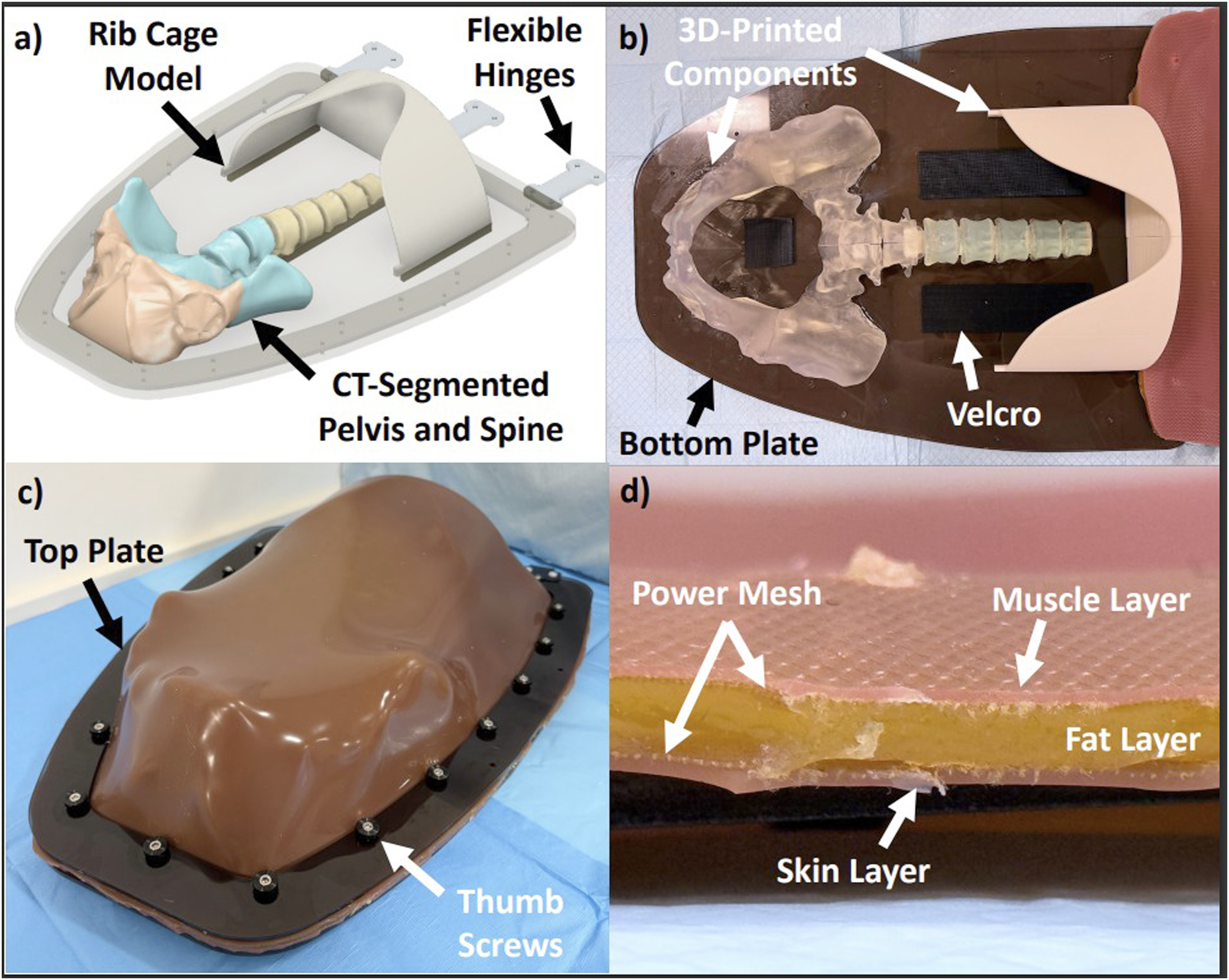
The base frame consists of two pieces of 50 cm x 50cm cast acrylic boards (McMaster-Carr, Elmhurst, IL, USA) that are laser cut (TroTec Speedy 400, Marchtrenk, Austria) with cutouts. On the bottom board, the 3D-printed parts are fixed with adhesives for airtight sealing. Velcro strips for attaching FLS modules are also added (Figure 1B). Screws fix a phantom skin to its underside of the top board (Figure 1C), and a 3D-printed hinge aligns the two boards together.
To replicate the abdominal wall, platinum-cure silicone rubber (EcoFlex 00-30, Smooth-On, Lehigh County, PA, USA), silicone gel, mesh fabric, and silicone pigments are used to differentiate layers of skin, fat, and muscle (Figure 1D). The skin is molded using a 42” x 28” tray with a thickness of 8.5 mm, in range of human skin. 7 The total raw materials cost is ∼$311, with ∼$20 for each reusable skin replacement.
Insufflation Testing
To test for insufflation, a Veress needle is inserted at Palmer’s point, replaced with a 12 mm trocar, and a measuring rod was inserted at the center to record pre-and post-insufflation heights (Figure 2A). MASE then undergoes insufflation (Karl Storz SCB Thermoflator, Tuttlingen, Germany) for 90 seconds. Post-insufflation max height is measured across five attempts (Figure 2A and B), and the internal pressure is recorded in mmHg. Insufflation and da Vinci SP Testing. (A) MASE with a 5 mm trocar inserted at Palmer’s point; (B) MASE after insufflating for 90 seconds; (C) FLS peg transfer and suturing modules secured in MASE; (D) da Vinci SP robot inserted in insufflated MASE; (E) Camera footage of attending surgeon practicing peg transfer in MASE; (F) Camera footage of resident practicing suturing in MASE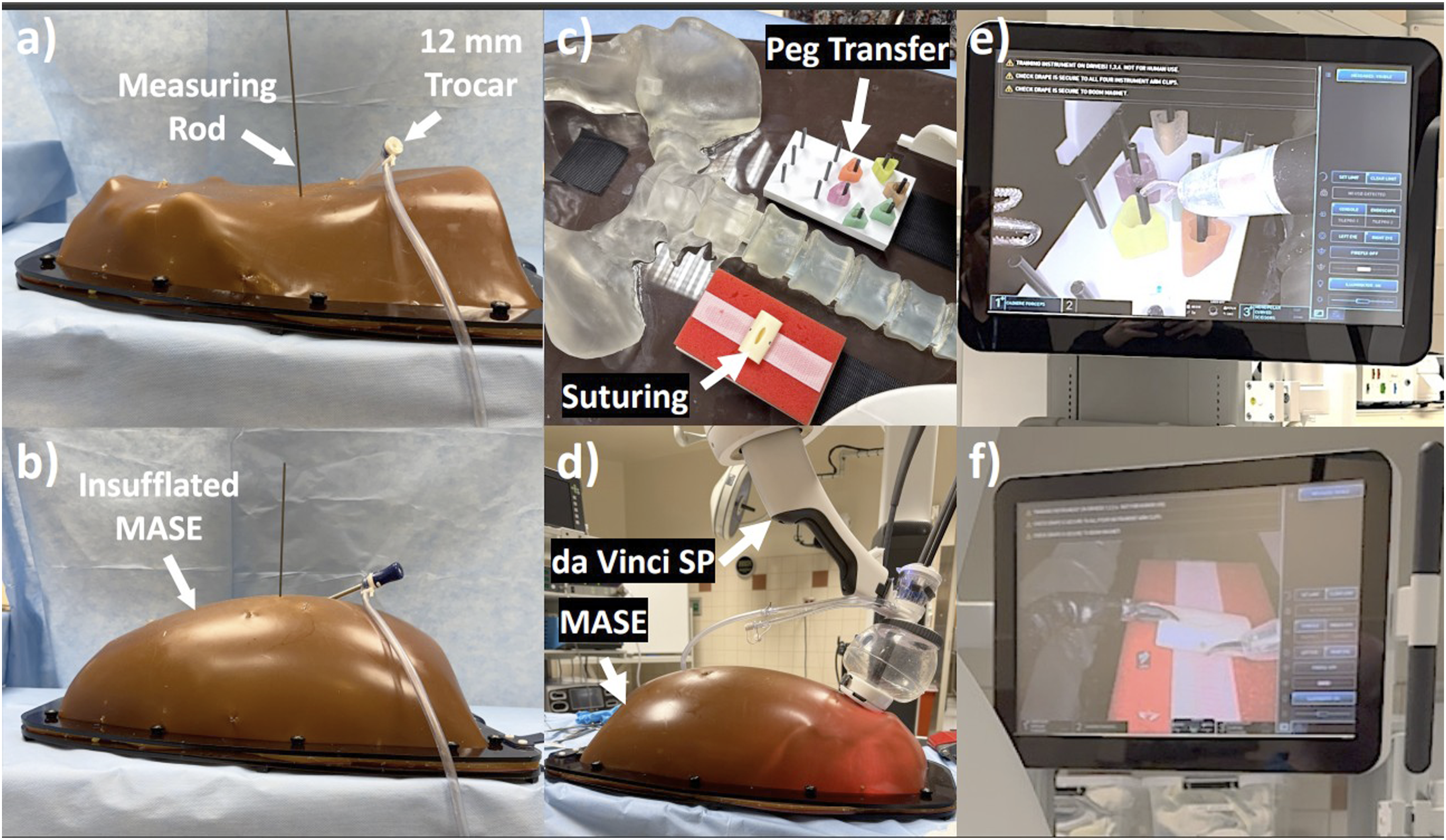
Insufflation Testing
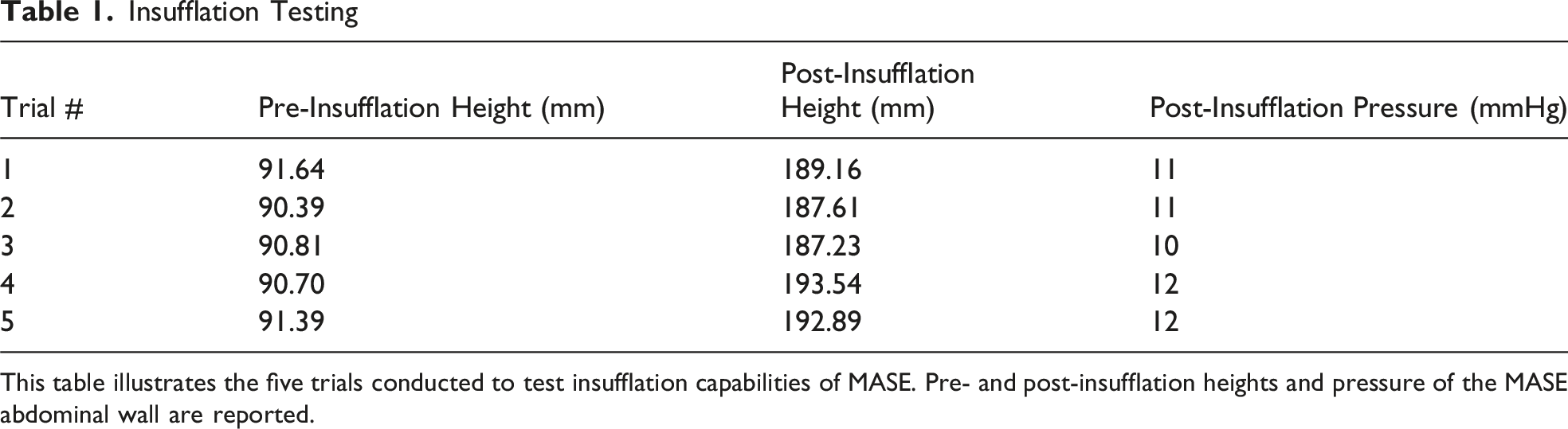
This table illustrates the five trials conducted to test insufflation capabilities of MASE. Pre- and post-insufflation heights and pressure of the MASE abdominal wall are reported.
da Vinci SP Demonstration
A proof-of-concept insufflation test on the da Vinci Single Port (SP) Robot (Intuitive Surgical, Sunnyvale, California, USA), was conducted. FLS peg transfer and intracorporeal knot tying modules are affixed onto the inner Velcro (Figure 2C). The da Vinci SP was docked onto MASE and insufflated through the robot port (Figure 2D). An attending robotic surgeon and a general surgery resident both completed peg transfer and intracorporeal knot tying inside the insufflated MASE with ease (Figure 2E and F).
A single skin model was reused throughout testing and demonstration. The port incisions were sutured closed, and new port entry points were created. The model was still able to withstand internal insufflation pressure, reaching up to 12 mmHg.
Conclusion
The proposed methodology utilizes cost-efficient materials to produce a patient-specific and insufflatable MASE but can be further developed to replicate anatomical realism. Future work will include the development and incorporation of the retroperitoneal space, integrating synthetic and/or ex-vivo organs (liver, gallbladder, bowel, etc.) within MASE, validating the mechanical properties of the skin model, and validating the educational functionalities of MASE through resident training.
Footnotes

Author Contributions
Funding
The authors received no financial support for the research, authorship, and/or publication of this article.
Declaration of Conflicting Interests
The authors declared the following potential conflicts of interest with respect to the research, authorship, and/or publication of this article: Dr. Sabino Zani is a consultant for Asensus, Medtronic, and Intuitive Surgical, and Dr. Katharine Jackson are consultants for Medtronic and Intuitive Surgical.