
Abstract
Introduction:
Lactational mastitis is common in lactating women, with Staphylococcus aureus as the most commonly isolated agent associated with infectious lactational mastitis. Currently, there are no evidence-based guidelines for antimicrobial treatment due to barriers in obtaining pharmacokinetic data from lactating women. To overcome this barrier, a suitable large animal model is needed. Goats are an ideal translational model for human mastitis due to their anatomical and physiological similarity to humans. The objective of this pilot study was to assess if goats would develop clinical mastitis following intramammary inoculation with a clinical human isolate of S. aureus with the goal of establishing an alternative in vivo model for future research. The hypothesis was that the infected mammary gland half would show similar clinical signs to women with mastitis and demonstrate a similar local immune response when compared to the control mammary gland half.
Methods:
One half of the mammary gland of two healthy lactating does was inoculated with a clinical human isolate of S. aureus. The other half of the mammary gland was sham inoculated with sterile buffered saline. Physical examinations, mammary gland assessments, and sterile milk samples were collected every 12 hours post inoculation. At 96 hours post inoculation, the goats were euthanized, and the mammary glands were examined for pathological changes.
Results:
Goats did not develop systemic signs of disease following inoculation. Focal infected mammary gland changes included warmth, swelling, redness, discoloration, and reduced milk production; the other mammary gland half remained normal throughout the study period. S. aureus was enumerated from only the infected mammary gland half. The microscopic findings of the infected half showed neutrophilic inflammation and cell necrosis consistent with acute mastitis.
Discussion:
This pilot study demonstrated lactating does can develop clinical signs like those observed in women. Goats have the potential to be a promising animal model to study infectious lactational mastitis.
Introduction
Lactational mastitis affects one in four women during the first 26 weeks postpartum, with Staphylococcus aureus as the most commonly isolated agent associated with infectious lactational mastitis.1–4 In women, diagnosis of mastitis is made symptomatically, with women experiencing localized, unilateral breast tenderness, redness, decreased milk output, and occasionally systemic signs, such as fever, fatigue, and body aches.4–6 Conservative treatment is recommended initially, but if symptoms persist for 24–48 hours, or a patient develops systemic signs, antimicrobials are started.1,6,7 Initial antimicrobial intervention often targets S. aureus because of its high prevalence (32–44%) in mastitis cases.1,4,6 However, there is no current consensus or evidence-based guidance for antimicrobial treatment of infectious lactational mastitis.2,6,8 One of the main obstacles to development of antimicrobial treatment guidelines is the challenge of performing pharmacokinetic trials in lactating women. There are no data available regarding antimicrobial drug concentrations in mastitic breast milk. One way to overcome this challenge is to investigate a suitable animal model to study infectious lactational mastitis. Such an animal model would share similar mammary gland anatomy, milk physiology, mastitis symptoms, and local immune response with women.
Historically, rodents have been used to study lactation, physiology and pharmacokinetics for human health. 9 Rodent mammary gland anatomy and milk secretion physiology are quite different from humans. 10 The human breast is a single pair of mammary glands, divided into 15–20 lobes of parenchyma, separated by dense fibrous connective tissue and varying amounts of adipose tissue.11–13 Each of the lobes drains into a lactiferous duct, then a sinus and terminates at a nipple with a single opening.11,12 Mice have five pairs of mammary glands and rats have six pairs, extending from the neck to the inguinal region surrounded by adipose tissue with a little amount of fibrous connective tissue. 10 Rodents have a single lactiferous duct per gland, greatly reducing the volume of milk produced during lactation, making pharmacokinetic trials and milk drug analysis challenging. 10 Pigs have also been used as models for human research, but to the research team’s knowledge, pigs have not been used for lactation studies for women. Pigs have a variable number of mammary glands, ranging from 12–18 pairs that run along their ventrum.13–15 Different from women, their teat has two external openings.14–16 In addition, obtaining serial milk samples from sows can be challenging due to the complex milk ejection reflex and short period of milk flow (10–20 seconds). 17
Small ruminants, particularly goats, are an attractive translational animal model to study infectious lactational mastitis in women. Goats have a similar mammary gland anatomy and milk secretion physiology when compared to women. Goats also have a single pair of mammary glands with branching duct systems separated by dense fibrous connective tissue and adipose tissue; milk drains into a similar lactiferous duct, then a sinus and terminates in a teat with a single opening.18,19 All mammals secrete milk through a combination of merocrine and apocrine secretion methods. Some mammals rely more heavily on one method compared to another. Women and goats predominantly secrete milk through apocrine secretion methods, while most other livestock secrete milk through merocrine methods.20,21 This similarity in physiology is an important advantage for pharmacokinetic or diagnostic studies. Additionally, goats produce consistent, larger milk volumes than rodents that can facilitate drug concentration analysis and serial milk sample collection.
Mastitis is a common disease in the dairy goat industry, with similar clinical signs observed in women. Goats with clinical mastitis demonstrate unilateral mammary gland tenderness, redness, swelling, abnormal milk secretions, and they can develop systemic clinical signs, such as fever, lethargy and anorexia. S. aureus is also a common pathogen responsible for clinical mastitis in the dairy goat industry. To the research team’s knowledge, goats have not been explored as a translational animal model to study infectious lactational mastitis in women. The overall objective of this project was to conduct a pilot study to assess if healthy goats would develop clinical mastitis following experimental intramammary inoculation with a clinical human isolate of S. aureus obtained from infected breast tissue. The overall hypothesis was that the infected mammary gland half would show similar clinical signs to women with mastitis and demonstrate a similar local, histopathologic response. This was accomplished through a disease induction trial, followed by a histopathological comparison of inoculated and control mammary tissues.
Materials and Methods
Animals used
Two mid-age (4–5 years of age), mid-lactating dairy goats (Saanen) sourced from a private goat dairy in North Carolina were enrolled in this study. Health screening through physical examination and serology for Coxiellia burnetti and Brucella were performed prior to the study. Milk from each half of the mammary gland was collected aseptically from each half of each goat for culture to ensure the goats did not have S. aureus mastitis. The goats were transported from the farm of origin and housed at the North Carolina State University College of Veterinary Medicine laboratory animal facility. This study was approved by NC State IACUC Protocol #24-440 and BUA Protocol #2025-02-972.
Experimental design
Each goat served as their own control. Following a 24-hour acclimation period and baseline physical examination, the right half of the mammary gland was inoculated with S. aureus. The left half of the mammary gland was inoculated with sterile PBS and served as a negative control. After disease inoculation, physical examinations were performed and aseptic milk samples for milk culture were collected every 12 hours. Goats were hand milked twice a day, following sterile milk collection. The study lasted for 96 hours, and the goats were euthanized at the end of the study period for mammary gland tissue collection.
Staphylococcus aureus preparation and disease inoculation
A human clinical strain of S. aureus obtained through the University of North Carolina’s Clinical Microbiology Laboratory was used in this study. The isolate was from breast abscess aspirate and was designated as methicillin-sensitive. To prepare S. aureus for inoculation from the stock solution, a loop was inoculated onto a 5% Sheep blood agar plate (Remel, Lenexa KS) and incubated overnight at 37°C. Following incubation, 3–5 S. aureus colonies were inoculated into sterile Brain Heart Infusion (BHI) broth (BD Inc, Frankline Lakes, NJ) and incubated at 37°C for 6–7 hours on a shaking incubator at 195 rpm. This was then centrifuged, supernatant was removed, and the bacterial pellet was washed with 1× PBS. This was done in triplicate. After the third wash, the bacterial pellet was resuspended in 5 mL of 1× PBS and the final concentration of the resuspended pellet was adjusted until 0.5 McFarland standard was met, equivalent to approximately 1.5 × 10⁸ cfu/mL. One milliliter of this culture was used as the disease inoculum in each goat.
For intramammary inoculation, the goats were restrained with a halter. The right teat was aseptically prepared, and a teat cannula (Jorgensen Labs, Loveland, CO) was sterilely introduced into the teat canal. The S. aureus inoculum was then administered into the teat (Fig. 1). After removal of the teat cannula, the right teat and right mammary gland were massaged to assist in diffusing S. aureus throughout the mammary gland. The left half of the mammary gland served as the negative control. The left teat was similarly aseptically prepared, and a teat cannula was sterilely introduced into the teat canal. Sterile PBS (1 mL) was administered into the teat through a similar method as previously described, with subsequent left teat and left mammary gland massaging.
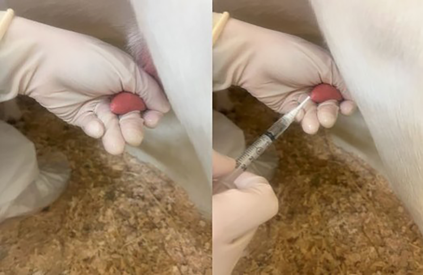
Sterile intramammary inoculation of the left half (negative control) with sterile PBS and the right half (diseased) with a clinical human S. aureus isolate.
Physical examination and mammary gland assessment
Prior to inoculation and every 12 hours following inoculation, goats received a physical examination. This included assessing systemic body temperature, heart rate, respiratory rate, rumen contraction rate, mucous membrane color, and the presence of hyperemic mucous membranes or injected sclera. The mammary gland was also assessed, first for any visual abnormalities (discoloration or swelling) and then palpated. While palpating the mammary gland, each half was assessed for warmth, firmness, pain, or swelling. Physical examinations and mammary gland assessments were conducted by the same two individuals (J.H. and W.M.) at each time point and notes were recorded.
Sterile milk sample collection and milking
After each physical exam and mammary gland assessment, a sterile milk sample from each half of the mammary gland was collected. The left half was collected before the right half. Both teats were dipped with an iodine-based pre-dip milking solution. The teat was cleaned with a dilute chlorohexidine solution, and the tip of the teat was cleaned with alcohol. The teat canal was stripped three times and then a sterile milk sample was collected into a test tube. The tube was placed on ice until it was brought to the laboratory for culture (about 20 minutes). Following sterile milk sample collection in each half, the goats were hand milked into a stainless steel bucket. At the conclusion of milking, each teat was dipped with an iodide-based postdip solution. Each teat had their own respective predip and postdip solution. Sterile milk sample collection and hand milking were done every 12 hours during the study period.
Staphylococcus aureus enumeration
To determine the burden of S. aureus in each half, the collected sterile milk sample underwent enumeration. Briefly, the milk samples were serially diluted (10−1 to 10−7) and 100 uL of each dilution was plated in triplicate on Mannitol Salt Agar (Thermo Scientific, Waltham, MA). Plates were incubated at 37°C for 24–48 hours. Following incubation, yellow colonies or colonies present on yellow agar were counted, with optimal plate counts being between 30 and 300 colonies. To confirm these colonies were S. aureus, a coagulase test was performed. Briefly, 8–10 colonies from countable plates were streaked on a blood agar plate. A control blood agar plate was also prepared, with half of the plate streaked with a S. epidermidis control (ATCC 14990, Microbiologics, St Cloud, MN) and the other half with a S. aureus control. Both plates were incubated overnight at 37°C. The following day, 2–3 colonies grown on the blood agar plates were transferred to a test tube containing 0.5 mL of reconstituted coagulase plasma (BD Inc, Frankline Lakes, NJ). The inoculum was mixed, vortexed and incubated at 37°C for 4–12 hours. Any clot formation was indicative of a positive test result and confirmed that the isolate was S. aureus.
Postmortem examination and histological evaluation
Postmortem examination of the mammary glands was performed at 96 hours post inoculation. Gross changes were evaluated throughout both right and left halves of the mammary glands including the gland cistern, distal secretory parenchyma and proximal secretory parenchyma. Representative mammary tissue samples from each portion were collected in 10% neutral buffered formalin and underwent routine H&E staining for histopathological evaluation.
Statistical analysis
Due to the pilot nature of the study and small sample size (n = 2), no formal statistical analyses were performed. Findings were interpreted descriptively.
Results
Physical examination and mammary gland assessment
In this pilot study, two mid-lactation does were enrolled. The does were S. aureus negative upon initial milk culture from both mammary gland halves prior to the start of the trial. Intramammary inoculation of the human S. aureus isolate was performed with no complications. During the study period, the does did not have abnormal physical examination findings, and they continued to eat well and drink normally. However, their mammary gland appearance, palpation and milk volume subjectively did change 12 hours post inoculation. One does developed a warm, swollen, erythematous and sensitive right half of her mammary gland (Fig. 2). The other does had dark discoloration, teat swelling, warmth and decreased milk production in the right half of her mammary gland (Fig. 3). For both does, the left half of the mammary gland remained normal.
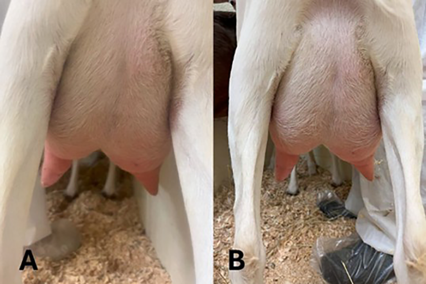
Images of mammary gland changes. Panel
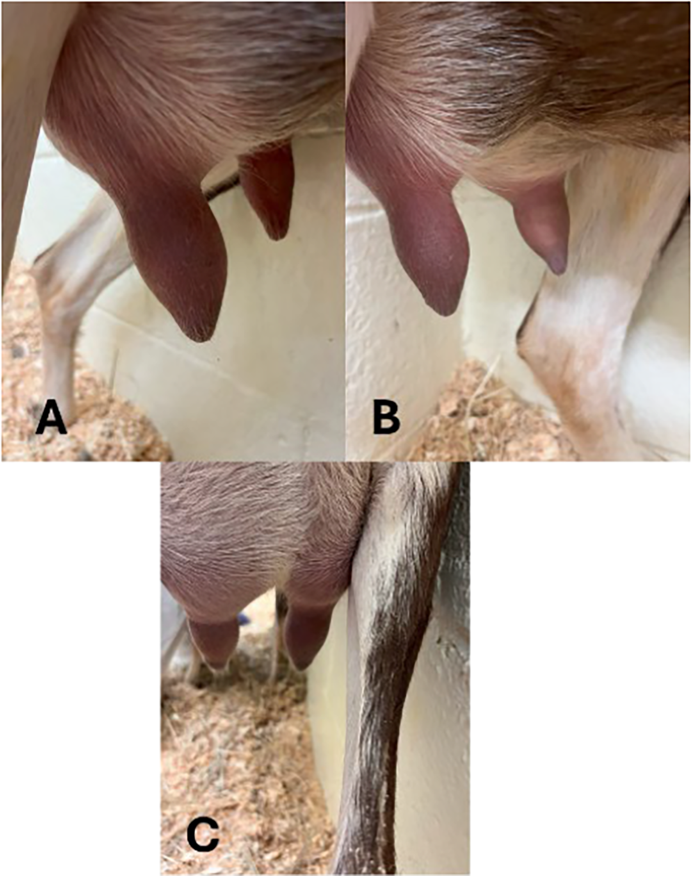
Images of mammary gland changes.
Staphylococcus aureus enumeration
Prior to inoculation, and every 12 hours during the study period, a sterile milk sample was collected from each half and underwent S. aureus enumeration. There was no S. aureus enumerated in the left mammary gland half of either goat at any time point. There was S. aureus growth noted in the right mammary gland half of both does starting at 12 hours post inoculation and it persisted throughout the study period (except for the 72 hour time point in one goat) (Fig. 4).
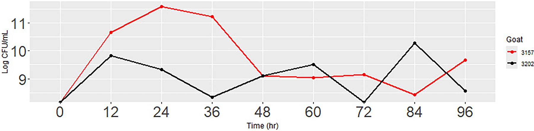
Log cfu/mL growth of S. aureus from the right half of the mammary gland of lactating does (n = 2). There was no growth in the left half.
Postmortem examination and histopathological findings
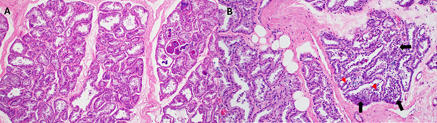
On postmortem examination, in one doe, there were subtle gross changes of swelling and redness noted in the teat canal of the right mammary gland half. Histological examination of the inoculated mammary glands in both does reveal multifocal, mild to moderate inflammation across the gland cistern and the distal and proximal secretory parenchyma. This inflammatory response is characterized by neutrophilic infiltration within the alveolar lumens, accompanied by necrotic, sloughed glandular epithelial cells with mild, multifocal lymphoplasmacytic and neutrophilic inflammation in the fibrous connective tissue stroma between the alveoli (Fig. 5). The lactiferous ducts near the gland cisterns of both inoculated glands contained mild neutrophilic and histiocytic inflammation with few sloughed necrotic epithelial cells. The control mammary gland from one doe had a few areas of acute necrotic alveoli near the gland cistern, likely related to milking trauma. Additionally, mild to moderate neutrophilic inflammation was observed in the alveoli at the proximal secretory portion of the control mammary gland. However, this inflammation is likely unrelated to S. aureus infection due to the lack of bacteria detected in milk samples. No remarkable microscopic changes were noted in the control mammary gland of the other doe.
Discussion
To the best of our knowledge, this is the first time goats have been studied as a potential animal model to study infectious lactational mastitis in women. Through this pilot project, the research team was able to demonstrate that with a local, intramammary inoculation of a clinical human S. aureus isolate, the goats showed focal mammary gland changes similar to those found in women with S. aureus mastitis. Additionally, on histopathology, evidence of neutrophilic inflammation was present only in the infected half of the mammary gland.
In goats, each half of the mammary gland is separate, and they do not communicate with one another. This allows for a unique opportunity in which each animal can serve as their own negative control, reducing the number of animals that would be utilized in a disease induction trial. After inoculation, the goats remained systemically normal, with no changes in body temperature, attitude, or appetite. In this study, the inoculum of 1.0 × 10⁸ cfu/mL was utilized based upon experimental models to study S. aureus mastitis infections in goats.22,23 In the article by Ma et al., goats were inoculated with either 104 or 108 cfu/mL of a clinical goat isolate of S. aureus. 22 Both inoculation groups demonstrated changes in body temperature, with the group inoculated with 108 cfu/mL showing a peak and average temperature of 40.5°C (105°F) at 12 hours post induction. 22 In the study by Fasulkov et al., goats were inoculated with a clinical bovine S. aureus isolate at 1.5 × 10⁸ cfu/mL. In this study, of the six goats used, only one developed an increased body temperature (41°C, 105.9°F) at 48 hours after inoculation. This is different from this pilot study, where neither goat had any significant elevation in body temperature. This could be because a clinical human isolate of S. aureus was used, our inoculation burden was too low or a combination of both factors.
Focal mammary gland changes were observed, with goats demonstrating warmth, swelling, sensitivity, redness, or decreased milk production and discoloration of the mammary gland. In the study by Ma et al., the inoculated mammary gland halves became warm 24 hours after inoculation, with a gradual decrease in milk production and visible milk clots in milk. 22 In the study by Fasulkov et al., at 48–72 hours post inoculation, the infected mammary gland halves were warm, swollen, painful, and reddened. 23 While we did not see any gross changes in the milk during the pilot trial, the clinical signs were similar among all three studies. The clinical signs observed in the pilot trial are consistent with clinical signs of mastitis observed in women. 6
As stated previously, rodents have been used as mastitis models for both women and cattle. There is a bit of variation in the scientific literature regarding the severity of clinical signs following intramammary bacterial inoculation to induce disease. The variation is due to the type of bacteria used to inoculate the disease, the type of solution in which it is administered, and whether a bacterial component rather than live bacterial organisms is used. 24 A few mice studies have used different origins of S. aureus to study mastitis; depending upon the source and the inoculum, some of the diseased groups showed significant drops in body temperature, systemic signs and death.24–26 In these studies, focal changes to the mammary gland (warmth, redness, swelling, pain) are not clinically observed ante-mortem, making it challenging to understand if mice and women share similar clinical signs with mastitis. Unplanned fatality was not observed in the pilot study. The research team is interested in determining if a higher disease burden of the clinical human isolate would result in more significant clinical signs and alterations in body temperature, mentation, and feeding.
In this pilot trial, S. aureus was not detected prior to inoculation in either half of the mammary gland. Following inoculation, S. aureus was enumerated starting at 12 hours post inoculation and throughout the study period. It was also only enumerated from the infected half of the mammary gland, confirming that the two halves do not communicate with one another and each goat can serve as their own control. This contrasts with mice, where different groups of mice are needed for the disease model and control group. 24 It is assumed that the S. aureus enumerated from the sterile milk samples in the pilot trial is the same clinical human isolate used for the disease induction; however, to be certain, additional confirmatory testing such as whole genome sequencing would need to be performed on both the clinical human isolate and the S. aureus cultured during the study period.
In this pilot trial, the mammary glands were collected at the end of the study period. Differences between the control and infected half were significant; the infected half demonstrated neutrophilic inflammation in the alveolar lumen with necrotic, sloughed epithelial cells at 96 hours post inoculation (Fig. 5). This is expected to occur due to S. aureus’s stimulation of the innate immune response and recruitment of neutrophils, as well as the presence of α-toxin. In cattle, histopathology of the mammary tissue following disease inoculation with S. aureus showed vacuolar degeneration of epithelial layers, epithelial erosions and ulcers throughout the ductal system and a rapid accumulation of neutrophils in the first 96 hours post inoculation consistent with our findings in goats. 27 In mice, similar histopathology is observed following disease induction. In Breyne et al., mice were inoculated with 3.2 × 10³ cfu/mL of a standard S. aureus; at 24 hours postinoculation, a significant influx of neutrophils was noted.25,28 In the study by Bramley et al., mice were inoculated with different strains of S. aureus at approximately 109 cfu/mL. There were large amounts of neutrophils observed at 2 hours post inoculation in the blood vessels and mammary tissues. Large areas of necrosis and abscess formation were also noted. Additionally, cocci were noted to be present in the mammary tissue and phagocytized at varying time points. 26
This was a pilot trial with many limitations. First, only two goats were infected. This is a very small sample size. Larger studies are needed to confirm our findings. Because of the small nature of the trial, interpretation of results should be taken with caution until a larger trial can be conducted. In our disease induction model, an intramammary inoculation approach was utilized. This does not mirror natural infection, but it ensures that a consistent amount of S. aureus was deposited in the mammary tissue. Currently, the underlying pathogenesis of infectious mastitis has not been clearly elucidated. Inflammation and alternation in the milk microbiota potentially play a role. 6 In women, nipple trauma, poor infant latch, blocked ducts, breast pump use, hyperlactation and a history of mastitis are thought to be risk factors.4–6,29 The skin around the nipple, or the infant’s mouth, may be a source of S. aureus, which is able to breach the mammary tissue through the cracked nipple skin and damaged teat canal. Milk is a rich media for bacteria to grow. 7 The development of mastitis may be exacerbated by milk stasis. 7 In goats, common risk factors for the development of mastitis include mammary gland injury, poor milking hygiene, abrupt weaning of kids, and stress. 18 Like women, infectious bacteria gain access to the mammary gland through cracked or damaged skin around the teat orifice and invade the teat canal. 18 Another limitation in this pilot study was the lack of blinding. The individual who performed the intramammary inoculation performed the physical examinations and mammary gland assessments. To have stronger outcome data, a future, large-scale study would include a randomized mammary gland half inoculation with blinded physical examination and mammary gland assessment. Additionally, milk volume was not quantified in this study. This would have allowed for an objective understanding of milk production following disease inoculation.
Conclusions
The overall objective of this pilot study was to determine if lactating goats would demonstrate similar clinical signs and histopathology changes following disease induction with a clinical human isolate of S. aureus. This small study provides a foundation for future research, such as assessing disease outcomes with larger inoculation burdens and characterizing inflammatory markers systemically and in the milk. This model has the potential to improve care, reduce premature cessation of breastfeeding, and enhance quality of life for lactating women and infants.
Authors’ Contributions
In this proposal, the overall idea and experimental design were developed by J.H. and D.F. The sample collection and analysis were conducted by J.H., M.S., W.M., P.S., D.F., and A.S. P.S. assisted with postmortem examination and histopathology interpretation. A.S. provided consultation regarding similarities to women’s health. Article was written by the combined efforts of J.H., M.S., W.M., P.S., D.F., and A.S.
Footnotes
Acknowledgments
The research team would like to thank Holly Grove Dairy for their gracious contribution of the lactating does that were enrolled in this study. Additionally, the research team would like to thank Sarah Harvey and Zach Norman for their assistance with sample collection and bacterial enumeration. Finally, the research team would like to acknowledge the laboratory animal group at NC State University for their excellent care and husbandry of the goats.
Author Disclosure Statement
No competing financial interests exist.
Funding Information
No funding was received for this article.