






Abstract
Introduction
Gastroesophageal reflux disease (GERD) is one of the most prevalent gastrointestinal diseases in the United States. Although considered benign, if untreated, GERD can cause complications ranging from esophagitis to esophageal adenocarcinoma. 1 Since the advent of proton pump inhibitors (PPIs), medical therapy has significantly reduced the complications of GERD. 2 However, PPIs now carry their own adverse side effects, and anti-reflux surgery is recommended for those who do not tolerate acid suppression. 1
Robotic or laparoscopic fundoplication are the surgeries of choice for an anti-reflux procedure. 3 Advancements in technology have led to the implementation of new techniques such as the Stretta procedure, ablation of the lower esophageal sphincter (LES), and most recently magnetic sphincter augmentation (LINX™ device; Ethicon, Raritan, NJ, USA). 1 Our institution presents a rare case of a patient who had durable success with the LINX device but ultimately had a recurrence of symptoms after 8 years, which we attribute to device failure.
Case Report
A 36-year-old male patient presented to the office with a recurrence of GERD symptoms causing significant functional impairment. The patient originally developed heartburn in the epigastrium, belching, and food regurgitation at the age of 20. He underwent diet and behavioral modification, twice-daily PPI therapy, 2 failed Stretta procedures, all with incomplete relief. At the age of 28, he underwent a laparoscopic LINX implantation procedure (size 13 device). Other than mild dysphagia, he was symptom free until age 36.
In terms of imaging, computed tomography of the chest was unremarkable. Upper endoscopy (EGD) showed nonspecific gastritis, and esophageal manometry showed a reduced lower esophageal resting tone and a small hiatal hernia (Supplemental Table). Without PPI use, pH testing showed a positive acid exposure time (8.6% total, upright 8%, supine 10.6%) and positive deMeester (35.1) scores. Gastric emptying studies were normal. Esophagram revealed a well-positioned LINX that remained static despite swallowing and peristalsis of the esophagus, which suggested a malfunction of the device (Fig. 1, Supplemental Video 1).
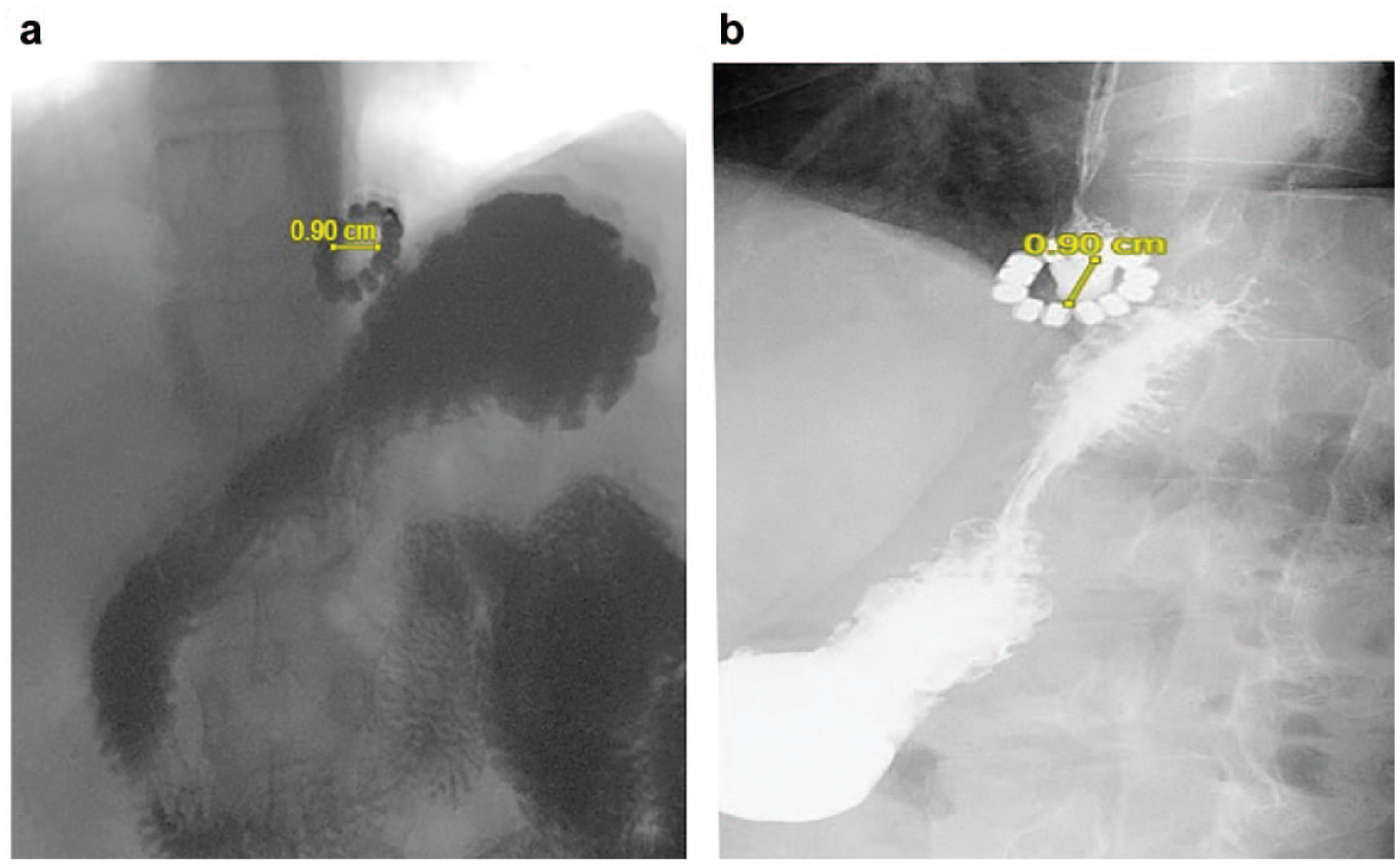
Preoperative esophagram demonstrating a static LINX™ device (Ethicon, Raritan, NJ, USA). (a) Device during swallowing of contrast. (b) Device after completion of the swallow; measurements represent the inner diameter of the LINX on the short axis.
After reviewing the diagnostic testing, the case was reviewed at a multidisciplinary committee for benign esophageal disease, which featured a magnetic sphincter augmentation device expert regarding the LINX. Through discussion, the LINX was suspected to be resting in the “open” position. The hiatal hernia seen on manometry was believed to be too small to be the predominant cause of the patient’s symptoms. When the patient originally developed symptoms at age 20, he was not noted to have a hiatal hernia. Moreover, recurrence of the same symptoms at age 36 was attributed to the device malfunction. Dilation was suggested as an initial solution to break up the scar tissue to increase mobility. Despite serial dilations to 20 mm and a follow-up EGD, and a 2-month period of observation postdilation, no clinical improvement was seen.
Having failed more conservative measures, informed consent was obtained, and the patient underwent a robotic LINX removal, small hiatal hernia repair, Nissen fundoplication, and EGD (Supplemental Video 2). Similar to the 1-stage laparoscopic LINX removal outlined in the 2017 publication by Asti et al., 4 we performed a 1-stage robotic removal using a combination of monopolar and ultrasonic dissection to remove the device and all 13 beads. Intraoperatively, we found the device placed intra-abdominally, on the gastroesophageal junction, encased with a fibrous capsule. After removal, a robot-assisted Nissen fundoplication was performed. Postoperatively, the patient tolerated the procedure well.
In the following months since the removal surgery, the patient endorsed significant relief of symptoms. The patient did suffer from a brief period of dysphagia after an episode of overeating in the early postoperative period. However, with diet modification, all of the previous symptoms were found to have resolved by his 6-month follow-up visit.
Discussion
Since its first use in humans in 2008, through initial Food and Drug Administration approval in 2012, the LINX has risen in popularity as an alternative to fundoplication. 5 The proposed benefits of the device include a good safety profile and efficacy. 5 To date, the literature regarding the LINX mostly describes dysphagia and GERD as the common complications.4,6 Our case is unique in that we describe and postulate how capsular fibrosis can be a potential cause of device malfunction of the LINX. Not only does our case provide long-term follow-up after LINX implantation, but it also illustrates how fibrosis can lead to a recurrence of reflux and describes a successful removal of a LINX robotically.
At a 5-year follow-up, Ganz et al. 6 reported that although 7 patients had the LINX explanted, only 1 did so due to persistence of GERD. The same study endorsed that no malfunctions of the device occurred in the 85 patients who were followed. The study also suggested that 98% of patients were cured of moderate or severe reflux at the 5-year interval. To date, previous papers4,6 have provided 5-year follow-up for patients or described explantation of the LINX after 5 years of therapy. Our case is an example of a patient with an 8-year follow-up duration prior to relapse.
The notion that capsular fibrosis hinders the mobility of the LINX is antagonistic to the initial feasibility porcine study, which demonstrated how individual beads were able to approximate. Despite the presence of capsule fibrosis after 40 weeks on implantation, the authors postulated that there was little significance. 7 The subjects in the initial study also maintained a higher LES pressure despite the fibrosis. Conversely, our patient had a lower LES pressure at the time of LINX failure. When looking at the static LINX on the esophagram (Fig. 1) coupled with the fibrosis seen in the operation, we theorize that over time, the foreign-body reaction causes an accumulation of capsular fibrosis. Once a threshold is surpassed, the fibrosis surrounding the device can force the device to be paradoxically “open,” thus allowing reflux to occur. Overall, we believe the dynamic nature of the LINX promotes continued capsular growth, which over time may lead to a device malfunction.
The original studies looking at LINX complications often compared LINX to the Angelchik device, an esophageal collar popular in the 1980s, 8 and the gastric band, a gastric collar for weight loss. 4 Some authors postulated that either technical error (band sizing) or fibrosis would lead to stenosis of the esophagus, which would cause dysphagia, stricturing, or erosion. 8 Subsequent follow-up studies have shown that although dysphagia is a common complaint, stricturing and erosion are rare. 5 This is likely due to the ability of the LINX to expand and retract, unlike the previous devices. It is possible that due to the dynamic nature of the LINX, clinically significant fibrosis takes longer to develop than previously described.7,8 Since our patient experienced success for 8 years, it is possible to expect future complications from longer-term use of the LINX.
Although LINX removal has been described in the literature, our case is the first to suggest capsular fibrosis as the predominant cause of failure. While this is only 1 case and 1 hypothesis, causes of device failure are likely multifactorial. Recurrence of symptoms may also involve components of device sizing, dysmotility, or pseudoachalasia. 8 Ultimately, our opinion is that if fibrosis plays a larger role in magnetic sphincter augmentation device failure, future research could generate alternative techniques to combat the problem.
Supplemental Material
sj-pdf-1-inv-10.1177_15569845241266245 – Supplemental material for Capsular Fibrosis as a Suggested Cause of Failure of Magnetic Sphincter Augmentation
Supplemental material, sj-pdf-1-inv-10.1177_15569845241266245 for Capsular Fibrosis as a Suggested Cause of Failure of Magnetic Sphincter Augmentation by Pamela Emengo, Daniel Nicastri and John Jacob in Innovations
Footnotes
Acknowledgements
We thank Dr. Gillian for his technical expertise and guidance in LINX troubleshooting and removal.
Declaration of Conflicting Interests
The authors declared no potential conflicts of interest with respect to the research, authorship, and/or publication of this article.
Funding
The authors received no financial support for the research, authorship, and/or publication of this article.

Supplemental Material
Supplemental material for this article is available online.
References
Supplementary Material
Please find the following supplemental material available below.
For Open Access articles published under a Creative Commons License, all supplemental material carries the same license as the article it is associated with.
For non-Open Access articles published, all supplemental material carries a non-exclusive license, and permission requests for re-use of supplemental material or any part of supplemental material shall be sent directly to the copyright owner as specified in the copyright notice associated with the article.
