
Abstract
Introduction
Traumatic brachial plexus injuries most commonly affect healthy young individuals. A number of treatment options are available, including nerve repair, nerve graft, and nerve transfer. In a literature review, Garg and colleagues demonstrated excellent outcomes associated with nerve transfers, with 96% of patients with nerve transfers achieving elbow flexion of M3 or greater, and 74% of patients with dual nerve transfers achieving shoulder abduction of M4 or greater. 3 These findings mirror those of a number of small series that present excellent outcomes for nerve transfers, specifically the triceps to axillary nerve transfer1,5,7 and the ulnar to musculocutaneous nerve transfer.6,8,9 Despite these positive findings, the literature continues to correlate increased age with poor outcomes for brachial plexus reconstructions.2,4,10-12
We present a 74-year-old male who was presented with brachial plexus palsy after a ski accident. Triceps to axillary nerve transfer, spinal accessory to suprascapular nerve transfer, and ulnar to musculocutaneous nerve transfer were performed with excellent functional outcome.
Case Report
A previously healthy and active 74-year-old right-hand dominant retired physician was injured in a life-threatening skiing accident. He sustained a head injury, fractures of the mandible, left radius and left upper ribs, a pneumothorax, phrenic nerve injury, and a deep laceration on the neck. On regaining consciousness, he was noted to have a severe left brachial plexus palsy. Early treatment for his left upper extremity injury included open reduction and internal fixation of his left radius fracture and extensive physical therapy to maintain passive range of motion.
The patient was presented to our facility 2 months post-injury having noted little-to-no return of proximal muscle function in the left upper extremity. Physical examination revealed marked disuse atrophy of the left parascapular musculature, the deltoid, biceps, brachialis, and brachioradialis. Passive range of motion demonstrated a 20° loss of passive shoulder abduction but full passive internal and external rotation. Gross motor testing revealed no motor function in the supraspinatus, infraspinatus, and deltoid, and only a flicker of contraction in the biceps brachialis and brachioradialis. Triceps and function distal to the elbow was otherwise normal. Sensory examination demonstrated absence of light touch in the axillary nerve distribution and dysesthetic sensation in the lateral antebrachial cutaneous nerve distribution. Sensation in the median and ulnar nerve distributions was normal. The working diagnosis was an upper trunk injury and possible C5 root avulsion.
Review of an outside magnetic resonance imaging (MRI) demonstrated a large amount of artifact about the supraclavicular brachial plexus with inadequate visualization of the rootlets. Computed tomography (CT) myelography at our institution demonstrated marked degenerative disease, foraminal stenosis with osteophytes, and cord compression at the C5 and C6 levels, but no evidence of root avulsion.
Electrodiagnostic examination demonstrated complete denervation of all muscles innervated by the upper trunk, but normal trapezius, rhomboid, and serratus function. The patient was recommended to return for a follow-up clinical and electromyographic study in 5 weeks to rule out any further recovery, but understood surgery would likely be necessary. At clinical follow-up 13 weeks post-injury, there was no recovery of shoulder abduction, external rotation, or elbow flexion. Triceps and all musculature distal to the elbow were strong. Electrodiagnostic studies demonstrated no conductivity or electromyographic function at any C5 or C6 innervated muscle distal to the serratus. There was incomplete denervation of the triceps with good motor unit preservation, decreased recruitment, and terminal collateral sprouting. Given the lack of electromyographic and clinical recovery, brachial plexus reconstruction was recommended and scheduled.
At surgery 16 weeks post-injury, the supraclavicular plexus was explored, and a large neuroma of the upper trunk was excised. The C5 and C6 roots, though physically intact, were not thought to be ideal candidates for long nerve graft reconstruction because of the severe foraminal compression noted on the CT myelogram. Spinal accessory to suprascapular nerve transfer was performed, followed by ulnar to biceps motor branch nerve transfer as described by Oberlin et al. 8 The patient was subsequently repositioned in lateral decubitus, and a long-head triceps-to-axillary nerve transfer was performed through a posterior approach. 5
At 6 months, he reported return of elbow flexion, and at 8 months, he had return of shoulder abduction. By 14 months post-op, he had regained the ability to touch his opposite shoulder and could actively abduct to 40°. Electrodiagnostic studies showed evidence of axonal regeneration and nascent potentials in the biceps, deltoid, infraspinatus, and supraspinatus. At 3 years post-op, he could abduct to 90° with strong resistance, limited only by arthritis in the glenohumeral joint. He could externally rotate to neutral against resistance, again only limited by glenohumeral arthritis. His shoulder abduction had recovered Medical Research Council (MRC) grade 4 strength, as had his biceps, which could flex to 140°. At 9 years post-op, his shoulder abduction retained M4 strength, and his elbow flexion strength had increased to M4+; however, his glenohumeral arthritis had progressed, limiting abduction to 65°. At 11 years post-op, he could abduct to 65° and forward flex at M4 strength, limited only by painful arthritis (Figure 1). Elbow flexion was M5− at both the biceps and brachialis, and bulk and tone were nearly symmetrical with the opposite side (Figure 2). Eleven-year electrodiagnostic studies demonstrated further improved motor unit recruitment in the supraspinatus, infraspinatus, and posterior deltoid. Following return of shoulder and elbow function, the patient returned to skiing recreationally (Figure 3).
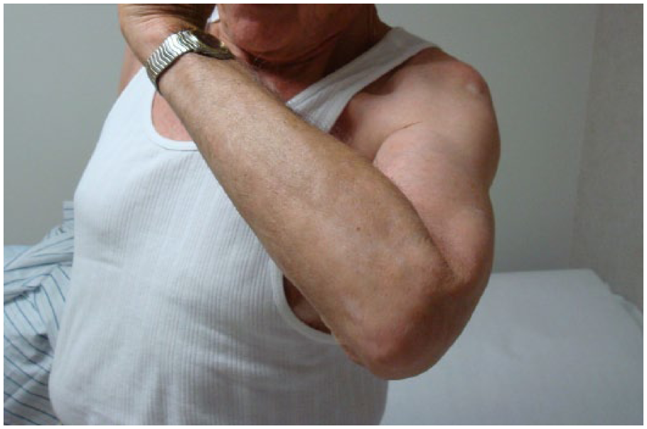
At 11 years post-op, the patient could abduct to 65 degrees and forward flex at M4 strength, limited only by painful arthritis.
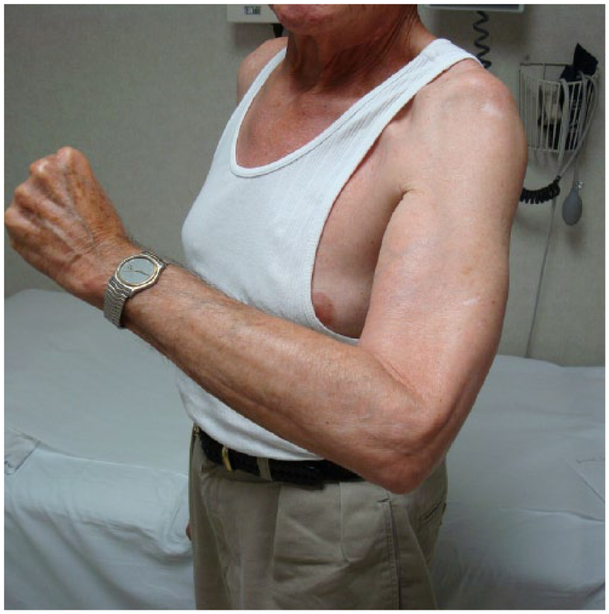
At 11 years, elbow flexion was M5− at both the biceps and brachialis, and bulk and tone were nearly symmetrical with the opposite side.
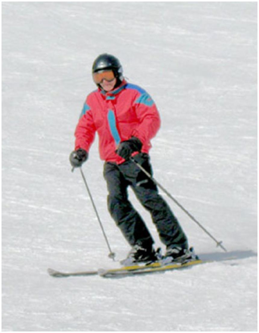
Following return of shoulder and elbow function, the patient returned to skiing.
Discussion
A number of case series have demonstrated poor outcomes for nerve repair and reconstruction in elderly patients. In an analysis of 194 musculocutaneous nerve reconstructions, patients younger than 20 demonstrated significantly superior biceps/brachailis power than those older than 40. 11 In a review of 146 axillary nerve repairs (age range, 9-72 years), Bonnard and colleagues demonstrated a statistically significant downward trend in the proportion of successful outcomes with increasing age. Eighty-three percent of patients younger than 20 regained M4 deltoid strength or better, compared with only 63% of those older than 35 and 61% of those older than 40. 2 Similarly, in a retrospective study of 33 axillary nerve repairs by Wehbe et al, 8 of 14 patients younger than 25 had favorable results compared to only 8 of 19 older than 25. 12 In 2009, Terzis et al looked at axillary nerve reconstructions in posttraumatic plexopathy patients and found that people younger than 20 years obtained superior deltoid power (mean = M3.18) than those older than 20 years (mean = M2.70). 10
Poor outcomes have also been associated with older patients receiving nerve transfers. In 2012, Lee et al found that deltoid strength after triceps to axillary nerve transfer correlated negatively with the age of the patient (age range, 16-79 years). Eleven of 12 patients aged 39 years or younger had successful results, compared with only 5 of 9 aged 40 years or older. Four of the 5 patients who did not achieve M3 abduction were older than 50, and no patients older than 50 years regained useful deltoid strength (M3 or greater). The study included 2 patients above 70, aged 72 and 79, who each recovered M2 and 20° and 50° of shoulder abduction, respectively. 4 It has been suggested that this trend may be related to a decreased capacity for nerve regeneration in older patients. 2
There are very little data available to evaluate the outcomes of nerve transfers in patients older than 70. We can attribute our patient’s outcome to several factors. Time from injury to surgery was only 16 weeks. Although age has been identified as an independent risk factor in the literature, shorter time from injury to surgery is well established as one of the most important factors in determining outcomes of nerve repairs.2,4,10 Furthermore, the patient was an avid skier and was otherwise in excellent health, with a body mass index of 20, which Lee et al associate with superior outcomes. 4 This case questions the widely held dogma that older patients who receive nerve transfers fare poorly. We do not advise routinely withholding nerve transfers for healthy older patients with traumatic brachial plexus palsy. A larger scale, multi-center approach may help to elucidate nerve transfer outcomes in older patients.
Footnotes
Ethical Approval
This study was approved by our institutional review board.
Statement of Human and Animal Rights
All procedures followed were in accordance with the ethical standards of the responsible committee on human experimentation (institutional and national) and with the Helsinki Declaration of 1975, as revised in 2008.
Statement of Informed Consent
Informed consent was obtained from all patients for being included in the study.
Declaration of Conflicting Interests
The authors declared no potential conflicts of interest with respect to the research, authorship, and/or publication of this article.
Funding
The authors received no financial support for the research, authorship, and/or publication of this article.