
Abstract
Keywords
Introduction
Purpura fulminans (PF) is a rare condition characterized by rapidly progressive skin necrosis and disseminated intravascular coagulation (DIC). It presents with symmetric purpuric lesions commonly located in lower and upper extremities. Thrombotic occlusion of small and medium-sized vessels can also affect end organs leading to multi-organ failure (MOF) and death. PF can occur secondary to a severe coagulopathy, viral infection, or an acute sepsis. It is most frequently seen in pediatric subset of patients in the context of meningococcemia, but there are reports showing it can occur at any age. 4
There is no specific treatment for this condition. Advances in early recognition and aggressive supportive management have helped decrease the mortality rate, but more than 50% of these patients must undergo surgical debridement with partial or total limb amputations. Evidence about timing of surgical debridement is controversial. Some studies had favored early surgical treatment, but recent evidence shows that this does not necessarily correlate with greater survival rates. In some cases, it is best to delay surgical debridement until necrosis demarcation to maintain maximum limb length.3,7 These studies must be interpreted with caution, as they are small studies with a few number of patients. Most surgical reconstructive procedures are described for pediatric patients, with few of them addressing the role of surgery in adult and elderly population. These patients may have a lower ability to overcome aggressive procedures and require a different approach. The goal in these patients is to preserve functionality without causing a new systemic inflammatory insult.
We report a case of an 86-year-old patient diagnosed with PF who was successfully treated with delayed debridement, partial amputation, and finger salvage with semiocclusive dressings.
Case Report
An 86-year-old diabetic patient, with a nonalcoholic fatty liver disease, was admitted to the intensive care unit with the diagnosis of sepsis secondary to cholangitis. Initial laboratory workup showed positive blood cultures to multi-sensitive Escherichia coli and Enterococcus gallinarum. Coagulation studies only revealed an increase of the international normalized ratio of 2.2.
He was initially managed with ventilatory assistance, vasoconstrictive agent (norepinephrine, maximum infusion rate 1.3 µg/kg/min) requirements and antibiotic therapy with ceftriaxone and metronidazole. During the following days, his condition continued to worsen and he developed purpuric lesions in his 4 extremities (Figure 1). Upon evaluation by vascular surgery, doppler ultrasound revealed no radial artery blood flow at the level of the wrist. Further investigation with plethysmography demonstrated no signal within any of his fingers. Dermatology and orthopedic consultations diagnosed PF.
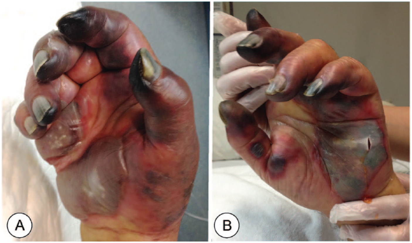
Initial presentation of hand lesions, 1 week after admission.
He underwent platelet transfusions, heparin administration, high-volume hemofiltration, and hemodialysis for 13 days. Surgical debridement was delayed until delimitation of necrosis. No intra-compartment measurements were performed, as there were no clinical signs of compartment syndrome during the patients evolution.
One month after admission, demarcation of skin necrosis in his hands demonstrated autoamputation of multiple digits (Figures 2A and 2B) and only superficial involvement of his lower extremities. Surgical debridement was performed, with revision amputations to his left index finger and middle finger, and his right thumb, middle finger, and ring finger. Due to his comorbidities, no flap coverage was performed and instead semiocclusive dressing was used to cover his left thumb, ring finger, and little finger. Two simple Tegaderm films were used to cover the fingers dorsal and volarly, without compression (Figures 2C and 2D). Dressing changes were performed routinely to the amputation sites in 48-hour intervals. Semiocclusive dressings were not removed until 6 weeks in the thumb, ring finger, and little finger (Figure 3).
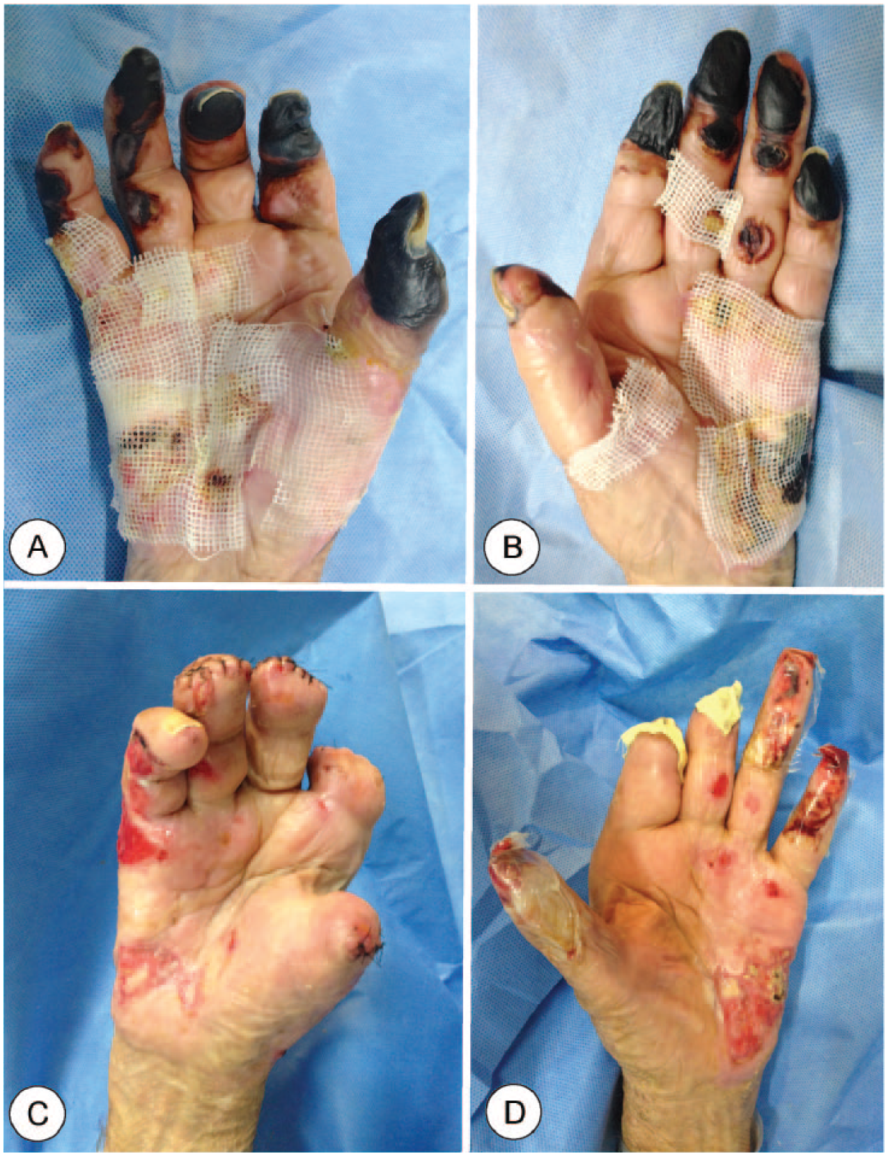
Evolution of skin lesions 4 weeks after admission.
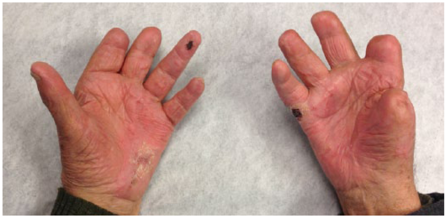
Physical examination at follow-up.
Two months after admission he was discharged. One of his amputation sites developed a minor infection and recovered with antibiotic treatment.
Discussion
PF presents as a sudden onset of painful cutaneous lesions characterized by well-defined erythematous macules and purpuric patches, with vesicle and bulla formation. PF distinguishes from other purpuric entities by a rapid progression of skin lesions to irregular areas of central blue-black hemorrhagic necrosis and a surrounding erythematous border. Most patients have severe systemic involvement, shock, and MOF. Mortality rates vary from 20% to 50%, and 25% of patients undergo 4 limb amputations. 9 There is no known incidence or prevalence of this condition.
Three forms of PF are described, including (1) neonatal (severe form of hereditary deficiency of protein C and S), (2) idiopathic or chronic (caused by a latent viral illness), and (3) acquired (commonly associated with a severe infection causing a consumption coagulopathy). Acquired or infectious PF generally presents with a premonitory illness, which later develops into systemic compromise, DIC, and soft tissue necrosis involving distal extremities and end organs. The pathogenesis of sepsis-induced PF is not completely understood, but endothelial cell injury and cell dysfunction appear to play a critical role.
Treatment of infectious PF is based on supportive management in an intensive care unit with ventilator assistance, aggressive fluid resuscitation, vasopressors, blood product transfusions, and an early recognition of the underlying cause. Evidence of specific therapies for PF is being studied. Fresh frozen plasma is used in sepsis, but may be insufficient in DIC. It is recommended to add platelet concentrate and cryoprecipitate transfusions for severe thrombocytopenia (platelet count <50°000/µL) and hypofibrinogenemia (fibrinogen concentration <1 g/dL). 1 Heparin infusion showed a 12% mortality reduction in sepsis and infectious DIC patients. 12 Recombinant human soluble thrombomodulin has shown promising evidence for patients with sepsis-induced coagulopathy without increasing the risk of bleeding. 11 Evidence of the use of activated protein C and high-dose antithrombin have failed to show a decrease in the mortality rates and are not currently administered.8,10 The use of vasopressor drugs itself has shown to increase the risk of acral necrosis, especially in patients with sepsis, DIC, autoimmune vasculitis, and endocarditis, among others. These drugs must be used with caution under these circumstances.
During the past years, improvement in survival rates has helped change the role of surgery to one that focuses in preserving and restoring functionality for PF patients. Early interventions and prophylactic fasciotomies have not shown to prevent loss of the extremities. 3 It appears that only in patients with compartment syndrome, fasciotomies are able to restore blood flow. In most of cases, loss of circulation is secondary to thrombosis of small vessels. There is no correlation between survival rates and preservation or sacrifice of a limb. This is why surgical debridement is delayed until necrosis delimitation. Salvage of 1 limb makes the difference to these patients with multiple amputations. 7 It is important to consider that up to 7% of PF patients can present with compartment syndrome. Clinical screening and measurement of intracompartment pressures are recommended, when there is high clinical suspicion. 2
Reconstructive strategies for pediatric patients have shown good results, including skin grafts, local flaps, and free flap coverage. There are few reports of adult patients with PF. Most of them focus on medical management of this condition, without addressing sequelae. These patients often have multiple comorbidities, immunosuppression, and lower response to systemic injury. Therefore, surgical management must be adjusted to avoid triggering a second inflammatory hit. In these patients, delayed surgical debridement is crucial, with amputation being limited only to evident zones of deep necrosis.
Management of fingertip injuries with an artificial barrier created with semiocclusive material has been described in several publications. 5 Recent evidence has proven it is safe even in fingertip amputations with exposed bone. 6 In our patient, the use of semiocclusive dressings in 3 of his digits allowed for healing without shortening. No complications were observed.
Our case illustrates the importance of early recognition of infectious PF, its underlying cause, and timely supportive management with antibiotic treatment. It also illustrates how delayed surgical intervention avoids an early added operative insult to a critical patient. He also favored from delimitation of necrosis, which allowed for less aggressive resection and a good functional outcome.
Footnotes
Ethical Approval
This study was approved by our institutional review board.
Statement of Human and Animal Rights
The procedures followed were in accordance with the ethical standards of the responsible committee on human experimentation (institutional and national) and with the Helsinki Declaration of 1975, as revised in 2008.
Statement of Informed Consent
The authors have omitted any identifying information in this case report to the reported patient.
Declaration of Conflicting Interests
The author(s) declared no potential conflicts of interest with respect to the research, authorship, and/or publication of this article.
Funding
The author(s) received no financial support for the research, authorship, and/or publication of this article.