
Abstract
Introduction
Carpal tunnel syndrome (CTS), or compression of the median nerve at the wrist, is the most commonly diagnosed peripheral compression neuropathy occurring in an estimated 1% of the general population. 1 Operative release is generally recommended for more severe nerve compression. 1 Typically, these are cases that have either failed conservative measures or present with moderate or severe changes on nerve conduction studies. Open carpal tunnel release (OCTR) of the transverse carpal ligament for the treatment of CTS has been widely used since 1951 and is considered the criterion standard. 2 However, over the past 2 decades, endoscopic carpal tunnel release (ECTR) has been shown to have equivalent efficacy and more rapid recovery. There is some evidence for decreased minor complications with endoscopic techniques, such as pillar pain and wound dehiscence. However, it should be noted that a higher risk of major complications such as nerve injury exists with endoscopic techniques.3-5 Regardless of the technique used, surgical division of the transverse carpal ligament allows for the restoration of median nerve function by lowering the intracarpal canal pressure through decompression. 6
The mechanism by which the carpal canal’s morphology changes following both OCTR and ECTR has been previously studied through the use of various imaging modalities, although few have compared the 2 methods directly in a single study. Gartsman et al 7 and Viegas et al 8 used plain radiographs to assess the morphologic changes of OCTR and ECTR, respectively. Richman et al 4 analyzed the outcomes of OCTR through magnetic resonance imaging (MRI), whereas Ablove et al, 9 Kato et al, 10 and Momose et al 11 used MRI to evaluate ECTR. Aslani et al 12 compared ECTR and OCTR outcomes using computed tomography. All the above studies concluded that carpal tunnel release (CTR), regardless of the technique used—open or endoscopic—results in an increase in carpal canal volume. However, 2 different mechanisms through which this change occurs have been presented. Some authors believe the volumetric increase to be a result of anteroposterior (AP) expansion, whereas others claim it is as a result of medial-lateral (radioulnar) widening of the bony carpal arch. Our study will determine through MRI analysis the morphologic changes that occur in both OCTR and ECTR, thereby clarifying any differences that exist between the 2 operative techniques. We hypothesize that there will be no significant difference in the morphologic changes seen following ECTR and OCTR.
Materials and Methods
Nineteen patients with CTS were enrolled into the study from October 2004 to February 2005. All patients failed an appropriate trial of conservative therapy (3-6 months) with wrist splints and activity modification. All patients in the study had both a clinical and an electrodiagnostic diagnosis of CTS. All patients had electrodiagnostic findings of moderate to severe CTS. This study was performed with 2 surgeons, one who performs all of his CTRs with an endoscopic technique and one who performs all of his CTRs with an open technique. Eleven patients underwent single portal ECTR (Agee technique), 13 and 8 patients underwent longitudinal short scar OCTR (no repair of palmar fascia). These patients were not randomized. They were sequentially selected from each surgeon’s practice if they had a clinical and electrical diagnosis of moderate to severe CTS and had failed conservative therapy and were willing to participate in the study. Postoperatively, all patients were placed in a bulky dressing of gauze and kerlix with a small palmar-based wrist splint that was worn for 2 weeks with the digits free. No patients were sent to physiotherapy postoperatively, and all were allowed to move immediately postoperatively in their wrist-based splint. Exclusion criteria included pregnancy, inflammatory arthropathy, previous wrist surgery, contraindications for MRI, mental illness, and an unstable home address.
Magnetic resonance imaging studies were performed preoperatively and 6 months postoperatively on treated and nontreated wrists using a knee coil (General Electric 1.5 Tesla MRI Scanner). Imaging and analysis protocols for carpal tunnel analysis followed well-defined published guidelines.14-16 Measurements obtained by MRI analysis included completion of transverse carpal ligament release, volume of carpal tunnel, diameter (carpal arch width) of carpal tunnel, median nerve to carpus distance, intertendinous distance, Guyon’s canal diameter (transverse distance), and Guyon’s canal AP distance.
The proximal border of the carpal canal was defined by appearance of the flexor carpi radialis tendon lying outside the carpal canal palmar to the distal pole of the scaphoid. The distal border of the carpal canal was defined by the radial divergence of the flexor pollicis longus tendon from the other flexor tendons. All computer-assisted measurements were completed by a single observer.
The volume within a 3-cm segment of carpal canal was measured for both hands of each patient (treated and untreated) both preoperatively and 6 months postoperatively. Carpal tunnel diameter was measured from the hook of the hamate to the beak of the trapezium (Supplemental Figure 1). The carpal arch angle was measured as the intersection of a line between the palmar ulnar and dorsal radial poles of the trapezium and a line drawn through the hook of the hamate (Supplemental Figure 1). Median nerve volar migration was measured as the difference in the distance preoperatively and postoperatively between the epicenter of the median nerve and the carpus (Supplemental Figure 2). The intertendinous distance was measured at the same level as was for median nerve volar migration and carpal tunnel diameter to determine whether the tendons diverge or converge as a result of the procedure (Supplemental Figure 2). Paired t tests were used to test preoperative and postoperative differences in the same patient, and Student t tests were used to test for differences between groups.
Results
Postoperatively, all patients stated resolution of their compression neuropathy. On MRI, the transverse carpal ligament was released completely in all patients in both the OCTR and the ECTR group. On imaging, no other abnormal findings were found that may have contributed to median nerve compression symptoms.
Volume of the Carpal Tunnel
At the 6-month postoperative interval, there was a significant increase in the volume of the carpal canal compared with preoperative values for both the OCTR and the ECTR group (Table 1). Canal volume in the treated hands of the OCTR group was 7.55 ± 1.66 cm3 preoperatively and 8.69 ± 1.66 cm3 postoperatively, resulting in a 15% increase in canal volume. Canal volume in the treated hands of the ECTR group was 7.06 ± 1.0 cm3 preoperatively and 7.90 ± 1.0 cm3 postoperatively, resulting in an 11.9% increase in canal volume. There was a significant difference between preoperative and postoperative volume, regardless of the technique used (P = .0001), and no significant difference in postoperative volume between open and endoscopic release (P = .2120).
Comparison of the Preoperative and Postoperative Volume of the Carpal Canal in Both OCTR and ECTR.
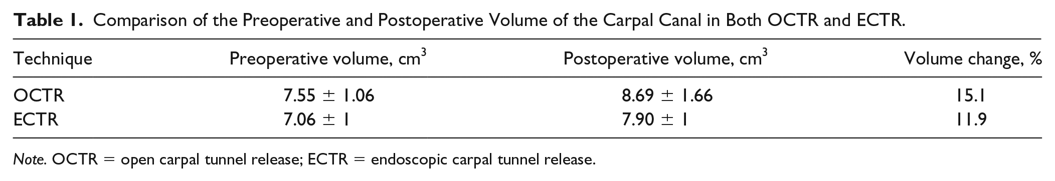
Note. OCTR = open carpal tunnel release; ECTR = endoscopic carpal tunnel release.
Transverse Distance of Carpal Tunnel
At the 6-month postoperative interval, there was no significant change in the transverse diameter of the carpal tunnel compared with preoperative values for both the OCTR and the ECTR group (Table 2). Transverse distance of the canal in the OCTR group was 21.77 ± 2.08 mm preoperatively and 22.58 ± 2.73 mm postoperatively, resulting in a 3.7% increase in transverse carpal canal diameter (P = .076). Transverse distance of the canal in the ECTR group was 21.34 ± 1.98 mm preoperatively and 21.83 ± 1.92 mm postoperatively, resulting in a 2.2% increase in transverse carpal canal diameter (P = .070). There was no significant difference between preoperative and postoperative distance, regardless of the technique used, and no significant difference in postoperative transverse carpal canal distance between open and endoscopic release (P = .259).
Comparison of the Preoperative and Postoperative Transverse Distance of the Carpal Canal in Both OCTR and ECTR.
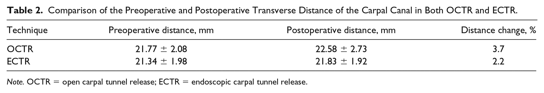
Note. OCTR = open carpal tunnel release; ECTR = endoscopic carpal tunnel release.
Median Nerve to Carpus Distance (AP Distance)
At the 6-month postoperative interval, there was a significant increase in the median nerve to carpus distance compared with preoperative values for both the OCTR and the ECTR group (Table 3). The AP distance in the treated hands of the OCTR group was 9.70 ± 1.87 mm preoperatively and 12.37 ± 3.70 mm postoperatively, resulting in a 27.5% change in AP distance (P = .009). The AP distance in the treated hands of the ECTR group was 10.04 ± 1.14 mm preoperatively and 11.18 ± 1.59 mm postoperatively, resulting in an 11.3% change in AP distance (P = .001). There was a significant difference between preoperative and postoperative AP distances, regardless of the technique used, and no significant difference in postoperative AP distance between open and endoscopic release (P = .065)
Comparison of the Preoperative and Postoperative AP Distance of the Carpal Canal in Both OCTR and ECTR.
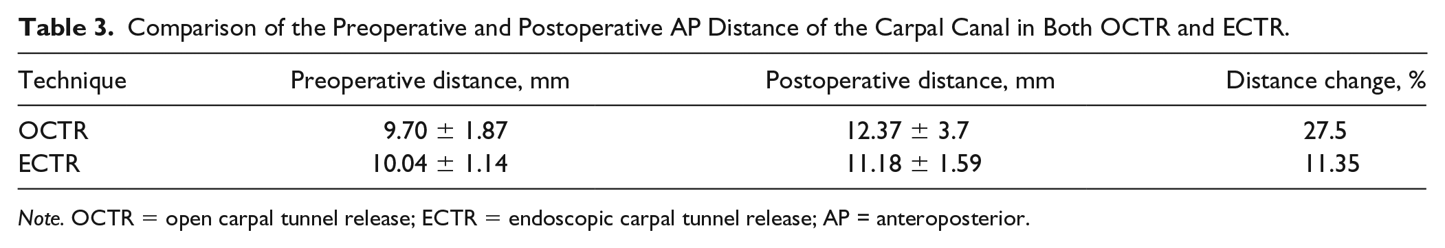
Note. OCTR = open carpal tunnel release; ECTR = endoscopic carpal tunnel release; AP = anteroposterior.
Intertendinous Distance (Divergence of Tendons)
At the 6-month postoperative interval, there was no significant change in the intertendinous distance compared with preoperative values for both the OCTR and the ECTR group (Table 4). Intertendinous distance in the treated hands of the OCTR group was 19.93 ± 1.67 mm preoperatively and 20.05 ± 1.82 mm postoperatively, resulting in a 0.6% change in distance (P = .475). Intertendinous distance in the treated hands of the ECTR group was 20.0 ± 1.97 mm preoperatively and 20.7 ± 2.14 mm postoperatively, resulting in a 3.5% change in distance (P = .180). There was no significant difference in postoperative intertendinous distance between open and endoscopic release (P = .25).
Comparison of the Preoperative and Postoperative Carpal Canal Intertendinous Distance in Both OCTR and ECTR.
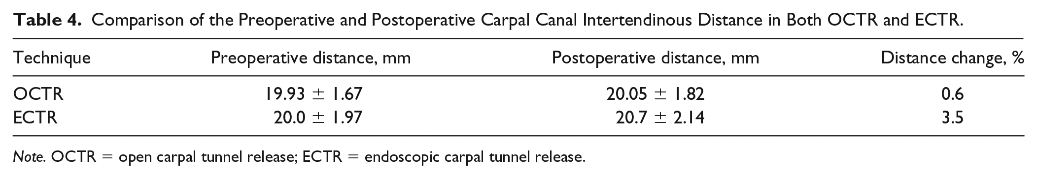
Note. OCTR = open carpal tunnel release; ECTR = endoscopic carpal tunnel release.
Guyon’s Canal Transverse Distance
At the 6-month postoperative interval, there was no significant change in the transverse distance of Guyon’s canal compared with preoperative values for both the OCTR and the ECTR group (Table 5). Guyon’s canal transverse distance in the treated hands of the OCTR group was 9.70 ± 1.50 mm preoperatively and 9.55 ± 1.82 mm postoperatively, resulting in a −1.56% change in distance (P = .967). Guyon’s canal transverse distance in the treated hands of the ECTR group was 8.49 ± 0.92 mm preoperatively and 8.42 ± 1.19 mm postoperatively, resulting in a −0.8% change in distance (P = .800). There was no significant difference between preoperative and postoperative Guyon’s canal transverse distances, regardless of the technique used, and no significant difference in postoperative Guyon’s canal transverse distance changes between open and endoscopic release (P = .92).
Comparison of the Transverse Distance of Guyon’s Canal in OCTR and ECTR.
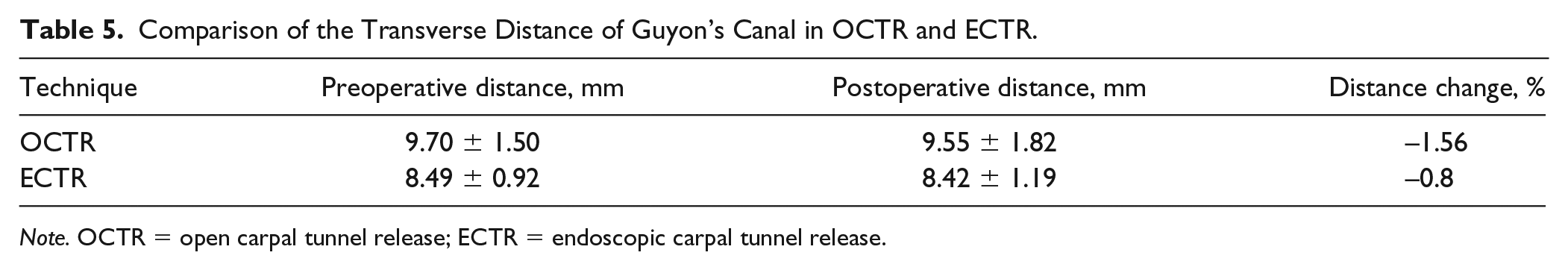
Note. OCTR = open carpal tunnel release; ECTR = endoscopic carpal tunnel release.
Guyon’s Canal AP Distance
At the 6-month postoperative interval, there was a significant difference in the Guyon’s canal AP distance compared with preoperative values for both the OCTR and the ECTR group (Table 6). Guyon’s canal AP distance in the treated hands of the OCTR group was 5.93 ± 0.67 mm preoperatively and 7.35 ± 0.91 mm postoperatively, resulting in a 23.9% change in distance (P = .009). Guyon’s canal AP distance in the treated hand of the ECTR group was 5.40 ± 0.97 mm preoperatively and 6.43 ± 1.01 mm postoperatively, resulting in a 19.1% change in distance (P = .0003). A paired t test showed a significant difference between preoperative and postoperative Guyon’s canal AP distances, regardless of the technique used, and no significant difference in postoperative distance between open and endoscopic release (P = .327).
Comparison of the AP Distance of Guyon’s Canal in OCTR and ECTR.
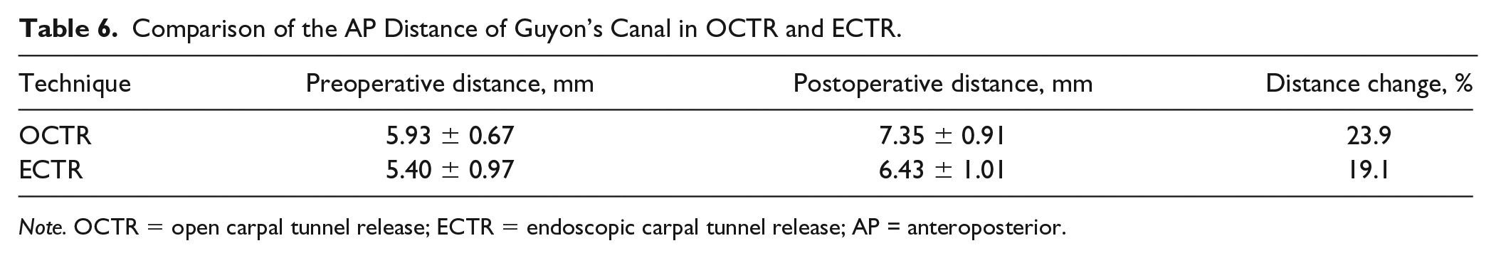
Note. OCTR = open carpal tunnel release; ECTR = endoscopic carpal tunnel release; AP = anteroposterior.
Discussion
Endoscopic carpal tunnel release and OCTR are the most common procedures used to resolve the symptoms of CTS when patients have failed conservative management. Various studies have defined the outcomes of ECTR and OCTR as clinically equivalent for subjective symptom relief and morphologic changes of the median nerve and carpal canal.9,12,17 Other studies have reported that ECTR is a superior method to OCTR, as endoscopic technique results in a shorter recovery time than OCTR.3,18
As discussed previously, prior studies have been unable to obtain clearly reproducible results regarding the mechanism by which decompression of the median nerve occurs. Gartsman et al 7 attributed the increase in carpal volume to carpal arch widening. Viegas et al 8 supported the same conclusion, claiming the volumetric increase resulting from ECTR was a result of the widening of the bony carpal arch, but was unable to comment on volumetric alterations or changes in the median nerve as these findings cannot be determined by plain film radiography. The results of Richman et al 4 support a different conclusion, claiming the increase in volume was a result of an increase in AP dimensions of the carpal canal in OCTR and described no changes to the carpal arch width. This conclusion is further supported by Ablove et al and Kato et al, who both studied ECTR with MRI. Both concluded that while ECTR did increase the carpal canal volume, no significant difference was found in the bony carpal arch width preoperatively and postoperatively.9,10 In 2014, Aslani et al 12 attributed the volumetric change associated with both OCTR and ECTR mainly to an increase in the AP distance of the canal and, to a lesser degree, an increase in carpal arch width. 12
Our study found a statistically significant postoperative increase in carpal canal volume, median nerve to carpus distance (AP carpal canal distance), and the AP distance of Guyon’s canal in both OCTR and ECTR. However, no significant change in intertendinous distance or the transverse distance of Guyon’s canal was shown in either technique. Furthermore, surgical release of the transverse carpal ligament resulted in no change in carpal canal transverse distance, contrary to the findings of Gartsman et al and Viegas et al. In addition, we found no significant postoperative differences between OCTR and ECTR for any of our parameters, suggesting that both techniques may result in an anatomically equivalent outcome. Our study suggests that it is in fact the AP distance of the carpal tunnel that increases, and it is this increased dimension that results in an increase in the volume of the carpal tunnel.
As noted above, we found that the AP dimension of Guyon’s canal also increases following CTR, regardless of the technique used. It is possible that patients at high risk of further peripheral nerve compression syndromes or who have a component of compression of the ulnar nerve at Guyon’s canal may actually derive some benefit at the level of the ulnar nerve from decompression of the carpal tunnel via release of the transverse carpal ligament.
Our study did not demonstrate any significant change in the intertendinous distance following CTR with either technique. There are some studies in the literature that demonstrate high rates of trigger fingers following CTR with the theory that release of the transverse carpal ligament changes the entrance angle of the flexor tendon to the A1 pulley, potentially predisposing to trigger finger.19,20 Although we assessed any change in intertendinous distance, we did not assess any change in the angle of entrance of the flexor tendons to the A1 pulley.
This study had several limitations. The number of subjects for both cohorts was small and there was no power analysis performed. It is possible that a morphologic difference exists between ECTR and OCTR that is clinically relevant, and our study is underpowered to detect it. There was a 4% difference in postoperative carpal tunnel volume increase between OCTR and ECTR. This was not statistically significant, although it is possible that it is clinically significant. In addition, the study was not randomized and included patients from 2 different surgeons. Follow-up was limited to a 6-month postoperative interval. Further follow-up study would be required to determine whether the morphologic changes to the carpal canal are maintained long term past the 6-month interval that we investigated. It should be noted that there are newer imaging modalities available, such as ultrasound techniques and MR neurography. The MR-based protocol used in this study represents the most up-to-date imaging protocol at our institution at the time that this study was performed.
We conclude that no significant differences in anatomical outcome appear to exist between ECTR and OCTR. Both surgical techniques result in a significant increase in the volume of the carpal tunnel secondary to an increase in the AP dimension. Furthermore, both ECTR and OCTR result in a significant increase in the AP dimension of Guyon’s canal (19% and 24%, respectively).
Supplemental Material
Supplemental_Figures – Supplemental material for Morphologic Analysis of the Carpal Tunnel and Median Nerve Following Open and Endoscopic Carpal Tunnel Release
Supplemental material, Supplemental_Figures for Morphologic Analysis of the Carpal Tunnel and Median Nerve Following Open and Endoscopic Carpal Tunnel Release by Blair R. Peters, Amanda M. Martin, Brett F. Memauri, Hardy W. Bock, Robert B. Turner, Kenneth A. Murray and Avinash Islur in HAND
Footnotes
Supplemental material is available in the online version of the article.
Ethical Approval
This study was approved by the Health Research Ethics Board, and informed consent was obtained from each patient.
Statement of Human and Animal Rights
This article does not contain any studies with human or animal subjects.
Statement of Informed Consent
Informed consent was obtained from all individual participants included in the study.
Declaration of Conflicting Interests
The author(s) declared no potential conflicts of interest with respect to the research, authorship, and/or publication of this article.
Funding
The author(s) disclosed receipt of the following financial support for the research, authorship, and/or publication of this article: This research was supported by a grant from the Health Sciences Centre Foundation.
References
Supplementary Material
Please find the following supplemental material available below.
For Open Access articles published under a Creative Commons License, all supplemental material carries the same license as the article it is associated with.
For non-Open Access articles published, all supplemental material carries a non-exclusive license, and permission requests for re-use of supplemental material or any part of supplemental material shall be sent directly to the copyright owner as specified in the copyright notice associated with the article.