
Abstract
The persistent carotid-vertebrobasilar anastomoses are arterial communications between the anterior and posterior circulations due to the persistence of embryological connections. We here present an extremely rare instance of a transclival persistent carotid-vertebrobasilar anastomosis in a 10-month-old infant, which does not fit into any of the traditionally described categories, such as the trigeminal artery, hypoglossal artery, or proatlantal artery.
Introduction
The persistent carotid-vertebrobasilar anastomoses are arterial communications between the anterior and posterior circulations due to the persistence of embryological connections,1,2 These connections manifest in several forms, each with varying degrees of frequency. The most common among them is the trigeminal artery, which originates at the expected location of the meningohypophyseal trunk. It follows a trajectory that is largely consistent with that of the trigeminal nerve, and may connect to the basilar artery or continues directly in a cerebellar artery. Another frequent occurrence is the hypoglossal artery, which is a regular vessel present in every human being. It typically originates from the neuromeningeal trunk of the ascending pharyngeal artery and travels along the hypoglossal canal. In certain instances, the hypoglossal artery may take over the supply to the whole or part of the posterior circulation, in which case it is referred to (inappropriately) as a “persistent” hypoglossal artery. The proatlantal artery is a hypertrophied connection between the occipital artery and the vertebral artery. It is worth noting that while this connection is present in a significant number of individuals, it only becomes the dominant contributor to the vertebrobasilar system in a select few. Another described entity is the otic artery, which has been reported in a few case reports. However, the majority of these reports are thought to be variants of the persistent trigeminal artery, rather than a distinct entity. 3
We herein describe an extremely rare transclival persistent carotid-vertebrobasilar anastomosis which defies classification within the traditional categories established thus far. In this report, we will delve into possible embryological hypotheses to explain this rare phenomenon.
Case report
The case involves a 10-month-old male with a history of a nasal skin mass, which was later proven to be a dermoid cyst. He underwent an MRI of the brain which revealed the presence of an enlarged vessel that coursed anterior to the vertebral bodies, connecting with the basilar artery by coursing through the right aspect of the inferior clivus in the occipital anterior synchondrosis, between the basioccipital and lateral occipital ossification centers (Figure 1). Further reconstructions confirmed that the vessel has a separate course from the hypoglossal canal, jugular foramen, or foramen magnum. Additionally, the hypoglossal canals and the jugular foramina were present and did not contain any abnormally enlarged vessels passing through them (Figure 2).
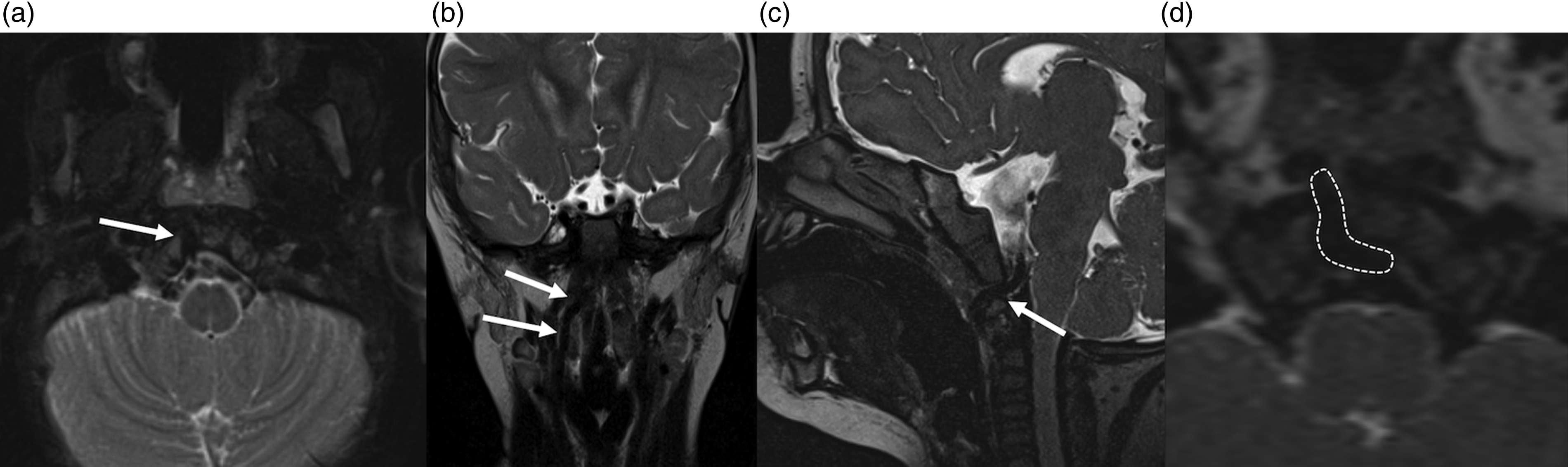
Axial T2 (a), coronal T2 (b), sagittal CISS (c), and axial reconstructions from CISS (d) showing the unusual course of the vessel supplying the basilar artery coursing in the anterior occipital synchondrosis (arrows in a, b, and c and dashed in d).
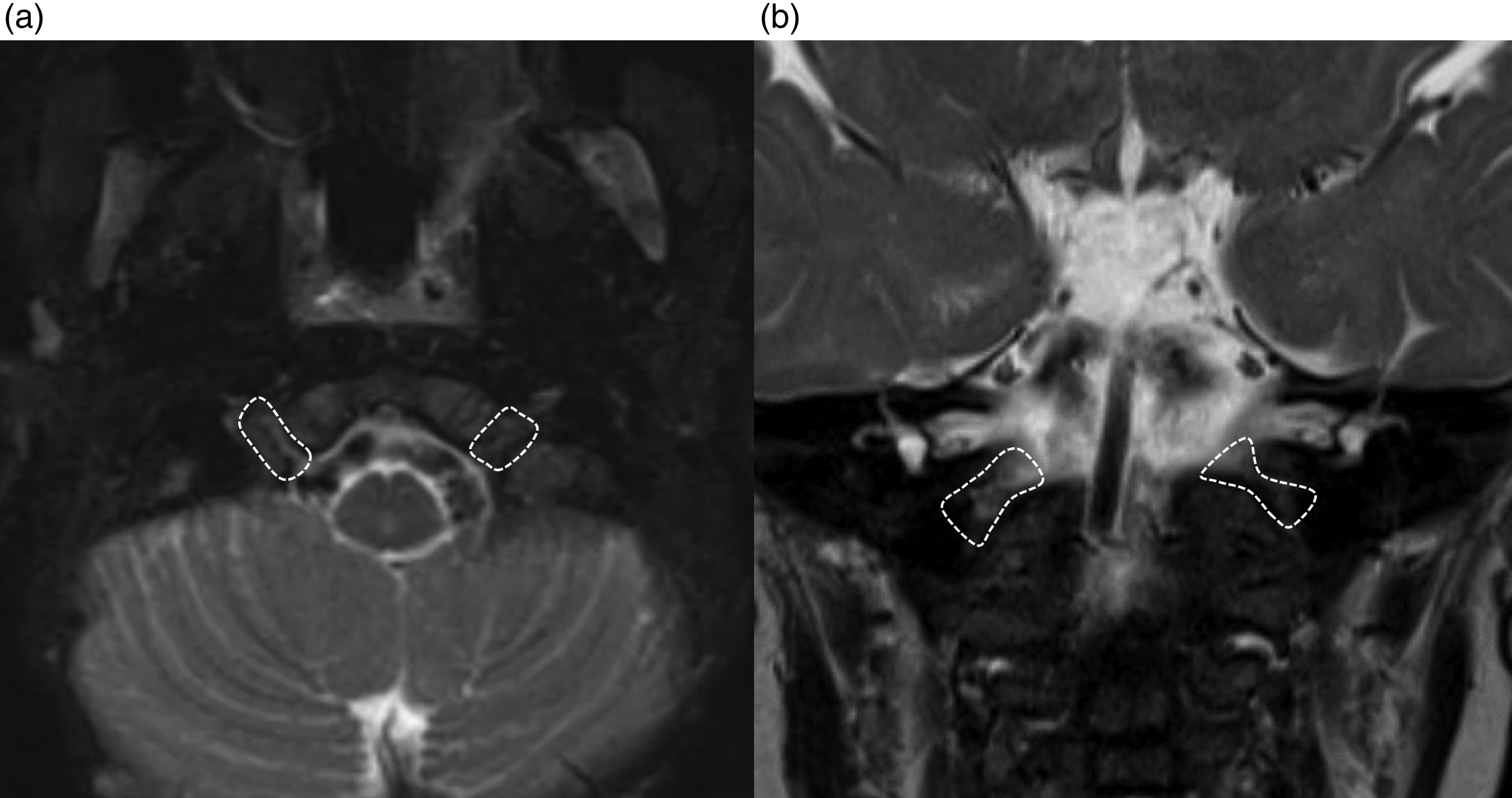
Axial T2 (a) and coronal T2 (b) images showing the normal hypoglossal canals (a) and jugular foramina (b) without abnormally enlarged vessels passing through them.
Discussion
This report demonstrates a very unusual pathway of blood flow to the basilar artery circulation through a vessel coursing through the clivus. This anatomical variant does not conform to the well-known established carotid-vertebrobasilar anastomoses such as the proatlantal artery, which passes through foramen magnum or the C1 foramen, the hypoglossal artery, which passes through hypoglossal canal, or the trigeminal artery, which arises from the cavernous internal carotid artery. The vessel described in this report does not fit the description of the mythical otic artery, 3 or the course of the transjugular artery, considered a subvariant of the hypoglossal artery passing through the jugular foramen. 4
A case similar to this one has been previously reported. 5 From an embryological perspective, there is no known connection that occurs in this specific clival location between the branches of the external carotid artery and the vertebrobasilar system. 2 However, one way to interpret this variant is to consider it as an alternative vascular route utilizing small vessel connections that exist at some point during embryogenesis, which, in the vast majority of cases, later regresses. Specifically, we know that the ascending pharyngeal artery supplies the nasopharynx through its pharyngeal division; from there, the branches supply the mucosa, submucosa, and periosteum of the adjacent clivus. The clivus is also vascularized by meningeal vessels.
As an example of the vasculature in this region, we show a cone beam CT angiography 6 of a patient with a foramen magnum meningioma that, given the vascular demand, recruited a vascular supply from the adjacent branches arising from the ascending pharyngeal artery (Figure 3). As shown in Figure 3, the increased tumoral demand necessitated transclival branches from the ascending pharyngeal artery to supply the intracranial meningioma.
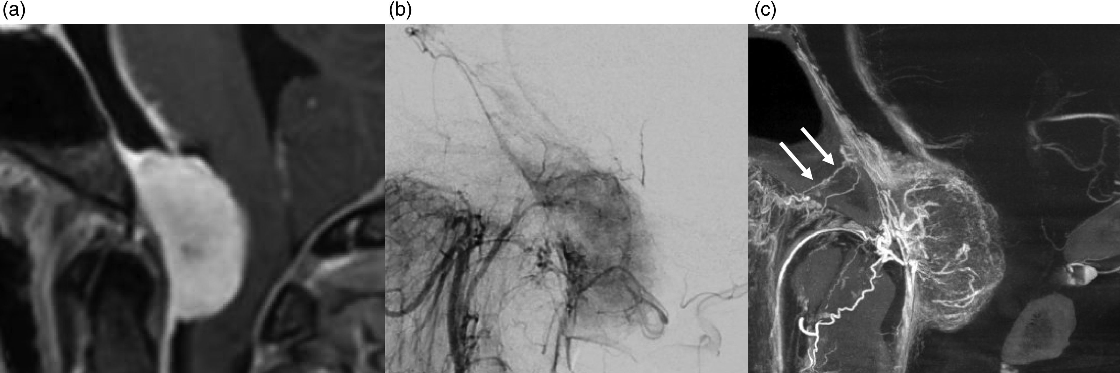
Sagittal T1 postcontrast (a), selective ascending pharyngeal artery lateral digital subtraction angiography (b), and sagittal MIP reconstruction of cone beam CT angiography (c) in a 40-year-old man presenting with a symptomatic foramen magnum meningioma in which enlarged transclival vessels supplied by the ascending pharyngeal artery were demonstrated (arrows in c).
Similar to how the hypoglossal branch of the ascending pharyngeal may supply a part of the posterior meninges, or how the accessory meningeal or the superficial temporal artery may supply part of the meninges, we anticipate that a pharyngeal branch of the ascending pharyngeal may take on this role as well. 7 In the inner aspect of the skull, if angiogenesis is promoted, it is possible that a potential synangiosis may develop between this meningeal vessel arising from the ascending pharyngeal artery and the basilar artery, ultimately resulting in a clear and developed anastomosis.
It is possible that the observed vessel is a regularly present embryonic intersegmental carotid-basilar anastomosis which is present only in a very transient stage of embryogenesis, and thus not depicted in the classic studies. 2 The presence of a canal within the clivus, through which vasculature traverses, has been previously documented in the literature. 8
To conclude, we report a rare case of a transclival persistent carotid-vertebrobasilar anastomosis that defies classification within the typical established categories.
Footnotes
Declaration of conflicting interests
The author(s) declared no potential conflicts of interest with respect to the research, authorship, and/or publication of this article.
Funding
The author(s) received no financial support for the research, authorship, and/or publication of this article.