
Abstract
We report three cases of aortoesophageal fistula (AEF), in which the patients remained free from catastrophic bleeding after endovascular stent-grafting. The three patients, who were not candidates for surgical repair because of their poor general condition and prognosis, underwent endovascular stent-grafting following the administration of antibiotics and were successfully managed; hemostasis was maintained for several months until their death. Although we did not find any conclusive evidence to support this strategy, our experiences suggest that endovascular stent-grafting of AEF is useful for maintaining hemodynamic stability.
Keywords
Introduction
Aortoesophageal fistula (AEF) is a rare but life-threatening condition that is associated with high mortality and morbidity. 1 There are a variety of etiologies of primary and secondary AEF. The optimal management strategy for, and the goal of the treatment of, the AEF differ depending on the etiology and/or the patient's general condition. Therefore, there is no established optimal management protocol, although several strategies have been described including open surgical repair, temporary control measures such as percutaneous embolization, the use of Sengstaken-Blakemore tubes, and esophageal-covered stents, endovascular stent-grafting, and various combinations of these.1-7 In this report, we present three cases of AEF involving patients who had remained free from catastrophic bleeding after endovascular stent-grafting.
Case Reports
Between November 2003 and May 2008, consecutive three patients with AEF were treated by endovascular stent-grafting at our institution. The materials and procedures used were approved by the Medical Ethics Committee of Kanazawa University, and written informed consent was obtained from each patient. The patient data, management strategies and outcomes are summarized in Table 1. None of the patients were candidates for open surgical repair because of their poor general condition, poor prognosis and/or possible infection within the mediastinum, although they were hemodynamically stable and the three patients and their families were eager to receive an aggressive treatment. So, we performed endovascular stent-grafting to avoid fatal bleeding from the AEF. Stent-graft placement was performed in an operating room under local anesthesia and systemic heparinization (100 U/kg). After surgical exposure of the unilateral common femoral artery, a 4F sheath (Medikit, Tokyo, Japan) was inserted into the femoral artery, and a 0.032 inch Radifocus guidewire (Terumo, Tokyo, Japan) was introduced and advanced across the target segment into the proximal descending aorta. After confirming the location of the true or false aneurysm on an arteriogram obtained using a 4F pigtail catheter (Medikit) inserted through the contralateral common femoral artery, a 0.035 inch Amplatz stiff guidewire (Cook, Bloomington, IN, USA) was introduced into the ascending aorta using the catheter exchange method. Transverse arteriotomy of the common femoral artery was performed, and an 18F sheath (Keller-Timmermans introducer sheath; Cook) was advanced into the target segment of the aorta over the guidewire. A stent-graft was deployed across the damaged segment of the descending thoracic aorta under road-mapping guidance, followed immediately by dilation with an occlusion balloon catheter (lock balloon catheter, Tokai Medical Products, Aichi, Japan). An aortogram was obtained just after the stent-grafting to confirm the disappearance of the false aneurysm in the descending thoracic aorta. The diameter and length of the stent-graft were chosen according to the aortic diameter in the landing zone, as measured on preprocedural computed tomography (CT) images. All of the successfully placed stent-grafts were Matsui-Kitamura stent-grafts, which are homemade stent-grafts composed of a self-expanding nitinol stent and a seamless, cylindrical woven graft made of polyester fabric that have been used in several Japanese institutions prior to the approval of a commercially available device. 8
Summary of the patient data, management strategies and outcomes
WBC/CRP, white blood cell count and C-reactive protein level just before stent-grafting. (–), not performed or not recognized
Three patients received one of the general broad-spectrum antibiotics by intravenous infusion immediately when an AEF was suspected. The same amount of broad-spectrum antibiotics was administered intravenously in the hospital for at least six weeks after stent-grafting and until clinical and laboratory parameters (fever, white blood cell count and C-reactive protein) were normal. After that, the antibiotic therapy was continued orally.
Case 1
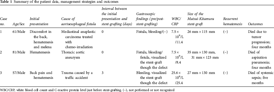
A 61-year-old man with anaplastic carcinoma in the posterior mediastinum was treated with chemo-irradiation. Three months after the start of the treatment, he suddenly felt discomfort in his back and presented with massive hematemesis and melena. He was diagnosed with AEF based on CT imaging (Figure 1a), emergency gastroscopic and bronchoscopic findings, and the stent-graft was deployed in the same manner as noted above. CT images obtained two months after the procedure revealed a completely sealed fistula and the absence of mediastinitis (Figure 1b). The patient experienced no hematemesis for four months after the procedure and died of progression and metastases from the mediastinal anaplastic carcinoma.
A 61-year-old man with an aortoesophageal fistula due to anaplastic carcinoma was treated with chemo-irradiation (Case 1). (a) Computed tomography (CT) image showing a mediastinal anaplastic carcinoma surrounding the aorta; the aortic wall was immediately adjacent to the air within the esophagus (arrowhead), and the esophageal mucosa was disrupted. (b) CT image obtained two months after the procedure revealing a completely sealed fistula and shrinking of the anaplastic carcinoma surrounding the aorta
Case 2
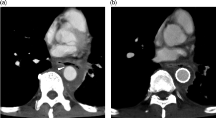
An 81-year-old man treated for severe chronic obstructive pulmonary disease and tuberculosis suddenly presented with massive hematemesis. He was diagnosed with AEF due to atherosclerotic descending thoracic aortic aneurysm (TAA) based on CT imaging (Figure 2a), emergency gastroscopic and bronchoscopic findings, and two stent-grafts were deployed to treat the two aneurysms in the same manner as noted above. CT images obtained one month after the procedure revealed a reduction in the size of the TAA (Figure 2b). A gastroscopic examination performed two months after the procedure demonstrated a dilated esophageal fistula and was able to directly visualize the stent-graft though the defect in the esophageal wall. The patient was followed-up for four months without hematemesis or mediastinitis and died of respiratory failure due to aspiration pneumonia.
An 81-year-old man with an aortoesophageal fistula due to a thoracic aortic aneurysm (Case 2). (a) Computed tomography (CT) image showing esophageal compression (arrowheads) by the descending thoracic aortic aneurysm aorta measuring approximately 75 mm in maximum diameter. (b) CT image obtained two weeks after the procedure showing the sealed and shrinking aneurysm (arrowhead)
Case 3
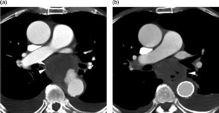
A 57-year-old man with poorly controlled diabetes mellitus and a history of cerebral infarction received a severe body bruise in a traffic accident. Despite the fact that he did not suffer any bone fractures or lung contusion on CT images, he experienced back pain after the accident. Four months thereafter, he presented with hematemesis and a septic state. Emergency gastroscopy demonstrated the esophageal fistula and a clot as well as oozing from the fistula. He was strongly suspected of AEF due to a trauma-related false aneurysm based on the CT imaging (Figure 3a) and gastroscopic findings, and the stent-graft was deployed as described above (Figure 3b). CT images obtained one month after the procedure demonstrated a reduction in the size of the false aneurysm (Figure 3c). Gastroscopic findings obtained two months after the procedure revealed expanding ulceration, with the stent-graft visible through the ulceration (Figure 3d). Bronchoscopic findings showed no abnormality. A subsequent open surgical repair was considered, but was considered to be too difficult by the surgeons due to the patient's mediastinitis and recurrent diabetic ketoacidosis. He experienced no hematemesis for five months after the procedure until his death, and died of systemic sepsis.
A 57-year-old man with an aortoesophageal fistula due to a post-traumatic aortic false aneurysm (Case 3). (a) Computed tomography (CT) image showing a false aneurysm of the descending thoracic aorta (black arrowhead) surrounding an area of ectopic gas collection, and a fistula (white arrow) located between the esophagus (white arrowhead) and aorta. (b) Intraoperative aortogram obtained before deployment of the stent-graft showing a false aneurysm of the descending thoracic aorta (arrowhead). (c) CT image obtained at one month after stent-grafting revealing shrinkage of the false aneurysm and remnant gas. (d) Gastroscopic image two months after stent-grafting demonstrated a dilated esophageal fistula and direct visualization of the stent-graft though the defect
Discussion
AEF, which arises from a variety of aortic or esophageal disorders, is associated with high mortality and requires immediate repair. The leading cause of primary AEF is erosion of a TAA into the esophagus. 2 Other causes include malignant mediastinal neoplasms (usually esophageal cancer), the ingestion of foreign bodies, inflammatory diseases of the mediastinum, trauma, radiotherapy, endovascular stent-grafting for thoracic aortic disease and aortic or esophageal surgery. 1 Among the present three patients, mediastinal anaplastic carcinoma that was treated with chemo-radiation, TAA and trauma were experienced in one case each.
The classic symptoms of AEF, which were described by Chiari, 9 are mid-thoracic pain, dysphagia and sentinel minor hematemesis followed by exsanguination after a symptom-free period. In most cases, the diagnosis of AEF is made after an episode of upper gastrointestinal bleeding. 1 Early diagnosis and prompt surgical management are crucial for survival, although diagnosis tends to be made late because of the above-mentioned non-specific symptoms and the low frequency of the condition. The management of AEF must focus on both the damage caused to the aortic wall to avoid massive hemorrhaging and esophageal ulceration to prevent infectious complications. Furthermore, various causes of AEF, such as TAA and foreign bodies, should be simultaneously managed whenever possible.
Although several treatment strategies have been described including open surgical repair, the variety of causes and the scarcity of AEF make it difficult to establish an optimal management protocol. The widely accepted surgical method for repairing AEF is excision of the damaged aorta combined with in situ aortic repair or extra-anatomical bypass, repair of the esophageal defect, and mediastinal debridement or drainage. This surgical method is associated with high operative and perioperative mortality rates because of contamination and/or the patients’ unstable hemodynamics.2,3 Recently, endovascular stent-grafting of AEF in high-risk patients has rapidly become an accepted alternative to surgical repair, and several successful cases involving this approach have been reported.4,5 General endovascular procedures are not complicated and are associated with high success rates because the damaged segment of the AEF is usually localized and located in the descending aorta (Zone 4), as was found in the present cases. The above method is less invasive, is associated with lower operative mortality than surgical repair, 6 and is an option for emergency or palliative control of aortic bleeding in cases in which the patient is not a good candidate for surgical repair. Although stent-grafting in fields that could become infected can lead to a high rate of infectious complications, the risk of graft infection in the short-term may be less than that associated with surgical graft interposition because stent-grafts are separated from such mediastinal tissues by the aortic wall. 5
The short-term results of endovascular stent-grafting of AEF to control bleeding are promising; however, the long-term outcome is not established. The risk of future mediastinitis and sepsis is increased because the fistula remains as it is. Therefore, when the patient's condition has stabilized, subsequent definitive open surgical repair of the esophageal defect and stent-graft removal are recommended. 6 However, patients with a poor prognosis due to an unresectable malignant tumor or other disorders, as was found in the present cases, are not candidates for the combined therapeutic approach, even if their condition has been stabilized. Our experiences suggest that even in such cases when patients and their families were eager to receive an aggressive treatment, controlling of aortic bleeding and prevention of fatal early exsanguination could be achieved by endovascular stent-grafting as a palliative therapy.
In conclusion, endovascular stent-grafting of AEF is feasible for the purpose of maintaining hemodynamic stability with acceptable perioperative mortality.