






Abstract
Purpose
To determine the effects of hypercholesterolemia in pregnant mice on the susceptibility to atherosclerosis in adult life through a new animal modeling approach.
Methods
Male offspring from apoE−/− mice fed with regular (R) or high (H) cholesterol chow during pregnancy were randomly subjected to regular (Groups R–R and H–R, n = 10) or high cholesterol diet (Groups R–H and H–H, n = 10) for 14 weeks. Plasma lipid profiles were determined in all rats. The abdominal aorta was examined for the severity of atherosclerotic lesions in offspring.
Results
Lipids significantly increased while high-density lipoprotein-cholesterol/low-density lipoprotein-cholesterol decreased in mothers fed high cholesterol chow after delivery compared with before pregnancy (p < 0.01). Groups R–H and H–R indicated dyslipidemia and significant atherosclerotic lesions. Group H–H demonstrated the highest lipids, lowest high-density lipoprotein-cholesterol/low-density lipoprotein-cholesterol, highest incidence (90%), plaque area to luminal area ratio (0.78 ± 0.02) and intima to media ratio (1.57 ± 0.05).
Conclusion
Hypercholesterolemia in pregnant mice may increase susceptibility to atherosclerosis in their adult offspring.
Introduction
Coronary heart disease is still the leading cause of morbidity and mortality in Western countries as well as in the developing world, contributing to approximately one-third of all deaths globally. 1 Atherosclerosis as the major etiology of cardiovascular disease continuously intrigues extensive studies on its complicated mechanisms and risk factors, among which dyslipidemia may be the pathophysiological basis. Numerous epidemiological studies confirm hyperlipidemia as the most important biological risk factor for atherosclerosis in many laboratory animal models of atherosclerosis-involving wild-type, naturally defective or genetically modified animals.2–4
Transgenic animals provide major advantages such as a clean genetic background and rapid induction of atherosclerosis. The apolipoprotein E (apoE) knockout mouse is the first widely used and one of the most well-characterized genetically manipulated models for atherosclerosis research. As a ligand for cell-surface lipoprotein receptors, apoE prevents atherosclerosis by clearing cholesterol-rich lipoproteins from plasma. 5 Homozygous apoE−/− mice have high plasma cholesterol levels up to five times higher than that of wild-type mice, and are associated with spontaneous development of extensive atherosclerosis. 6 Feeding with high-lipid chow leads to a further increase in total blood cholesterol levels in apoE−/− mice. 7
Embryonic and fetal development is a delicately regulated process. The pioneering work of Barker et al. 8 indicated that impaired growth and development in prenatal and early postnatal life may be an important risk factor for ischemic heart disease. In a morphometric postmortem analysis of atherosclerosis in fetuses and children, maternal hypercholesterolemia was demonstrated to be associated with a higher incidence of fatty streaks during the fetal period and a faster progression of atherosclerotic lesions after birth.9,10 Subsequent animal studies also suggested a relationship between maternal hypercholesterolemia and increased atherosusceptibility of the adult offspring.11–13 However, Madsen et al. 14 failed to observe similar results in adult apoE(+/−) progeny of apoE−/− mothers and concluded that hypercholesterolemia during pregnancy does not affect the development of advanced atherosclerosis in the adult offspring.
To solve the current discrepancy, the objective of the current study was to investigate whether maternal hypercholesterolemia in pregnancy would increase the susceptibility to atherosclerosis in adult offspring by feeding apoE−/− pregnant mice a high cholesterol diet.
Materials and methods
Mice and the experimental procedures
Healthy apoE−/− mice (20 female and 10 male) were purchased from the Model Animal Research Center of Nanjing University (Nan Jing, China). Mice were housed in temperature- and humidity-controlled rooms with a constant 12-hour light and 12-hour dark cycle. They were provided with food and water ad libitum (the experimental procedure was approved by The Third Military Medical University Ethics Committee). Female mice were caged with male mice at a ratio of 2 : 1 until pregnancy was confirmed by checking the presence of sperm in vaginal fluid under microscope examination each morning. Pregnant mice were randomly divided into two groups (n = 10 in each group), with Group R continuing to receive regular chow, and Group H starting to receive high cholesterol chow (21% milk fat, 0.2% cholesterol and 78.8% regular chow, provided by the Laboratory Animal Center of the Third Military Medical University, Chongqing, China) once their pregnancies were confirmed. Two healthy male offspring were randomly selected from each mother after weaning at 21 days of age. These male offspring from Group R or Group H mothers were further divided into four groups (n = 10 each group). After weaning, they were fed with either regular chow (Group R–R and Group H–R) or high cholesterol chow (Group H–H and Group R–H) until 14 weeks of age. Maintenance, care and application of experimental procedures were provided by certified personnel and veterinary staff in accordance with Chinese guidelines on experimental animal care.
Plasma lipid profile measurements
Blood samples of pregnant mothers were collected before pregnancy and after delivery. Blood samples of the offspring were obtained at 14 weeks of age. Mice were anesthetized by sodium pentobarbital (2%, 15 mL/kg, intraperitoneal injection) (Sigma, America). A total of 0.2 mL blood of each mouse was collected through tail bleeding into heparin-coated tubes (15 IU heparin/mL blood). Plasma was separated by centrifugation at 872 g for 15 min and was frozen at −80℃ until analysis.
Levels of total triglycerides (TG), total cholesterol (TC), high-density lipoprotein-cholesterol (HDL-C) and low-density lipoprotein-cholesterol (LDL-C) were measured using a colorimetric method with automated chemistry analyzer (Olympus Au2700, Japan) by the Department of Clinical Laboratory, Southwestern Hospital of the Third Military Medical University. The inter-assay coefficients of variation were 1.6% for TG, 3.6% for TC, 2.8% for HDL-C and 2.2% for LDL-C, respectively.
Abdominal aorta histology and atherosclerotic lesion assessment
After blood collection, while animals were still under anesthesia, mice thorax and abdominal cavities were opened. Tissue perfusion and fixation (at 76 mmHg) was performed through the left cardiac ventricle with phosphate-buffered saline (PBS, 137 mmol/L NaCl, 2.7 mmol/L KCl, 4.3 mmol/L Na2HPO4 and 1.47 mmol/L KH2PO4; pH 7.4) for 5 min followed by 4% paraformaldehyde in 0.1 mol/L PHEM buffer (60 mmol/L Pipes, 25 mmol/L HEPES, 10 mmol/L ethyleneglycotetraacetic acid and 2 mmol/L MgCl2; pH 6.97) for 5 min. The abdominal aorta was harvested and fixed in a 10% buffered formalin solution for 6 h. After fixation, the tissue was dehydrated in ethanol and xylene, paraffin embedded and then transversely sectioned at 5-µm thickness. Routine staining was performed with hematoxylin–eosin. For atherosclerotic lesion assessment, three sections (proximal, middle and distal) were selected in each aorta specimen and were examined under the microscope. Images were acquired by an attached digital camera and analyzed with image analysis software (Image Pro Plus 6.0, Media Cybernetics, Inc., MD, USA). Measurements, including blood vessel lumen area (LA, the internal area of blood vessel formed by the intima), plaque area (PA) and intima and media thickness, and the ratios of PA/LA and intima to media were taken.
Immunohistochemistry of matrix metalloproteinase-9
Mice aorta slides were deparaffinized, dehydrated, and the antigen was retrieved. The slides were subsequently rinsed with phosphate buffer three times. The endogenous peroxidase activity was blocked by incubating the slides with 3% H2O2 for 15 min. Then, the slides were incubated with the primary antibody (1 : 50, Wuhan, BOSTER) or PBS as a negative control for 60 min. The slides were further incubated with streptavidin–biotin peroxidase complex solution and finally DAB (3,3′ diaminobenzidine) solution for color reaction. The images were acquired by a digital camera (400X). The integrated optical density (IOD) value was measured by Image Pro Plus 6.0 image analysis software. The average optical density (AOD) value was calculated by an equation: AOD = IOD/blood vessel area.
Statistical analysis
Data were expressed by mean ± standard error. Statistical analyses were performed using SPSS 13.0 (SPSS Inc., Chicago, IL). Lipids levels before pregnancy and after delivery were analyzed with the paired t-test. Other analyses were made using the one way analysis of variance test (followed by Bonferoni’s post hoc analysis) or t-test if fewer than three groups were being analyzed. For all analyses, a p-value of less than 0.05 after correction was considered significant.
Results
Lipid levels of mothers before pregnancy and after delivery
Lipid levels of mother apoE−/− mice before pregnancy and after delivery.
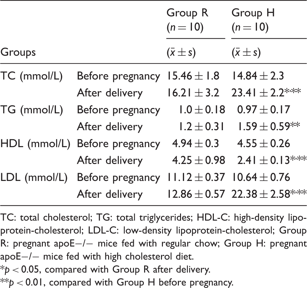
TC: total cholesterol; TG: total triglycerides; HDL-C: high-density lipoprotein-cholesterol; LDL-C: low-density lipoprotein-cholesterol; Group R: pregnant apoE−/− mice fed with regular chow; Group H: pregnant apoE−/− mice fed with high cholesterol diet.
p < 0.05, compared with Group R after delivery.
p < 0.01, compared with Group H before pregnancy.
Lipid levels of adult male offspring at 14 weeks of age
Lipid levels of adult male offspring at 14 weeks of age.
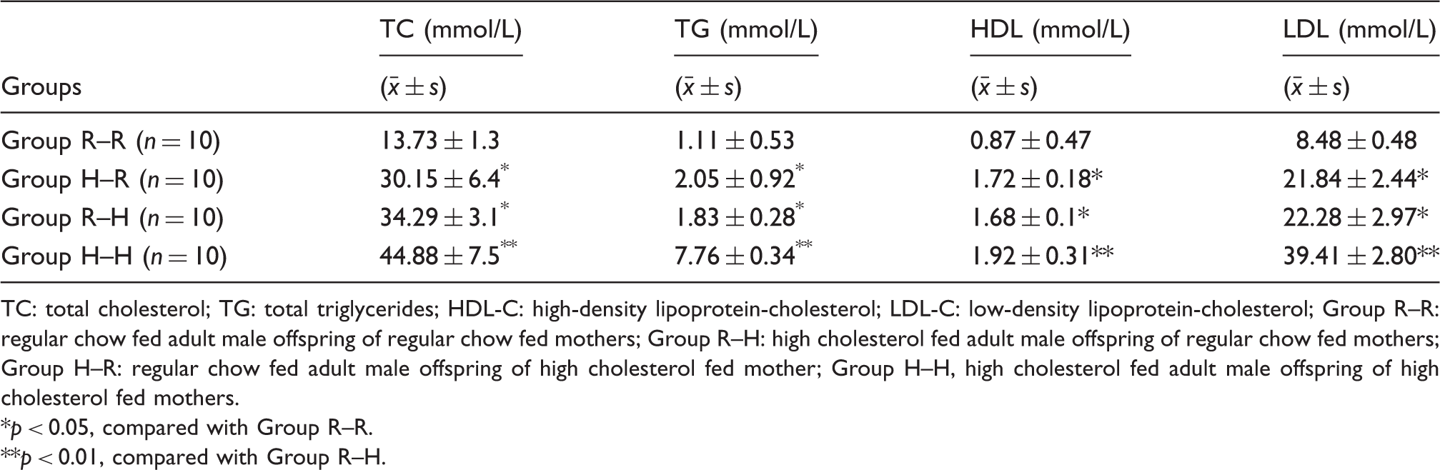
TC: total cholesterol; TG: total triglycerides; HDL-C: high-density lipoprotein-cholesterol; LDL-C: low-density lipoprotein-cholesterol; Group R–R: regular chow fed adult male offspring of regular chow fed mothers; Group R–H: high cholesterol fed adult male offspring of regular chow fed mothers; Group H–R: regular chow fed adult male offspring of high cholesterol fed mother; Group H–H, high cholesterol fed adult male offspring of high cholesterol fed mothers.
p < 0.05, compared with Group R–R.
p < 0.01, compared with Group R–H.
Atherosclerotic lesion assessment in adult male offspring
Atherosclerotic plaque was examined on three hematoxylin–eosin staining slices of abdominal aorta in all 40 adult male offspring. Atherosclerotic plaque formation can be divided into five stages: (1) intima LDL accumulation, (2) oxidation of LDL, (3) recruitment of macrophages, (4) transformation of macrophage into foam cell and (5) formation of a fibrous cap containing smooth muscle cell (SMC) as Tedqui and Mallat protocol.
15
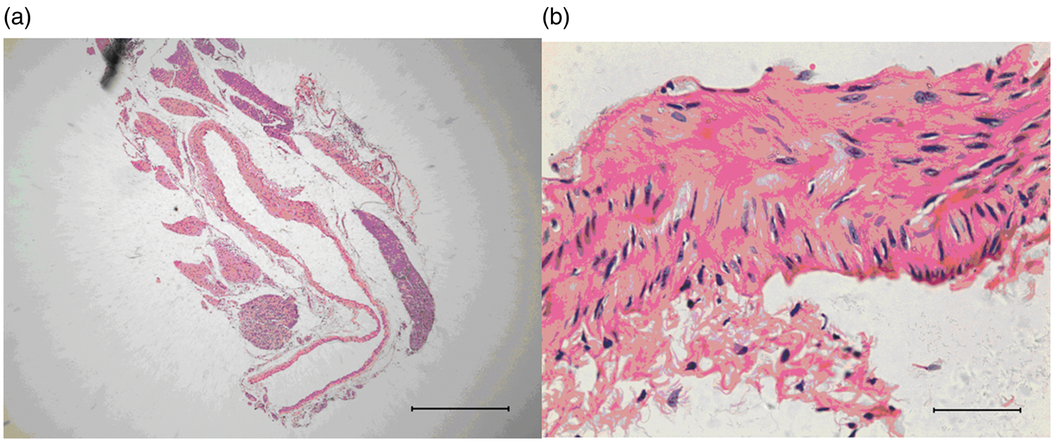
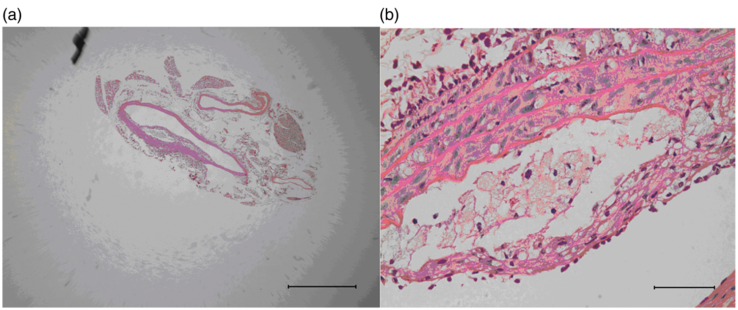
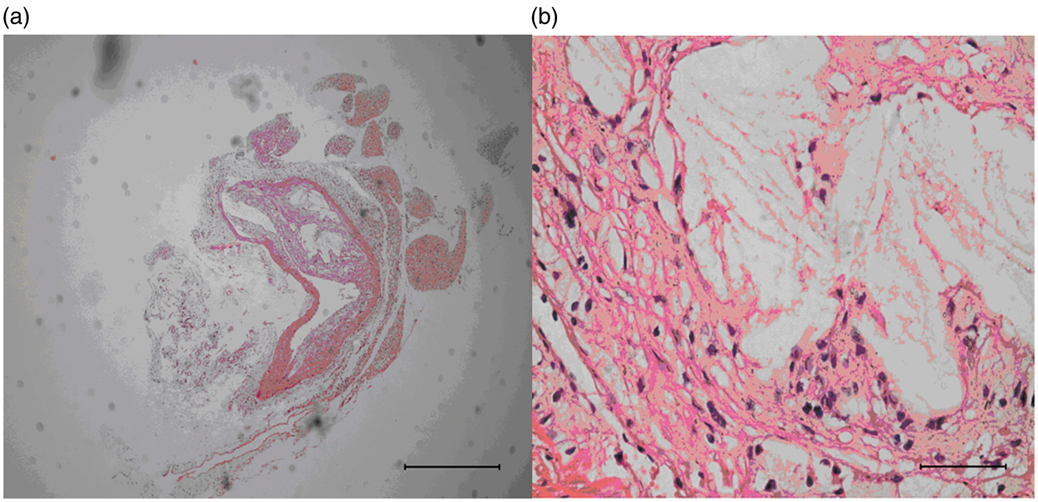
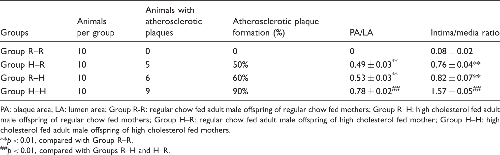
The intima was intact in Group R–R mice, and there was no evidence of atherosclerotic plaque formation (Figure 1(a) and (b)). Atherosclerotic plaques and sub-endothelial aggregate of macrophage foam cells were identified in five of 10 mice in Group H–R (Figure 2(a) and (b)) and in six of 10 mice in Group R–H (Figure 3(a) and (b)). In Group H–H, nine of 10 mice formed atherosclerotic plaques (Figure 4(a) and (b)). The atherosclerotic plaques were covered by an obvious fibrous layer with lipid cores inside formed mainly by lipid deposits. The atherosclerotic plaques protruded toward the lumen of the artery and caused vascular narrowing leading to disruption of the intima. Groups H–R and R–H indicated vascular narrowing as suggested by increased PA/LA and intima/media ratios (Table 3). Mice in Group H–H demonstrated the narrowest vascular lumen with the highest PA/LA and intima/media ratios (p < 0.01 vs. Groups H–R and R–H) (Table 3).
The intima was intact and there was no evidence of atherosclerotic plaque formation in Group R–R. Scale bars: (a) 500 µm and (b) 50 µm. Observed atherosclerotic plaque formation and sub-endothelial aggregation of macrophage foam cells in five of 10 mice in Group H–R. Scale bars: (a) 500 µm and (b) 50 µm. Observed atherosclerotic plaques formation and sub-endothelial aggregation of macrophage foam cells in six of 10 mice in Group R–H. Scale bars: (a) 500 µm and (b) 50 µm. Nine of 10 mice formed atherosclerotic plaque in Group H–H. The atherosclerotic plaque was covered by an obvious fibrous layer and with a lipid core inside formed mainly by lipid deposits. The atherosclerotic plaque protruded toward the lumen of the artery and caused vascular narrowing, leading to the disruption of intima. Scale bars: (a) 500 µm and (b) 50 µm. The percentages of atherosclerotic plaque formation, PA/LA values and the intima/media ratios of adult male offspring at 14 weeks of age ( PA: plaque area; LA: lumen area; Group R-R: regular chow fed adult male offspring of regular chow fed mothers; Group R–H: high cholesterol fed adult male offspring of regular chow fed mothers; Group H–R: regular chow fed adult male offspring of high cholesterol fed mother; Group H–H: high cholesterol fed adult male offspring of high cholesterol fed mothers. p < 0.01, compared with Group R–R. ##p < 0.01, compared with Groups R–H and H–R.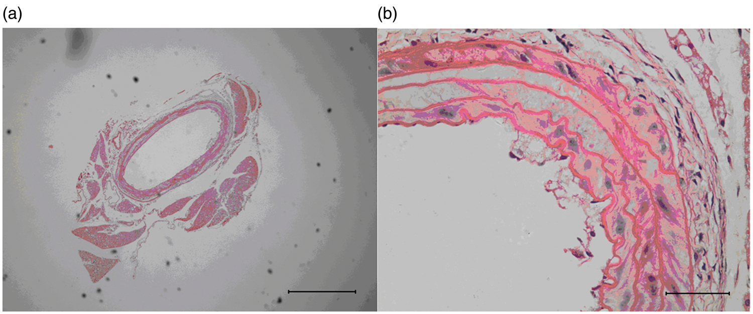
Immunohistochemistry of matrix metalloproteinase-9
The AOD value was semi-quantitatively assessed on aorta slides in all 40 male offspring. Matrix metalloproteinase-9 (MMP-9) was mainly expressed in endothelium cells and SMCs. The AOD value in Group H–H was significantly higher (0.043 ± 0.008, p < 0.01) than that in other groups. The AOD value was relatively low in Group H–R (0.028 ± 0.006, p < 0.01, compared with that of Groups H–H and R–H) and Group R–H (0.031 ± 0.005, p < 0.01, compared with that of Group H–H) and almost negative in Group R–R (0.009 ± 0.001, p < 0.01, compared with that of other groups) (Figure 5).
Aortic matrix metalloproteinase-9 expression of adult male offspring (streptavidin–biotin peroxidase complex method, 400X) in Group R–R (a), Group R–H (b), Group H–R (c) and Group H–H (d).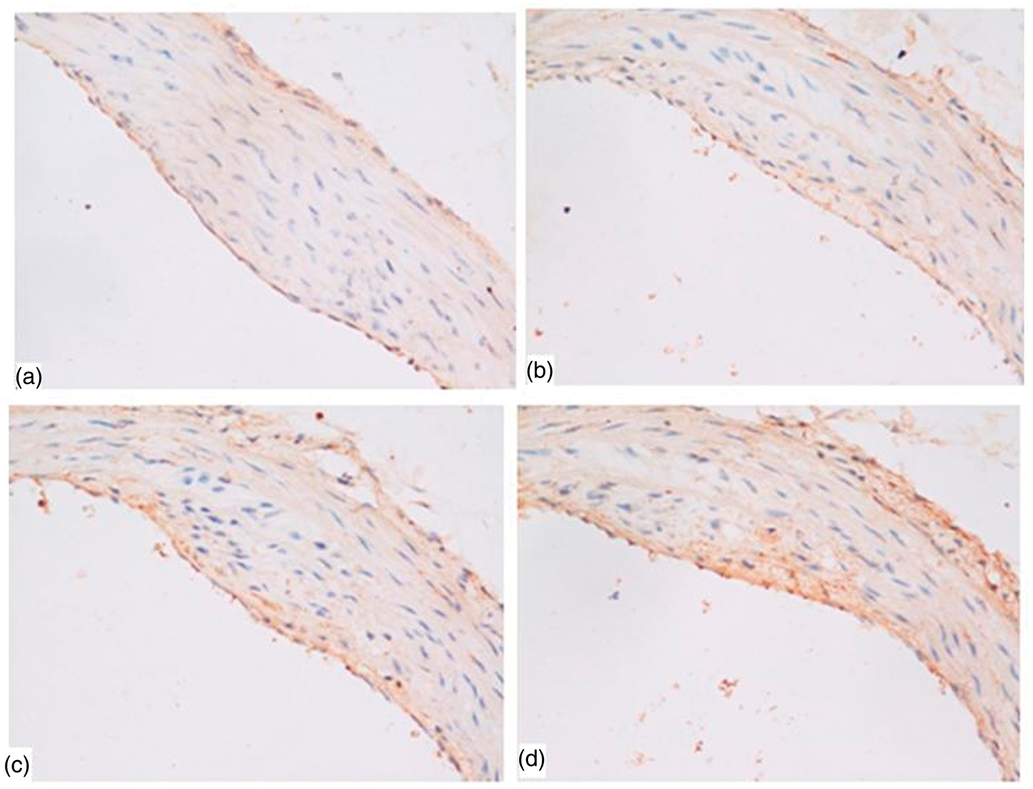
Discussion
The present study clearly demonstrated that both intrauterine exposure to maternal hypercholesterolemia and Western diet-induced hypercholesterolemia in apoE deficiency mice was capable of enhancing the susceptibility to atherosclerosis and associated with accelerated atherogenesis later in adult life. High cholesterol diets markedly raised maternal cholesterol levels as well as other lipid levels compared with those fed with a regular diet. In male offspring, prenatal high lipids in the maternal environment and postnatal high cholesterol diets similarly increased the expression of MMP-9 and the susceptibility to atherosclerosis. It was not unexpected that the male offspring, fed with a diet of high cholesterol, of high cholesterol diet treated mothers (Group H–H) had the highest lipid levels, MMP-9 levels and most severe atherosclerotic lesions.
Our results are in line with many previous studies which also investigated possible underlying mechanisms. Palinski et al. 11 suggested that pathogenic programming in utero increases the susceptibility to atherogenic risk factors later in life and both maternal cholesterol-lowering treatments and antioxidant interventions significantly reduce postnatal lipid peroxidation and atherosclerosis in their offspring in rabbits. In animal studies by Napoli et al. 12 carried out in LDL receptor-deficient mice and Alkemade et al. 13 in apoE−/− mice, atherogenic effects of maternal hypercholesterolemia were established. The possible mechanisms of maternal hypercholesterolemia priming for atherosclerosis may be related to the increased lipid peroxidation and formation of reactive oxygen species which affect several signaling pathways, including the NF-κB pathway, FasL and TNF receptor pathways as well as major moderators playing an essential role in regulation of these pathways, such as superoxide dismutase or fibroblast growth factor-binding protein.12,13 It is well-known that inflammation is involved in the basic pathological process of atherosclerosis and cardiovascular disorders. 16 Similar to oxidative stress, maternal inflammatory stress affects pro-inflammatory and anti-inflammatory gene transcriptions in the fetus. 17 In addition, in a more recent study, Alkemade et al. 13 suggested that other mechanisms might be related to the induced susceptibility for neointima formation in the offspring. Subsequent postnatal induction of hypercholesterolemia induces changes in the epigenetic profiles that may modulate gene expression patterns of the vasculature. These results demonstrated that both in utero programming and diet-inducted hypercholesterolemia affect histone methylation modifications and expression of accompanying lysine methyltransferases in vascular endothelial cells and SMCs. 18
However, our results seem to conflict with a previous study by Madsen et al., 14 who did not observe the influence of hypercholesterolemia during pregnancy on the development of advanced atherosclerosis in the adult offspring, regardless of sex. This discrepancy might be related to the difference in mice, time-points and animal procedures used in these two studies. Madsen et al. 14 studied 6-month old heterozygous apoE-deficient (apoE+/−) offspring of either apoE+/+ or apoE−/− mice all under an atherogenic diet from 6 weeks of age, whereas we used 14-week old homozygous apoE-deficient (apoE−/−) progeny with either high fat diet or regular chow fed pregnant apoE−/− mice from birth. It has been well-established that apoE−/− mice are more prone to atherosclerosis than apoE+/− mice, and a high-lipid diet quickens the progression of artery lesions.
The novelty of the current study is that it was conducted with a very clean genetic background (all animals were apoE−/− mice), and only compares one factor, that is, regular chow versus a high cholesterol diet of mothers during pregnancy and of offspring after birth. It clearly demonstrated the effect of diet during fetal development on adult atherosclerosis risk and spontaneous advanced lesion formation. However, only 50–60% of mice in Group H–R and R–H had atherosclerotic plaque formation mainly due to individual difference. The relatively short experiment length (14 weeks) might also be a reason. The animal model established in the present study may be useful for further studies to understand the pathophysiology of atherosclerosis and to provide new interventions for the prevention and therapy of coronary heart disease.
Footnotes
Conflict of interest
The authors declare that there are no conflict of interest.
Funding
This research was supported by the National Basic Research Program of China (2013CB531406); the National Science and Technology Support Program (2011BAI15B02) and the National High-tech R&D Program of China (863 Program) (2012AA020603).