
Abstract
We describe a patient who survived a ruptured abdominal aortic aneurysm without any surgical intervention. The patient had previously had endovascular repair of the aneurysm and surveillance of a stable persistent type II endoleak. This case highlights the difficulties surrounding type II endoleak, its natural history, and the ongoing controversies of its management.
Keywords
Introduction
A ruptured abdominal aortic aneurysm (AAA) is invariably a fatal condition. A recent review of 21,206 cases of ruptured AAA in the USA included 3796 cases that did not receive operative management; mortality for these patients was 100%. 1
To prevent AAAs progressing to rupture, endovascular aortic aneurysm repair (EVAR) is now the treatment of choice. Compared to open surgical repair, EVAR has lower rates of postoperative mortality. 2 However, this survival advantage is lost within 6–36 months, with large randomised controlled trials from the UK (EVAR 1), 3 USA (OVER),4,5 and the Netherlands (DREAM),6–8 demonstrating higher rates of medium-term morbidity and need for reintervention in patients with EVAR. Many of these complications are due to endoleak, where the aneurysm sac has continued perfusion despite endograft deployment. Type I and III endoleak and stent migration or kinking are all associated with increased risk of aneurysm rupture in post-EVAR patients. 9
Type II endoleak is due to backflow of collateral blood from aortic side branches into the aneurysm sac. Although this is the most common form of endoleak (estimates range from 8 to 44% of cases), 10 there is no consensus among experts regarding thresholds for surgical intervention. Many endoleaks resolve spontaneously within the months following EVAR, but controversy remains as to how to best manage those that persist. 10
We report a patient with a type II endoleak, who, despite receiving no surgical intervention, survived a ruptured AAA.
Case report
A 92-year-old woman presented to our emergency department with lower back and abdominal pain with associated nausea. Her medical history included chronic cardiac failure (CCF) secondary to aortic stenosis and mitral regurgitation, hypertension, dyslipidaemia, chronic atrial fibrillation (on warfarin), renal impairment, mild chronic obstructive pulmonary disease, osteoarthritis, and most significantly, AAA.
Four years before this presentation, she had undergone an EVAR for a 5.5-cm AAA. Complete angiography had showed good position, maintained visceral vessels, and no endoleak. There was no endoleak on her computed tomography angiography (CTA) at 1 month post-EVAR; however, at 12 months, there was evidence of type II endoleak, with 10-mm contrast pooling posterior to limbs of the graft and adjacent to a single lumbar vessel. This was despite a small reduction in sac measurements (5.3 cm). CTA scans over the next 2 years revealed a persistent lumbar vessel endoleak and further reduction in aneurysm sac diameter (5.0 cm).
However, on presenting to the emergency department at 4 years post-EVAR, contrast-enhanced computed tomography showed a substantially larger aneurysm sac (8.0 cm) with delayed phase images confirming a persistent type II endoleak with adjacent posterior aneurysm rupture (Figure 1).
CTA shows a large AAA sac with posterior contrast flush consistent with type II endoleak, periaortic stranding and large left retroperitoneal haematoma consistent with AAA rupture. AAA: abdominal aortic aneurysm; CTA: computed tomography angiography.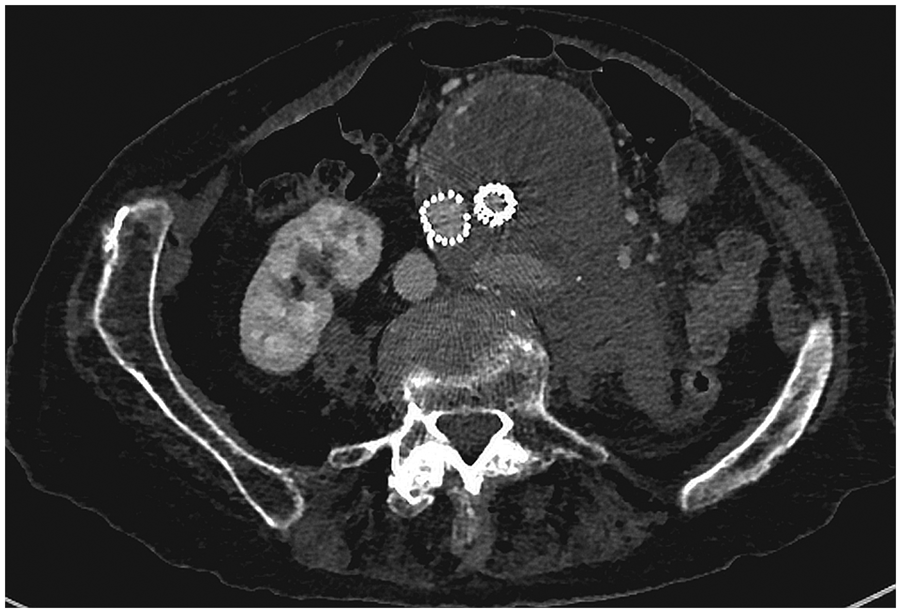
Given her age and comorbidities, it was decided that intervention would be inappropriate. The patient and her family were informed of her poor prognosis and agreed that comfort care would be provided. The day after presentation, to everyone’s surprise, the patient was sitting in bed, feeling well, and eating her breakfast. Accordingly, she was discharged into the care of her general practitioner, for ongoing conservative management.
There were no re-presentations with further deterioration of her aneurysm rupture. She was briefly readmitted with an exacerbation of her CCF 3 months later, resulting in discharge to a residential aged care facility. She lived in this facility for 3 months, but her CCF and fluid overload continued to worsen and was the eventual cause of her death.
Discussion
It is rare for a patient to have a ruptured AAA due to type II endoleak. In a recent review of 1515 patients with post-EVAR type II endoleak, only 14 cases (0.9%) went on to rupture. 10 To the authors’ knowledge, this is the first case report of a patient surviving such a rupture without surgical intervention.
It may be that the characteristics of a type II endoleak predisposed this patient to a more favorable outcome. Specifically, the backfilling pressures of the inferior mesenteric and lumbar arteries are low. As such, rupture is qualitatively different from a primary-ruptured AAA, with a slower bleed that is more easily tamponaded. There have been reported cases of chronic-contained primary AAA ruptures, where retroperitoneal bleeding is tamponaded and prevents rapid exsanguination. These patients can survive for a number of weeks without surgery. 11
Endoleak has been noted to be the Achilles heel of EVAR. Type II endoleaks have been particularly controversial with indications and methods of treatment-lacking consensus. Jones et al. 12 reported an association between persistent type II endoleak and risk of late rupture, whereas others13,14 describe a more benign course and no association. It has been suggested that type II endoleak with sac expansion should be an indication for intervention, but even the association between sac expansion and rupture is inconsistent (evident in the current case). 10
Conclusions
The current case illustrates the unpredictable nature of type II endoleak and again highlights the need for further research to inform the best practice in this area. It also raises the possibility that a type II endoleak resulting in low-pressure rupture is more likely to successfully tamponade, and less likely to be fatal, than a primary AAA rupture.
Footnotes
Conflict of interest
None declared.
Funding
This research received no specific grant from any funding agency in the public, commercial, or not-for-profit sectors.