






Abstract
Abstract
Objectives
Research in the field of lower extremity vascular disease has increased in popularity over the years. To adequately characterize and validate the effectiveness of vascular interventions, in vivo experimentation in large animals is required. Thus, it is necessary to find a method to detect the shape and density of blood vessels in the lower extremities that can evaluate and verify the treatment measures’ effectiveness and have high accuracy and repeatability. This study characterized factors that determined both the accuracy and overall value of digital subtraction angiography in lower limb arteriography using a canine animal model.
Methods
Six beagle dogs were anesthetized and immobilized on the motorized table. The femoral artery was accessed using an indwelling needle. A bolus of contrast agent was injected into the access site, and digital subtraction angiography with bolus chase technology was used to collect contrast images for analysis. At the end of the procedure, the anesthetized dogs were euthanized using an overdose of potassium chloride. After confirming the euthanasia of the dogs, the cadavers were taken to the experimental animal center of Xinjiang Medical University and processed by qualified institutional personnel.
Results
The final arteriographic images of the hind limbs from all six dogs were precise, and the branches of small vessels could be distinguished without any visible artifacts.
Conclusions
These results suggested that arteriography using digital subtraction angiography could reveal the shape and density of blood vessels in canine animal models. This method has great potential to significantly improve research related to limb ischemia due to its simple and reproducible results.
Keywords
Background
A peripheral arterial disease in the extremities can result in severe adverse consequences for limb function and quality of life. Arterial disease in lower extremities has exhibited increasing significance, in part due to the increased incidence of type 2 diabetes. One common clinical manifestation in diabetic patients with peripheral artery disease is the formation of foot ulcers, which is an important risk factor for amputation and death in diabetic patients. 1 At present, the main principle of diagnosis and treatment of peripheral artery disease is to reconstruct the peripheral artery circulation. However, most treatment protocols are still ongoing, and to date, there is little consensus on a specific treatment protocol. The use of animal models is an essential link in medical research to study the pathogenesis of diseases and optimize treatment plans. 2 Animal models that simulate acute and chronic ischemia of the lower limbs are established through ligation, transection, and embolization of arteries. These research models are gaining interest in the research community, both nationally and internationally, to improve diagnoses and treatment approaches for peripheral artery diseases. 3 Due to the ease and cost of experimentation, rabbits and rats are the most widely used animal models despite the small size of their blood vessels and skeletons relative to humans. Furthermore, though there are numerous limitations associated with simulation studies, simulation studies provide an ideal platform to observe and carry out research in this field.
The examination and evaluation of angiogenesis are essential when establishing a hindlimb ischemia model to study peripheral artery disease in canine models. Digital subtraction angiography (DSA), which is the “gold standard” for the clinical diagnosis of peripheral and cerebral vascular diseases, precisely visualizes vascular morphology and density. DSA is also the primary choice for the clinical diagnosis of peripheral vascular disease, assessment of residual vascular degree, and determination of treatment results. 4 DSA also is the most direct and reliable method to image peripheral blood vessels. This is the first study describing the feasibility and effectiveness of DSA in a canine hindlimb model to provide a theoretical basis of vascular surgery and operational reference for this method of in vivo analysis.
Methods
Design
The aim of this study was to establish successful angiographic imaging of canine animal models.
Time and place
This study was completed at Xinjiang Medical University over seven months.
Materials
Experimental animals
Six 12-month-old female beagles were selected with an average body mass of 15.7 ± 0.6 kg (a range of 15.0–16.0 kg), a body length of 55 ± 2 cm, and a tibia length of 11.0 ± 0.8 cm, indicating that the dogs were of approximately equal body size. All dogs were healthy and were obtained from an animal experiment center. Veterinarians randomly selected six beagles for the study. The experimenters were blinded to the selection of the dogs.
Additional information concerning the six dogs used in this study. The beagles were raised in an animal experiment center, and the adult dogs were housed individually in kennels constructed from concrete. Each kennel had a window, indoor heating and cooling, and was cleaned twice daily. The dogs were provided with tap water for drinking water and dog kibble, which was used by the breeders. The physiological welfare of the dogs was assessed and maintained before and after the experiment. Routine health welfare was provided by veterinarians who regularly provided physical exams and assessed the behavioral and psychological welfare of the dogs. The dogs received daily physical exercise in the form of walks and attention from their caretakers. At the end of the experiment, the dogs were euthanized.
Equipment
A Siemens axiom artis MP multi-functional angiography machine was used for imaging. An 18 G intravenous indwelling needle (1.3 mm × 30 mm, Suzhou Linhua Company) was used, and a 30 ml syringe was used to deliver an injection of lipanol (300 g/L, Beijing Beilu Company, Beijing, China) as the contrast agent. Specifically, 30 ml of lipanol and physiologic saline were mixed at a ratio of 2:1 and attached to the interface of the indwelling needle for injection into the artery.
Methods
Preoperative preparation
Before DSA, the dogs were fasted for 12 h to prevent life-threatening risks (e.g., aspiration) while under anesthesia. The area for the indwelling needle placement was shaved and prepped.
Anesthesia and positioning
Shutai (France Vick Co., Ltd., batch No.: 6smm 2017.12) and Sumianxin II (xylazine hydrochloride injection, Shengda animal drug Co., Ltd., Dunhua City, Jilin Province, batch No.: 20180401), which are commonly used in dogs due to the short induction time and high level of safety, were used as anesthetics. About 0.5 ml of each anesthetic was loaded into a syringe at the prescribed ratio of 1:1 and simultaneously injected intramuscularly in the quadriceps muscle. Each dog was placed on the motorized patient table after the successful administration of the anesthesia. A sterile bandage was used to fix the tongue to avoid suffocation caused by the tongue moving back into the pharyngeal region. The heart rate and respiration were monitored throughout the procedure.
Experimental procedure
Before initiation of the experiment, any objects that might have affected the C-arm movement and the motorized patient table were removed, and the starting and ending points of the moving track of the motorized table were determined. Two researchers, wearing lead aprons and other protective measures, were positioned on either side of the motorized table. The pulsating femoral artery on the upper surface of the inner thigh was identified by palpation and punctured through the skin using an 18 G indwelling needle, and the needle was fixed in position using tape. One researcher held the hind limb of the dog with both hands to prevent movement of the limb during radiography, which would adversely affect the image quality. The contrast agent was manually injected at an average rate of 3 ml/s by another researcher at the beginning of image acquisition. In the control room, an experienced imaging technologist controlled the motorized table and imaging exposure according to the flow of contrast agent in the hind limb blood vessels. The sequence subtraction image was carefully observed and recorded using a total exposure time of 15 s. If the image acquisition was not satisfactory, the contrast agent was administered a second time using the same protocol. At the end of the angiography, the indwelling needle was removed, and the puncture site was held under pressure for 15 min to avoid internal bleeding. See Figure 1 for the puncture location and contrast agent injection. Follow-up arteriography was carried out on the six dogs at one, two, and four weeks after the initial procedure. At the end of the procedure, the anesthetized dogs were euthanized with an overdose of concentrated potassium chloride. After confirming the dogs were euthanized, the cadavers were taken to the experimental animal center of Xinjiang Medical University and processed by qualified institutional personnel.
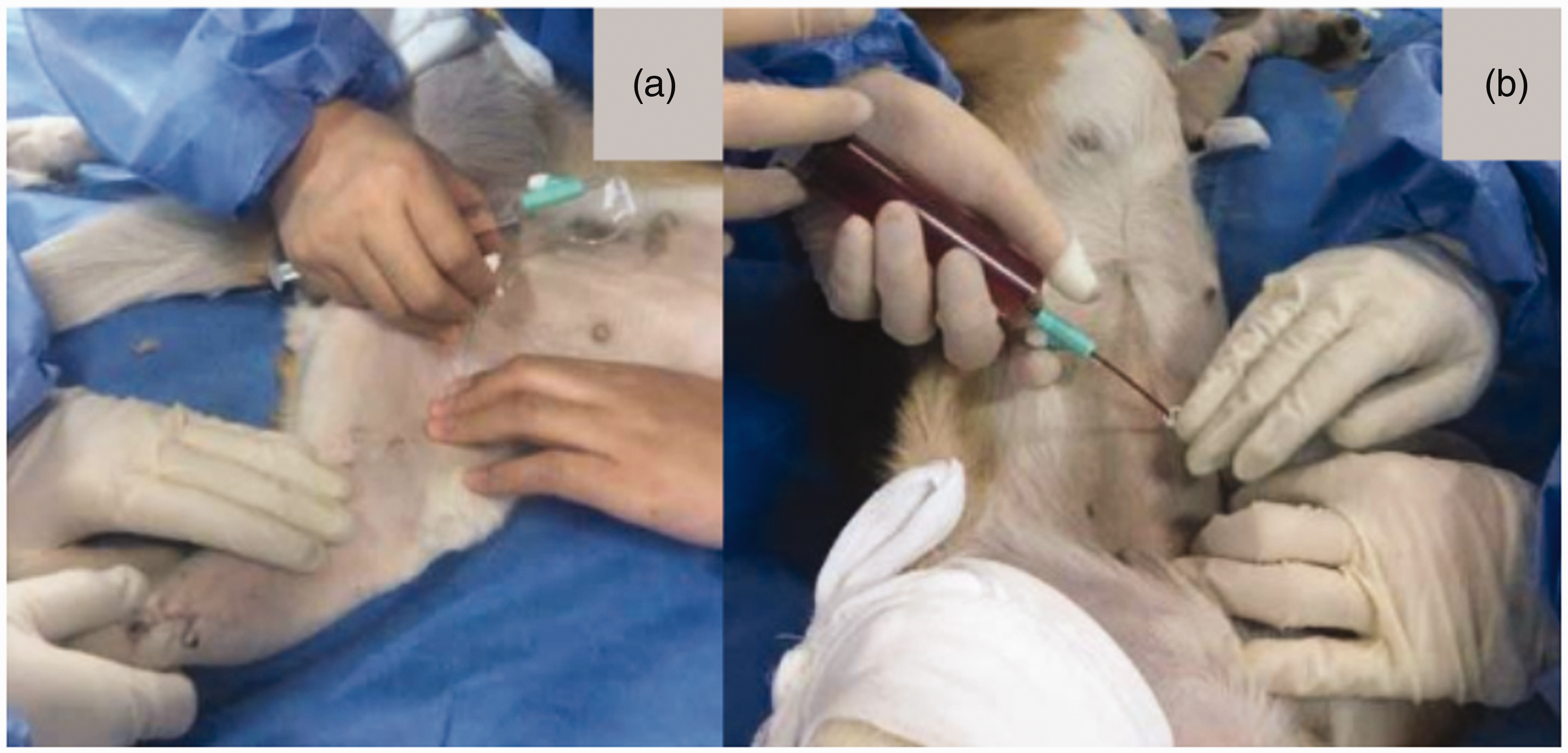
Photograph of the arterial puncture and contrast agent injection: (a) shows the location of the puncture point. The position of the upper segment of the femoral artery is superficial, and the artery is straight. Thus, the puncture conditions are ideal at this location; (b) shows the injection of the contrast agent in the direction of the course of the blood vessel.
Main observation indicators
The collected images were classified as excellent, good, and bad. The image acquisition is shown in Figure 2. The excellent category included clear images with no artifacts. The small blood vessels were distinctly visible (six to seven main hind limb arteries were visible). The good classification included clear images with only a few artifacts and some inadequate visualization of small blood vessels (four to five main hind limb arteries were visible). The bad image classification included blurred images or noticeable artifacts, and only the main blood vessels were identified (<4 main hind limb arteries were visible). The image quality was assessed by a deputy chief physician and an attending physician in the procedure room. And the images were graded after discussion.

Schematic diagram of angioplerosis developed on the arteriography: (a) shows the contrast agent beginning to fill along the main artery; (b) shows the image of the dog’s main hind limb artery, with a clear display of each branch of the major blood vessels, 1 is the posterior proximal femoral artery, 2 is the posterior middle femoral artery, 3 is the posterior distal femoral artery, 4 is the saphenous artery, 5 is the caudal tibial artery, 6 is the popliteal artery, and 7 is the cranial tibial artery; (c) shows the simultaneous images of arteries and veins, with a clear display of the local microcirculatory vascular density.
Results
Quantitative analysis of experimental animals
Six beagles were included in the analysis with no loss of subjects.
Image acquisition results
Twelve arteriography image sets were acquired from the six dogs for three times. Finally, thirty-six image sets were evaluated. The initial images obtained from the first dog were bad. For the first dog, the contrast agent was not diluted, which led to movement of the dog’s legs, resulting in blurred images of the blood vessels. After diluting the contrast agent and increasing the fixation of the hind limbs during the procedure, the angiography image quality for the 36 hind limbs from the six experimental dogs was excellent.
Effect of the procedure and prognosis
The average time for each procedure was 20 min, and no additional anesthetic was administered. The six dogs generally recovered within 30 min after the procedure ended, and no deaths occurred. There were slight swelling and subcutaneous hemorrhage at the puncture site of two dogs, which was caused by pressure applied to the puncture site for insufficient time. The swelling was absent one week later. The activity levels of the dogs resumed on the second day after normal eating. No functional or morphological abnormalities such as limping on the hind limbs were observed.
Discussion
Advantages of beagles as animal models
Animal models are indispensable in medical research, especially for procedures that are invasive, involve trauma, or have uncertain safety and, therefore, cannot be directly carried out on humans in clinical research. Thus, using an animal model to conduct preliminary research to verify safety and efficacy is necessary. 5 The beagle is the only standard experimental dog recognized worldwide because of its gentle temperament, stable genetic profile, uniform anatomic structure, and stable physiological parameters. Compared with rats and other small experimental animals, the beagle is closer to humans as a research model for skeletal and blood vessel structures, which is critical for comparison of research results between the experimental model and humans. 6
In the study of scoliosis, Mingming et al. 7 observed that the beagle animal model was similar to human clinical cases and exhibited typical scoliosis parameters when establishing a scoliosis model. Thus, the beagle animal model successfully and accurately simulated human clinical scoliosis. Also, the modeling protocol was simple, the success rate was high, and the damage to accessory structures around the spine was minimal.
Hongshuai et al. 8 used the beagle canine model to study hip joint stress by fixing one side of the forelimb and then increasing the load on the hind limb of the dog to simulate the load-bearing walking state in humans. Three-dimensional gait analysis was conducted to reveal that the changes in the mechanical parameters of the hind limb of the beagle conformed to the biomechanical changes that took place in the human hip joint. The study highlighted that the beagle animal model was useful to study the diseases of the human hip joint.
Kim et al. 9 and Nakamura et al. 10 studied a new type of internal fixation materials and found that the bone shape and specification of beagles were similar to humans and therefore could be used to test standard size screws and steel plates. Furthermore, they determined that the beagle has a muscle strength load that can reach that of humans and approximates the stress and stability exhibited by humans well.
In research of osteonecrosis of the femoral head and revision of the hip joint, Omoto et al. 11 and Feng et al. 12 reported that the hip joint and surrounding tissue of beagles were suitable to simulate human tissues. They claimed that the modeling protocol was simple, the success rate was high, and the imaging results were clear. The local bone mineralization rate after the protocol was similar, and the biological consistency was sufficient. In a follow-up osteotomy experiment in their study, beagles provided a more satisfactory bone model than small animals and were better able to recapitulate the simulation and visualization of the protocol.
In the field of vascular researches including the coronary, renal, hepatic artery, and leg circulation, there were several studies using dogs as experimental animal.13–17 Among them, Björk 17 used arterial venography on dogs’ legs to explore improvements in visualization of the veins.
The authors believe that, compared with sheep, orangutan, and other animals, dogs especially beagles are the best animal model for this field of research. They provide better representations of the experimental procedures and more closely simulate human skeletal, nervous, and vascular structures.
Operation and interference factors of DSA
DSA is a combination of angiography and computer technology. It utilizes contrast agent scanning and development of radiographs using digital processing, which removes unnecessary tissues from the image and produces distinct vascular morphology. At present, DSA technology mainly includes stepwise and segmentation technologies. The stepwise technology realizes the acquisition of a complete image and a dynamic image by controlling the movement and exposure of the motorized table. In contrast, segmentation technology uses the injected contrast agent and collects the image iteratively. The stepwise technology has the advantages of low contrast agent consumption and short exposure duration. 18 The technique used in this study was the stepwise technique for DSA.
DSA is regarded as the “gold standard” for diagnosing vascular diseases worldwide, especially for the diagnosis and treatment of peripheral artery diseases, such as diabetic foot and lower extremity arteritis. Accurate angiography of arterial circulation is an important foundation for diagnosis, reconstruction of blood circulation, and prognosis.19–21
Yoshinori et al. 22 performed DSA of the hepatic artery to diagnose and treat liver tumor diseases. Due to the excellent imaging quality, we could easily observe the morphology of the hepatic artery and its branches and the morphology and number of proliferative vessels in the tumors. These detailed images provide a more intuitive approach to treatment for tumor tissue resection.
Blagojevic et al., 23 Haigang et al., 24 and Abd-Elgawad et al. 25 highlighted the clinical diagnosis, treatment, and research of lower extremity artery disease, in which DSA technology could be used to accurately assess blood circulation and define the location of the affected vessels and affected areas of tissue. These capabilities play an indispensable role in the formulation of treatment plans, even in the case of amputation, since DSA can provide a more reliable reference standard, thus reducing the amount of amputated tissue and allowing as much limb function to be preserved as possible.
In a study of animal models, McCollough et al. 26 performed contrast imaging on beagles’ hearts by injecting a contrast agent into the central vein. According to the density of the contrast agent before and after the same cycle and the volume difference, the volume and ejection fraction of the ventricles could be quantitatively measured. The author indicated that DSA technology had the advantages of objectivity, a simple protocol, and repeatability.
Anesthesia and contrast agents are the two most important factors affecting the final image quality achieved with DSA.27,28 When DSA is performed, it is necessary that the examinee keep absolutely still relative to the motorized table. Good anesthesia can be used in place of sedation and analgesia to ensure a smooth procedure. However, deep anesthesia will lead to profound inhibition or even the death of the animal, such that normal and effective images cannot be obtained. However, light anesthesia can result in movement of the animal due to pain stimulation during the procedure, resulting in motion artifacts that adversely affect the image quality. 29 In this study, 1 ml of intramuscular anesthesia using a 1:1 ratio of Shutai and shumianxin II was used. The degree of anesthesia was appropriate, and the effective time of anesthesia was about 40 min, which resulted in a smooth procedural outcome. 30
The influence of contrast agents on the image quality of DSA is primarily affected by the concentration, dose, and flow rate. If the concentration of the contrast agent is too high, it will cause motion artifacts due to the stimulation of blood vessels in the dogs. If the contrast agent is administered at a concentration that is too low, this results in poor imaging of small blood vessels. 31 In this study, lipanol (300 g/L) was used as the contrast agent. During the initial procedure, the contrast agent was not diluted, resulting in the movement of the hind limbs of the dog during the injection due to irritation of the contrast agent, resulting in poor image quality. In subsequent trials, the concentration of the contrast agent was diluted at a ratio of 2:1 for lipanol and normal saline. No movement was observed in the dogs when the contrast agent was diluted, which resulted in the collection of adequate images that met the experimental requirements.
The use of a proper flow rate also is an essential factor in ensuring adequate image quality. For example, if the flow rate for contrast medium administration is too slow, the contrast medium will be diluted by the high-speed arterial blood flow in the artery. If the flow rate used to administer the contrast medium is too fast, it will result in increased pressure on the blood vessel, which could damage or even rupture the blood vessel. 32 In this study, a 30 ml syringe was used for manual injection of the contrast agent, and 30 ml of the contrast agent was pushed into the artery at a constant speed over 10 s. The average flow rate was approximately 3 ml/s. During the procedure, the flow of the contrast agent in the artery was tracked and imaged, and the quality of the collected images was satisfactory.
In this study, an 18 G indwelling needle was used instead of a catheter. The needle can directly puncture the femoral artery through the skin, which avoids the process of crossing the iliac artery in the routine procedure. This approach resulted in minimal stimulation, and damage to the blood vessel is convenient to perform and saves on cost. However, in the process of puncturing the femoral artery, the first beagle shook when the bolus of contrast agent was administered, resulting in poor quality angiographic images. To obtain high-quality images, the canine limbs were fixed in position, and a diluted concentration of the contrast agent was used. It is necessary to master the puncture tool, adjust the concentration of contrast agent, and control the contrast agent rate of injection to avoid possible massive bleeding and limb tremor or movement. It should be noted that there is a risk that the protocol will be adversely affected by the accidental removal of the needle. The images obtained by this method clearly show the path and morphology of the larger blood vessels. Also, the imaging of small blood vessels is sufficient, and the image quality meets the requirements needed for research experimentation. In addition, all dogs in this study successfully underwent three repeated arteriography sessions over two months. At the same time, some limitations in this experiment should be noted.
Conclusion
The beagle is the only standard experimental canine breed, and it is well-suited for use as a large animal model of human diseases. The use of DSA has a clear effect on the angiography of the arteries in the canine lower hind limb. It can display the distinct shape and density of the arteries of the lower extremity, and the image quality can completely meet the experimental requirements. The use of DSA also adequately recapitulates the research simulation of similar diseases. Moreover, the DSA technology is simple to operate and is highly repeatable. Thus, DSA can be used to simulate human lower extremity artery diseases in a canine experimental model. The verification of related theories is of great significance. However, appropriate measures should be taken during preoperative preparation, intraoperative anesthesia and immobilization, contrast agent regulation, and image processing to ensure that the quality of the final DSA images meets the experimental requirements.
Footnotes
Declaration of conflicting interests
The author(s) declared no potential conflicts of interest with respect to the research, authorship, and/or publication of this article.
Funding
The author(s) disclosed receipt of the following financial support for the research, authorship, and/or publication of this article: This study was funded by Natural Science Foundation of Xinjiang (Grant ID: 2017D01C267).
