
Abstract
Objective:
To evaluate the diagnostic performance of cystic artery colour Doppler ultrasound indices in differentiating acute from chronic cholecystitis and to assess their added value over clinical and grayscale findings.
Methods:
In this prospective study, 101 adults with clinically suspected cholecystitis underwent ultrasound before cholecystectomy. Grayscale features and colour Doppler measurements of cystic and hepatic artery peak systolic velocity and resistive index were recorded; histopathology was the reference standard. Logistic regression models using clinical and grayscale variables alone and then with cystic artery peak systolic velocity were evaluated with receiver operating characteristic analysis.
Results:
In total, 33 patients had acute and 68 had chronic cholecystitis. Acute cholecystitis showed more abnormal grayscale features, including increased gallbladder size, wall thickening and pericholecystic change. Cystic artery peak systolic velocity was higher in acute than chronic disease (40.8 ± 14.9 vs 26.1 ± 15.5 cm/s; p < 0.001), as was hepatic artery peak systolic velocity (74.0 ± 24.4 vs 60.2 ± 22.4 cm/s; p = 0.006), whereas resistive index did not differ meaningfully. Cystic artery peak systolic velocity showed fair discrimination (area under the curve 0.78; cut-off 31.5 cm/s; sensitivity 0.91; specificity 0.71); hepatic artery peak systolic velocity performed more modestly (area under the curve 0.67). Adding cystic artery peak systolic velocity to a grayscale-only model produced negligible improvement in overall performance.
Conclusion:
Cystic artery peak systolic velocity is a useful adjunct for distinguishing acute from chronic cholecystitis when grayscale findings are equivocal, whereas resistive index adds little diagnostic value. Routine Doppler assessment is unlikely to change decisions when grayscale ultrasound is definitive.
Keywords
Introduction
Acute cholecystitis has an important impact on the healthcare system and is responsible for approximately a third of all acute surgical emergency admissions.1,2 This is the highest frequency of any single separate disease constituting the causes of emergency admissions. The morbidity of this disorder is considerable, and associated with significant healthcare costs.3,4 To start appropriate therapy and offer the best care to the patient, a timely and precise diagnosis is crucial.5 –7
Biliary colic and chronic cholecystitis – typically managed electively – can mimic acute cholecystitis. An accurate diagnosis of acute cholecystitis at an appropriate time is advocated to reduce complications such as sepsis, gallbladder perforation, and abscesses and to improve outcomes and overall patient care. However, the surgical risk may be significant in critically ill patients. So, differentiating acute cholecystitis which needs urgent surgery from chronic cholecystitis or biliary colic has an important impact on patient management.
Acute pancreatitis, hepatic abscess, peptic ulcer disease, and right-sided pneumonia are among the countless frequently occurring aetiologies of epigastric or right upper quadrant pain, overlapping with the diagnostics of acute cholecystitis. Moreover, this clinical overlap emphasises the need for robust, non-invasive imaging tests to confirm the diagnosis.8,9 Ultrasonography is the most frequently used relatively inexpensive and available modality for imaging patients with suspected acute cholecystitis. In a recent meta-analysis of 40 studies, the pooled accuracy of Ultrasound for the diagnosis of acute cholecystitis was 83%, with a 71% sensitivity and 85% specificity. 10 The most sensitive sonographic sign is a positive sonographic Murphy sign in the presence of gallstones; wall thickening (>3 mm) and pericholecystic fluid are supportive findings, whereas distension and sludge are non-specific. Sometimes diagnosis of acute cholecystitis and deciding between urgent or elective surgery is critical in ill patients. So, investigating any non-invasive method to increase diagnostic accuracy is valuable. A few works have investigated the value of Doppler Ultrasound for the diagnosis of acute cholecystitis with promising results.11 –13 We aimed to investigate the added value of Doppler indices for the diagnosis of acute cholecystitis.
The arterial resistive index (RI) can be expressed as RI = (peak systolic velocity (PSV) – end-diastolic velocity (EDV))/PSV. 14 The RI can be used to indicate the relative resistance by the vessel wall to blood flow, and an increased RI has been proposed to be an indicator of increased vascular resistance caused by oedema and infection in the case of acute cholecystitis. The PSV of the cystic artery (CA) is important because of the high blood flow during the inflammatory process as the major reason for the higher PSV. 15 However, colour Doppler indices have not been used frequently in recent decades to discriminate between acute and chronic cholecystitis, although they had been proposed as indicators to distinguish between acute and non-acute inflammation. There is very limited published work on this topic, and the available results are inconsistent and sometimes ambiguous. Diagnostic thresholds for RI and PSV are not standardised in cases of acute cholecystitis. The main objective of this research was to examine the diagnostic efficacy of the colour Doppler ultrasonography indices such as the cystic or hepatic artery RI or PSV in ruling in or ruling out the diagnostics of acute cholecystitis in comparison with chronic cholecystitis. We also aimed to investigate if adding Doppler to the grayscale features will increase the overall accuracy of ultrasound. Therefore, the aim of this study was to evaluate the diagnostic performance of CA colour Doppler ultrasound indices in differentiating acute from chronic cholecystitis.
Methods
Study design and population
Ethical approval for this study was obtained from the institutional ethics committee and all patients provided written informed consent. Patients who were scheduled for cholecystectomy between April 2021 and September 2023 underwent grayscale and Doppler ultrasound within 2 days before surgery. Patients whose surgeries were cancelled or postponed more than 2 days for any reason or those with unsuccessful Doppler exams, patients with substantial comorbidities, cirrhosis, hepatocellular carcinoma, hepatic metastases, inadequate clinical data, current pregnancy, or trauma were excluded.
Patients were categorised as acute or chronic cholecystitis based on post-surgical histopathological reports. Histopathology served as the reference standard for acute vs chronic cholecystitis.
Ultrasonography protocol
After ⩾ 6 hours of fasting, ultrasound was performed in supine and right lateral decubitus positions utilising a Samsung RS-85 ultrasonography machine (Samsung Medison Co., Ltd., Seoul, Republic of Korea) and a 2.5–5.5 MHz curved-array probe 16 by one expert abdominal radiologist with 10 years’ experience. Ultrasound exams constitute grayscale, colour Doppler, and spectral Doppler investigations. Gallbladder longitudinal, anteroposterior, and transverse diameters, wall thickness, pericholecystic fluid or echogenic fat, presence of gallstones and impacted stone in the gallbladder neck, and sonographic Murphy sign were grayscale variables. On colour Doppler, the proportion of gallbladder wall length demonstrating colour flow was graded as 1/4, 2/4, 3/4, or 4/4 (entire wall). On Spectral Doppler RI and PSV of both cystic and hepatic arteries were measured. The CA was examined adjacent to the gallbladder neck and the hepatic artery at the porta hepatis. The Doppler angle was maintained between 30° and 60°. Each spectral Doppler variable was obtained three times and averaged.
This study adheres to STARD 2015; the completed checklist and a participant-flow diagram are provided in the Supplementary Material.
Clinical data collection and variables
Demographic data (age, sex), clinical findings (duration of symptoms, fever, tachycardia), and laboratory data (leukocytosis, neutrophil percentages, haemoglobin, and platelet counts, bilirubin, liver transaminases, Alkaline phosphatase, C-Reactive Protein) were extracted from medical records.
Pathology results
All pathologic specimens of patients who were subjected to cholecystectomy were reviewed by a senior pathologist. The acute phase was identified and labelled as acute cholecystitis. The suppurative form and acute on chronic forms of cholecystitis were also labelled as acute cholecystitis. All patients were categorised into two phases of disease which were chronic and acute cholecystitis for this study.
Statistical analysis
Statistical analyses were conducted using SPSS software version 25.0. For descriptive statistical analysis, demographic and clinical characteristics of the recruited patients were presented in terms of mean ± standard deviation (SD) or median (range) for continuous variables and number (percentage) for categorical variables. Comparisons of continuous and categorical variables between two groups were performed using the t-test or chi-square test, respectively. Non-parametric tests (Mann–Whitney U and Fisher’s exact) were used when assumptions for parametric tests were not met. The diagnostic utility of RI and PSV of cystic and hepatic arteries in forecasting acute cholecystitis was assessed using logistic regression models. With the maximum sensitivity and specificity, the ideal cut-off value of RI and PSV was found using receiver operating characteristic (ROC) curve analysis.
Multivariable logistic regression identified independent predictors of acute cholecystitis. Thereafter, a multivariable logistic regression including significant variables was fitted to identify independent indicators of acute cholecystitis. Using receiver operating characteristic (ROC) analysis, we evaluated diagnostic performance; the area under the curve (AUC) summarises discrimination by plotting sensitivity against 1 – specificity for various cut-off values. To assess the added value of Doppler ultrasound, two regression models were developed: one based solely on grayscale features and another incorporating both grayscale and Doppler parameters. The diagnostic performance of the models was compared using the AUC.
Results
One hundred and one patients were included in the study, 33 patients with acute cholecystitis and 68 patients with chronic cholecystitis based on histopathology (Table 1).
Demographic, clinical, and laboratory data.
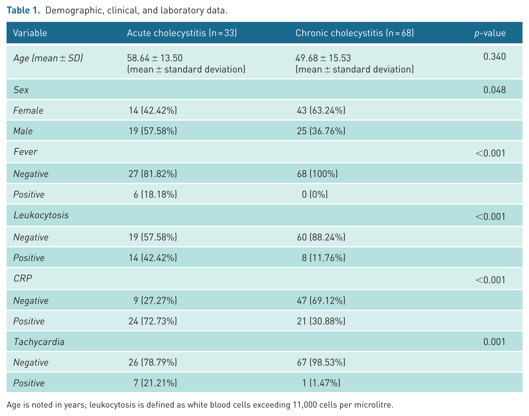
Age is noted in years; leukocytosis is defined as white blood cells exceeding 11,000 cells per microlitre.
There was no significant age difference (58.64 vs 49.48 years; p = 0.34), but sex differed significantly, with acute cholecystitis more prevalent in males (42.42% vs 63.24%; p = 0.048). Among clinical and laboratory variables leukocytosis, Neutrophil percentages, fever, tachycardia and a positive C-reactive protein (CRP) were significantly more prevalent in those with acute cholecystitis (p < 0.001).
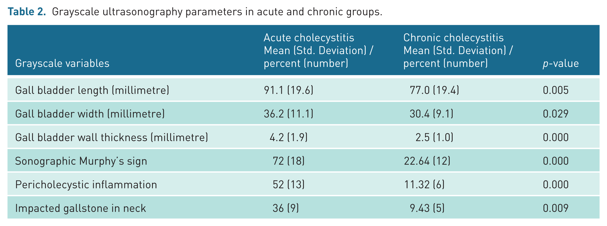
All grayscale variables were significantly more prevalent in the acute cholecystitis group. Gallbladder length was significantly greater in acute cholecystitis (91.1 ± 19.6 mm) compared to chronic cholecystitis (77.0 ± 19.4 mm; p = 0.005). Gallbladder width was also significantly greater in the acute group (36.2 ± 11.1 mm vs 30.4 ± 9.1 mm; p = 0.029). Similarly, gallbladder wall thickness was markedly higher in acute cases (4.2 ± 1.9 mm) than in chronic cases (2.5 ± 1.0 mm; p < 0.001). Pericholecystic inflammation, a hallmark of acute inflammation, was observed in 52% of acute cases but only 11.3% of chronic cases (p < 0.001) (Table 2).
Grayscale ultrasonography parameters in acute and chronic groups.
Doppler ultrasound demonstrated a significant association between the extent of intramural colour flow and the diagnosis of acute cholecystitis. Restricted colour flow at the gallbladder neck was more common in non-acute cases, whereas extension of flow through three-quarters of the gallbladder length was significantly associated with acute cholecystitis (p < 0.001) (Table 3).
Gallbladder wall vascularity.
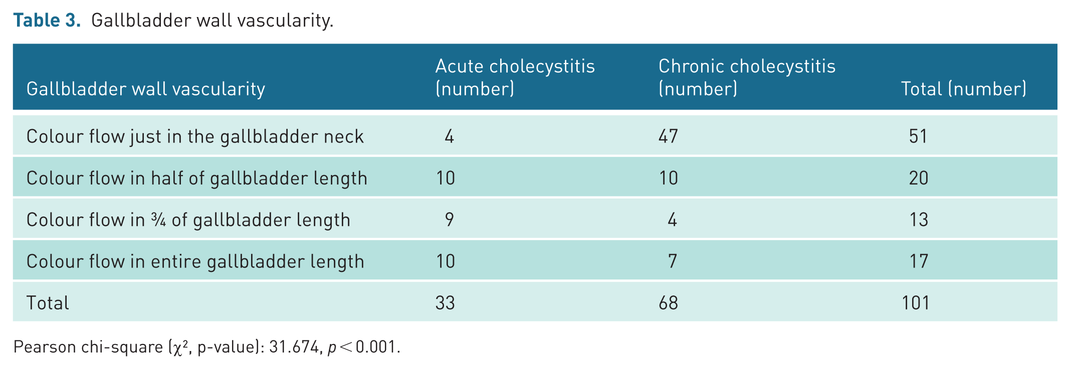
Pearson chi-square (χ², p-value): 31.674, p < 0.001.
A comparison of spectral Doppler ultrasound parameters between patients with acute and chronic cholecystitis revealed a statistically significant increase in the PSV of the CA in the acute group (40.76 ± 14.89 cm/s) compared to the chronic group (26.12 ± 15.51 cm/s; p < 0.001). Likewise, the PSV of the hepatic artery was significantly higher in patients with acute cholecystitis (73.97 ± 24.45 cm/s) than in those with chronic cholecystitis (60.16 ± 22.43 cm/s; p = 0.006). In contrast, no statistically significant differences were observed in the RI of either the cystic or hepatic arteries between the two groups. These findings suggest that elevated PSV values, particularly of the CA, may serve as potential Doppler sonographic indicators in the differential diagnosis of acute versus chronic cholecystitis (Figure 1, Table 4).
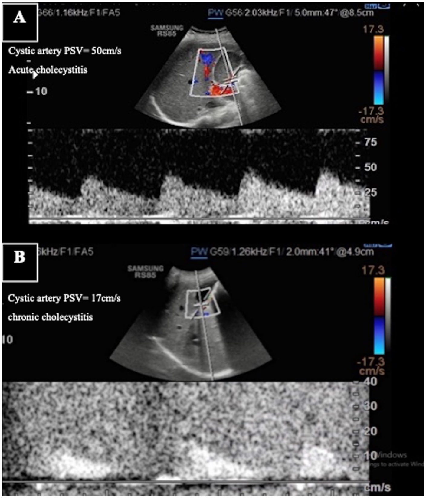
Doppler sonographic findings in acute and chronic cholecystitis. (a) Acute cholecystitis: Colour Doppler ultrasound showing increased vascularity with elevated cystic artery peak systolic velocity (PSV = 50 cm/s), consistent with active inflammation. (b) Chronic cholecystitis: Reduced vascularity and lower cystic artery PSV (17 cm/s).
Colour spectral Doppler ultrasonography indices of acute and chronic cholecystitis.
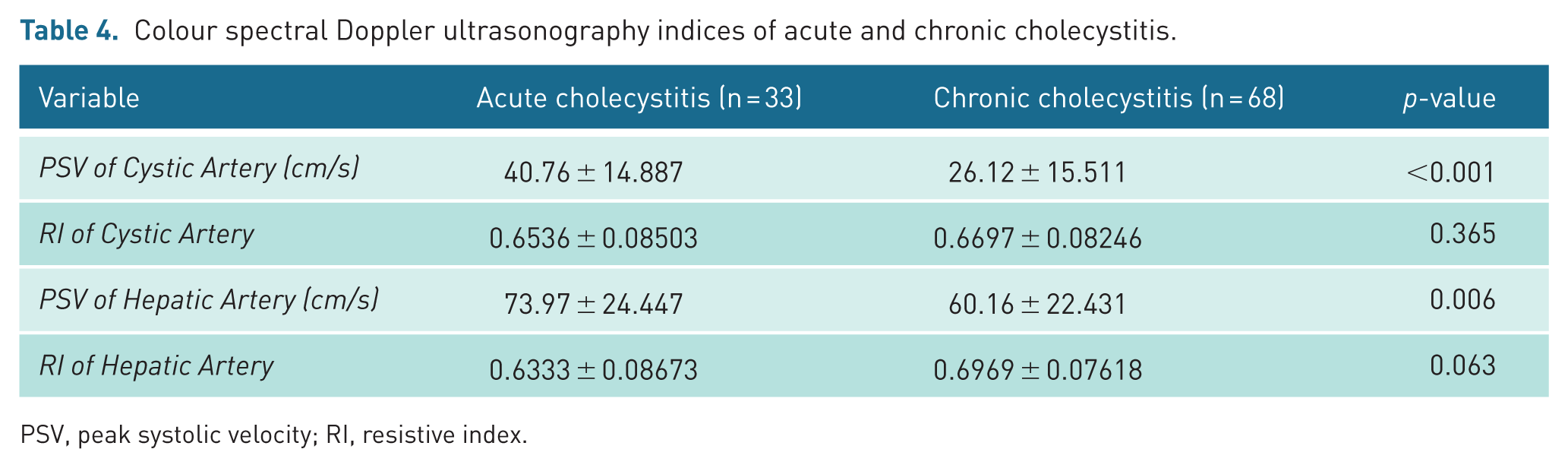
PSV, peak systolic velocity; RI, resistive index.
The AUCs of the CA and Hepatic Artery RI, were 0.428 and 0.428, respectively, which indicates weak diagnostic performance, while the AUCs of CA PSV and Hepatic Artery PSV were 0.779 and 0.674, respectively, indicating good and moderate discrimination. A cut-off of 31.5 cm/s for the CA, PSV resulted in a sensitivity, specificity, PPV, NPV, and accuracy of 0.909, 0.706, 0.6, 0.941 and 0.772 while a cut-off of 57.5 cm/s for hepatic artery PSV yielded sensitivity 0.970, specificity 0.882, PPV 0.799, NPV 0.983, and accuracy 0.910 (Figure 2, Table 5).
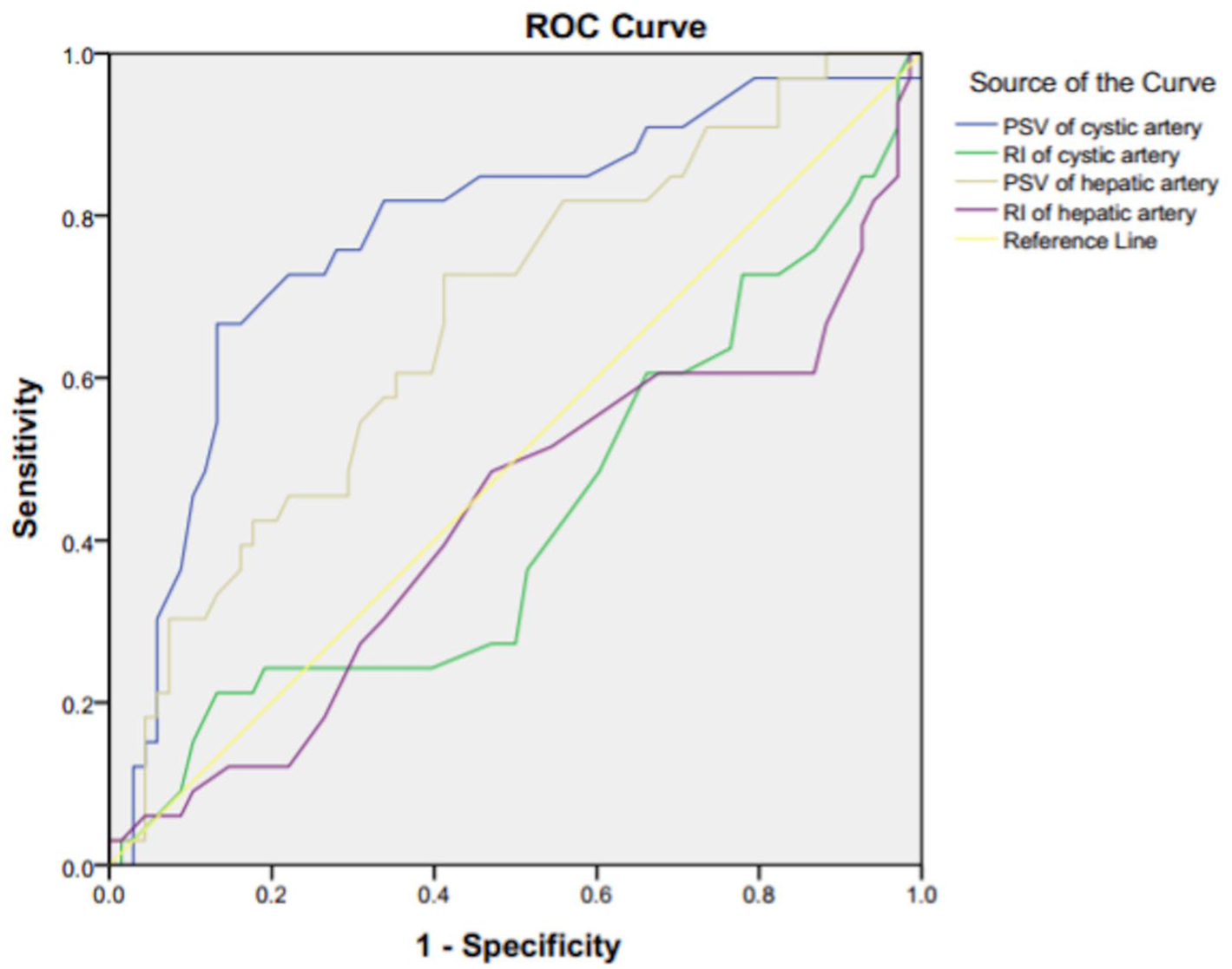
Receiver operating characteristic curves for cystic- and hepatic-artery peak systolic velocity (PSV) and resistive index (RI) in distinguishing acute from chronic cholecystitis; area under the curve values are shown with 95% confidence intervals.
Diagnostic performance of cystic and hepatic artery Doppler indices in differentiating acute from chronic cholecystitis based on ROC curve analysis.
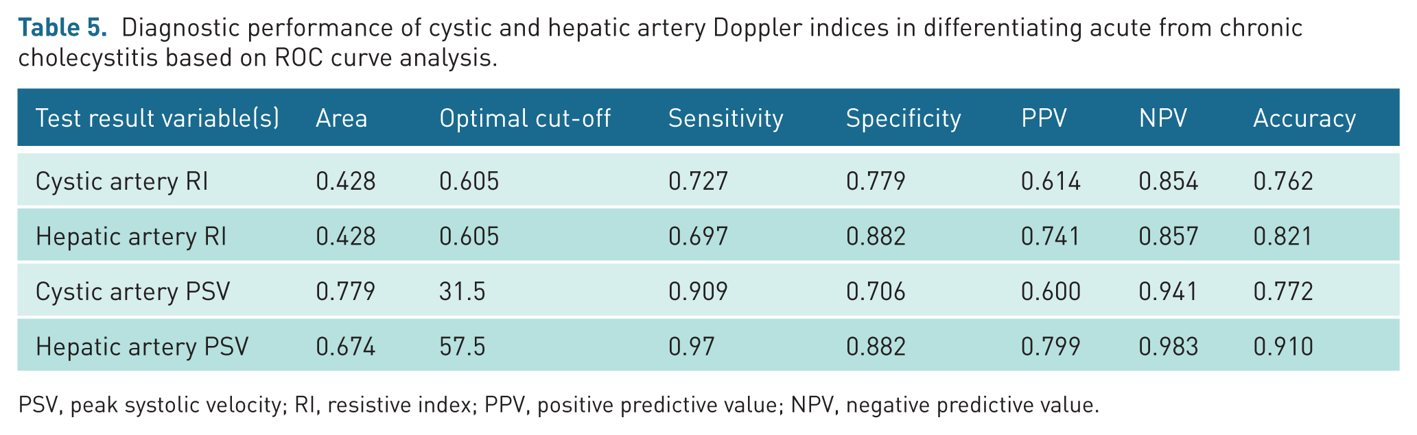
PSV, peak systolic velocity; RI, resistive index; PPV, positive predictive value; NPV, negative predictive value.
To evaluate the diagnostic efficacy of these Spectral-Doppler variables we conducted ROC curve analysis. (Figure 2, Table 5)
According to these results, CA RI and Hepatic artery RI are not useful for diagnosis, whereas CA PSV and Hepatic artery PSV show good and reasonable performance, respectively, in distinguishing chronic and acute cholecystitis.
The added value of considering CA PSV beside grayscale findings for the diagnosis of acute cholecystitis was examined using two logistic regression equations. CA PSV is not included in the first model, but it is included in the second model. (Supplemental Table S1)
Both models demonstrated an adequate fit to the data, as indicated by non-significant Hosmer–Lemeshow test results, an insignificant result that displays a good fit of the model to the data.
The comparison of predictive accuracy between the models with and without CA PSV provides insights into the minimal increase in diagnostic performance. The addition of CA PSV raises the model’s accuracy by only 0.1 percentage points, bringing it from 94% to 95%. This paucity of contribution is likely owing to the inherent robustness of grayscale variables, which overshadow the addition of a weak predictor such as CA PSV.
Discussion
The diagnostic work-up for acute cholecystitis is currently one of the most crucial parts of clinical practice as it is a very common disorder with a high potential for severe forms of complications. The present study aimed to evaluate the efficiency of the indices of colour Doppler ultrasound, namely the RI and PSV of the CA and hepatic artery, in acute cholecystitis distinction from chronic cholecystitis. Findings illustrated that PSV may be a promising index to use for the diagnosis of acute cholecystitis, while RI was weak.
In light of this, several investigations have previously examined the predictive utility in regard to hyperaemia of the gallbladder wall for the identification of acute cholecystitis. 17 Nevertheless, no research has examined the CA’s overall noteworthy corrected angle peak systolic velocity. Our research indicates that integrating colour Doppler ultrasound indices, particularly CA PSV, into the diagnostic protocol may provide a modest improvement in diagnostic yield – approximately 1%. While this enhancement is limited, it may still aid clinical decision-making when used alongside conventional grayscale parameters. The minimal contribution of Doppler indices is likely attributable to the strong diagnostic performance of grayscale variables, which may overshadow the incremental value of other predictors such as CA PSV.
Our results demonstrated significant differences in CA PSV between patients who have acute cholecystitis (40.76 ± 14.887 cm/s) and individuals who have chronic cholecystitis (26.12 ± 15.511 cm/s).
Out of all studied parameters, CA PSV demonstrated good diagnostic performance (AUC = 0.779) using ROC curve analysis (Figure 2). The optimal cut-off value was calculated to be 31.5 cm/s, yielding a sensitivity of 90% and a specificity of 70% while RI demonstrated poor diagnostic performance (AUC = 0.42).
These findings are in keeping with previous studies in which the authors have demonstrated that PSV could be a potential diagnostic marker. Also, Perez et al. 18 reported that the CA PSV was a differentiating low-cost, non-invasive diagnostic marker between acute cholecystitis and similar gallbladder disease with an AUC of 0.82, an optimum cut-off of 30 cm/s.18,19 The consistency of the findings across all studies suggests that PSV is a reliable diagnosis tool.
However, the modest usefulness we found differs from some early studies, such as that of Paulson et al, 15 who described RI as useful in the diagnostics of acute cholecystitis, with increased RI values suggesting increased vascular resistance because of inflammation. Differences in design, the populations studied and the quality of the ultrasound machine used might account for the discrepancies, and these could be addressed in what would be a welcome standardisation of future research.
Doppler ultrasound measurements might improve diagnostic accuracy of acute cholecystitis. Loehfelm and colleagues 12 conducted another large report to assess diagnostic performance related to elevated peak systolic HAv in patients with suspected acute cholecystitis. They reported that HAv ⩾ 100 cm/s was more accurate (69%) than individual grayscale markers of acute cholecystitis, including the presence of gallstones (51%) plus sonographic Murphy sign (50%).
Although it is imperative to learn about Doppler ultrasound velocities as our research and Loehfelm et al. 12 demonstrate, the value of CA PSV represents a more specific approach to the diagnosis of gallbladder-specific inflammation. Hepatic artery velocity was emphasised by Loehfelm et al – which is a useful measure but is likely influenced by a broad range of other hepatic and systemic diseases. CA PSV, however, is a more differentiated measure of the vascular changes present and is likely a more directly sensitive diagnostic criterion.
The first-line imaging technique for probable acute cholecystitis is ultrasonography because it is non-invasive, widely available, and low-cost. Our results underscore the diagnostic value of ultrasonography, particularly in conjunction with other sophisticated methods, including colour Doppler ultrasound. Classic parameters of grayscale ultrasound, including the thickness of the gallbladder wall and the existence of pericholecystic fluid, have shown statistically significant differences between the two groups (acute vs chronic cholecystitis) in the present study.
Our study, based on the preliminary work done by Mencarini et al, has utilised modern imaging advancements including colour Doppler ultrasound. We found that CA PSV is a useful parameter in diagnosing acute cholecystitis; however, it does not provide additional diagnostic value beyond traditional grayscale sonographic parameters. Doppler indices were not included in the study referencing Mencarini et al., and our paper pushes the field further instead of using an arbitrary cut-off point.
PSV provides an objective, quantitative parameter with minimal subjectivity. Our study demonstrates that the addition of colour Doppler parameters to grayscale ultrasonography does not significantly enhance diagnostic accuracy.
Limitations
This study does have some limitations, including our single-centre design and small sample size, which may limit generalisability. Another limitation is that a single radiologist performed all examinations, which may introduce observer bias. Multicentre research with increased patient cohorts, including multiple observers, may be more robust to yield generalisable data.
Second, the results may be less applicable to the general patient population if individuals with major incomplete clinical information and comorbidities were excluded. This is because these comorbid conditions, including cardiovascular disease or liver disease, may change the hemodynamics within the vasculature of the gallbladder and, consequently, the measured ultrasound parameters. Future studies on the diagnostic efficacy of the sonographic indices should also encompass a more heterogeneous patient population to minimise selection bias and offer a better grasp of the clinical utility of a priori sonographic indices in the diagnostics of acute cholecystitis.
Further research will also be required to implement our technique across diverse populations of basic scientists and clinical providers because the reproducibility of ultrasound measurements is directly related to inter-operator variability. Efforts need to be taken to standardise ultrasound protocols, including measurement techniques and Doppler settings, so results can be reproduced consistently across different field settings.
Conclusion
CA PSV is a useful, non-invasive adjunct for diagnosing acute cholecystitis, but it adds only a small incremental benefit beyond grayscale ultrasound. The RI found no usefulness in our study, but the presence of multiple ultrasonographic parameters does warrant further evaluation. Ultrasonography offers a myriad of approaches that remain an essential diagnostic tool in acute cholecystitis.
Supplemental Material
sj-docx-1-ult-10.1177_1742271X261434665 – Supplemental material for Diagnostic value of cystic artery colour Doppler ultrasound indices in acute cholecystitis
Supplemental material, sj-docx-1-ult-10.1177_1742271X261434665 for Diagnostic value of cystic artery colour Doppler ultrasound indices in acute cholecystitis by Alireza Abkhoo, Mina Behzadi, Narjes Mohamadzadeh, Niloofar Ayoobi Yazdi, Amir Ghasemlouei, Jayran Zebardast and Faeze Salahshour in Ultrasound
Footnotes
Acknowledgements
None.
Ethical considerations
This study was approved by the Research Ethics Committee of Imam Khomeini Hospital Complex, Tehran University of Medical Sciences, Tehran, Iran (approval reference IR.TUMS.IKHC.REC.1401.358). The study was conducted in accordance with the 1964 Declaration of Helsinki and its later amendments.
Doppler GB_Title Page.
Consent to participate
Not applicable. Only anonymised and de-identified ultrasound images are used, and no identifiable photographs of patients are included. According to institutional policy, specific written permission is not required in this situation.
Consent for publication
Not applicable. This article reports an observational study of a cohort of surgical patients and is not a case report.
Author contributions
All authors read and approved the final version of the manuscript.
Funding
The authors received no financial support for the research, authorship, and/or publication of this article.
Declaration of conflicting interests
The authors declared no potential conflicts of interest with respect to the research, authorship, and/or publication of this article.
Data availability statement
De-identified data and analysis code are available from the corresponding author upon reasonable request.
Use of AI-Assisted tools
AI tools were used only for language editing; all content was generated and verified by the authors.
Supplemental material
Supplemental material for this article is available online.
References
Supplementary Material
Please find the following supplemental material available below.
For Open Access articles published under a Creative Commons License, all supplemental material carries the same license as the article it is associated with.
For non-Open Access articles published, all supplemental material carries a non-exclusive license, and permission requests for re-use of supplemental material or any part of supplemental material shall be sent directly to the copyright owner as specified in the copyright notice associated with the article.