
Abstract
Background and aim
We determined to investigate the incidence and clinical impact of new cerebral microbleeds after intravenous thrombolysis in patients with acute stroke.
Methods
The THAWS was a multicenter, randomized trial to study the efficacy and safety of intravenous thrombolysis with alteplase in patients with wake-up stroke or unknown onset stroke. Prescheduled T2*-weighted imaging assessed cerebral microbleeds at three time points: baseline, 22–36 h, and 7–14 days. Outcomes included new cerebral microbleeds development, modified Rankin Scale (mRS) ≥3 at 90 days, and change in the National Institutes of Health Stroke Scale (NIHSS) score from 24 h to 7 days.
Results
Of all 131 patients randomized in the THAWS trial, 113 patients (mean 74.3 ± 12.6 years, 50 female, 62 allocated to intravenous thrombolysis) were available for analysis. Overall, 46 (41%) had baseline cerebral microbleeds (15 strictly lobar cerebral microbleeds, 14 mixed cerebral microbleeds, and 17 deep cerebral microbleeds). New cerebral microbleeds only emerged in the intravenous thrombolysis group (seven patients, 11%) within a median of 28.3 h, and did not additionally increase within a median of 7.35 days. In adjusted models, number of cerebral microbleeds (relative risk (RR) 1.30, 95% confidence interval (CI): 1.17–1.44), mixed distribution (RR 19.2, 95% CI: 3.94–93.7), and cerebral microbleeds burden ≥5 (RR 44.9, 95% CI: 5.78–349.8) were associated with new cerebral microbleeds. New cerebral microbleeds were associated with an increase in NIHSS score (p = 0.023). Treatment with alteplase in patients with baseline ≥5 cerebral microbleeds resulted in a numerical shift toward worse outcomes on ordinal mRS (median [IQR]; 4 [3–4] vs. 0 [0–3]), compared with those with <5 cerebral microbleeds (common odds ratio 17.1, 95% CI: 0.76–382.8). The association of baseline ≥5 cerebral microbleeds with ordinal mRS score differed according to the treatment group (p interaction = 0.042).
Conclusion
New cerebral microbleeds developed within 36 h in 11% of the patients after intravenous thrombolysis, and they were significantly associated with mixed-distribution and ≥5 cerebral microbleeds. New cerebral microbleeds development might impede neurological improvement. Furthermore, cerebral microbleeds burden might affect the effect of alteplase.
Introduction
Cerebral microbleeds (CMBs), small round hypointense radiological lesions detected on blood-sensitive MRI sequences, represent an established cerebral microangiopathy. Pathologically, CMBs mostly correspond to hemosiderin-laden macrophages in the adjacent vasculopathy that results from red blood cell leakage. Strictly lobar CMBs typically corresponds with cerebral amyloid angiopathy (CAA), whereas deep or mixed CMBs may indicate hypertensive arteriopathy,1,2 and they contribute clinical concerns of an increased risk of potential intracranial hemorrhage (ICH). Besides ICH, evidence from three single-center observational studies suggests that new CMBs were detected after intravenous thrombolysis (IVT) using recombinant tissue plasminogen activator (rt-PA), with rates that ranged from 4 to 5%.3–5 It is probably assumed that rt-PA plays a role in the etiology of new CMBs after acute ischemic stroke, although this has never been proven in a randomized controlled trial. The rates at which new CMBs emerge, the extent to which CMBs modify the effects of IVT, and the factors that affect CMBs development remain unclear.
Recently, the efficacy and safety of MRI-guided intravenous rt-PA have been demonstrated clearly in patients with unknown-onset stroke, and this therapeutic strategy is recommended in guidelines.6–8
As a prespecified sub-study of the THrombolysis for Acute Wake-up and unclear-onset Strokes with alteplase at 0·6 mg/kg (THAWS) trial, 8 we aimed to investigate the temporal dynamics of CMBs development by conducting three consecutive MRI assessments. Furthermore, we aimed to elucidate the clinical impact of CMBs by assessing associations with ICH and functional outcomes.
Methods
We analyzed the dataset of the THAWS trial; an investigator-initiated, multicenter, randomized, open-label, blinded-endpoint evaluation, controlled trial. The study design, inclusion and exclusion criteria, randomization procedure, and results have been described previously.8–10 In brief, to test whether alteplase at 0.6 mg/kg was effective and safe, the THAWS trial recruited 131 patients with acute ischemic stroke on awakening or with an unknown time of onset who had diffusion-weighted imaging (DWI)-fluid‐attenuated inversion recovery (FLAIR) mismatch (Supplementary Figure 1). The trial was approved by the local ethics committee or institutional review board at each participating center. Patients or their relatives provided written informed consent in accordance with ethical regulations.
Study population
We reviewed the imaging data from all the patients randomized in the THAWS trial. This study’s exclusion criteria were lack of images and artifacts on T2*-gradient-recalled echo (GRE) MRI. As a prescheduled, brain MRI, including T2*-GRE, was performed at baseline, at 22–36 h, and at 7–14 days. The details of clinical and imaging assessments, and the MRI protocol have been described previously.8,9
Assessments of cerebral microbleeds
MRI was performed using a 3 T (n = 61) or 1.5 T (n = 52) MRI scanner. The same field strength was used at all three time points. We assessed the presence, numbers, and anatomical distributions of CMBs at baseline. CMBs were identified as punctuate hypointense lesions <10 mm on T2*-GRE, according to consensus recommendations. 11 The numbers of CMBs in each location were assessed using standardized definitions.11,12 Thus, CMBs distributions were classified as strictly lobar, mixed, and deep (deep and infratentorial).11,12 All of the T2*-GRE MRIs were analyzed centrally using OsiriX MD, version 6.5.2 (Pixmeo SARL, Geneva, Switzerland), to attain side-by-side visual comparisons of the images at baseline and follow-up at 22–36 h and 7–14 days. New CMBs were defined as CMBs that were not present at baseline. Newly emerging small, hypointense lesions adjacent to the DWI, which indicated the acute infarct core, were not identified as new CMBs.3–5 Adjudication of CMBs was made by consensus of two raters (K.M, S.Y), a stroke neurologist with >15 years’ experience who were blinded to clinical information. In the case of disagreement, final consensus was reached involving a third experienced observer (M.I.). Assessments of other small-vessel disease (SVD) markers are described in the supplemental material.
Outcome measures
The outcome measures were (1) new CMBs on images obtained at 22–36 h and 7–14 days, (2) unfavorable functional outcomes, including modified Rankin Scale (mRS) score 3–6 at 90 days, the mRS score at 90 days, and change in the NIHSS score from 24 h and 7 days, (3) any ICH after IVT, including symptomatic ICH (sICH), parenchymal hematoma (PH), and remote parenchymal hematoma (rPH). The definition of sICH was an increase in NIHSS score by ≥4 from baseline and PH type II (PH-2) on MRI at 22–36 h after treatment initiation. 13 rPH was defined as an ICH remote from the symptomatic ischemic area. 14
Statistical methods
Analysis of outcome was performed in the intention-to-treat population for all patients with available information. Demographic, radiological, and clinical variables were compared between the two groups using the Fisher’s exact test for categorical variables, and the 2-sample Wilcoxon rank-sum test or Student’s t-test for continuous variables, as appropriate. NIHSS change was analyzed by analysis of covariance, where the model included treatment group as a factor and NIHSS at baseline as a covariate. Mixed-effect binary regression models adjusted for MRI field strengths as random intercepts were performed for evaluating the association of individual CMBs findings (i.e., presence, numbers, burden categories [0, 1, 2–4, and ≥5], distributions) with outcomes. Model was adjusted for age and sex. Interactions between treatment and each CMBs findings were also tested. We used Firth’s penalized likelihood to evaluate the impact of alteplase that had an association on new CMBs development with zero cells by conventional regression model. Since this work is considered as hypothesis generating, p-values were reported based on two-sided hypothesis without specific adjustment for multiplicity, and a p-value of <0.05 was used to determine statistical significance. Analysis was performed using STATA (version16, Stata Corp LP, College Station, TX, USA).
Results
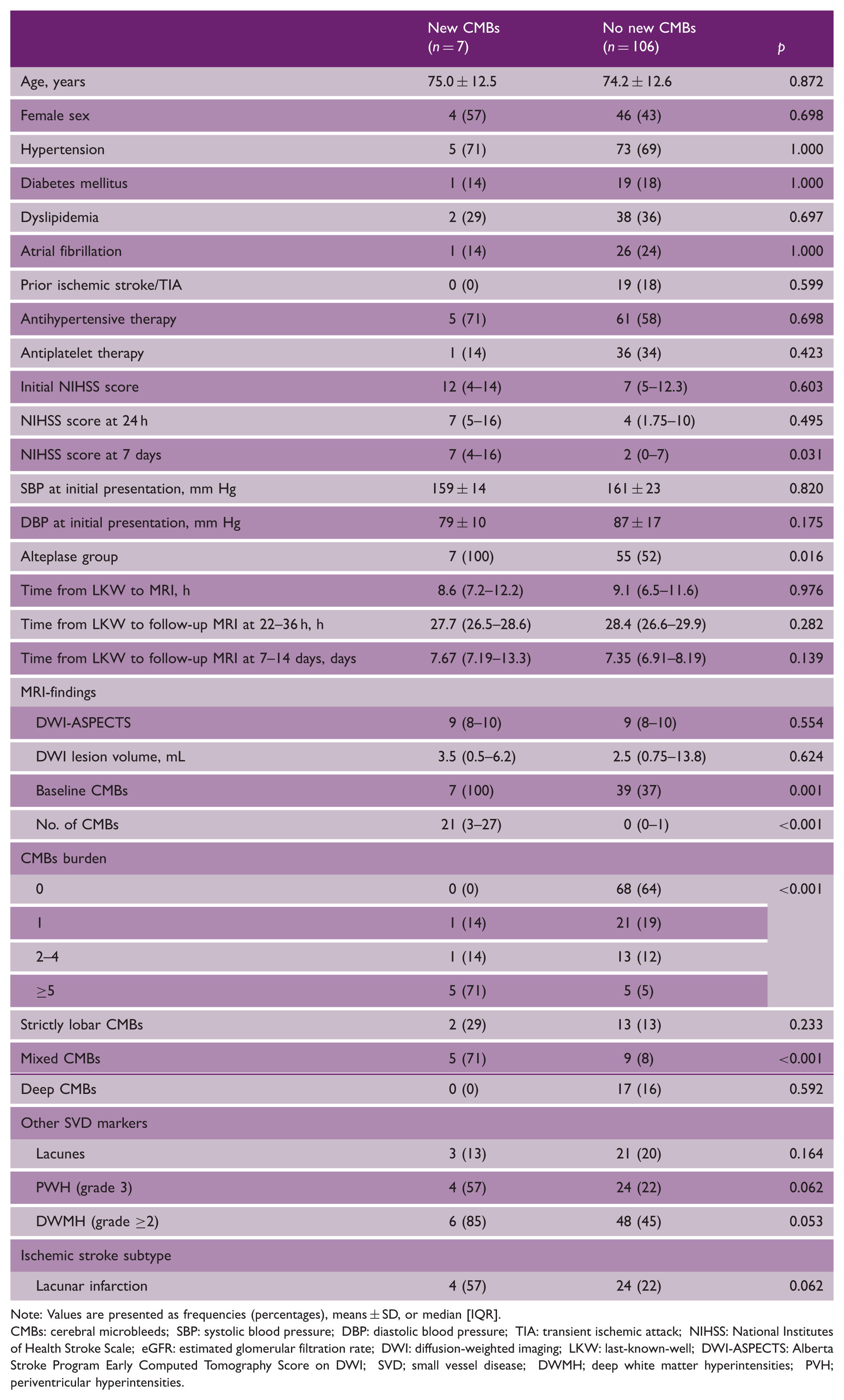
Of the 131 enrolled patients, 126 patients with follow-up data obtained within the allowance schedule were reported in the main publication. 8 Of these, 13 patients were excluded due to incomplete MRI examination including the movement artifacts. Thus, 113 patients were available for final analysis (47% women, mean age 74.3 ± 12.6 years, median NIHSS 7 [IQR: 5–13], alteplase : control = 62 : 51). The baseline characteristics of patients between the treatment allocation are shown in Supplementary Table 1. The prevalence of baseline CMBs was not different between the groups (28/62 with alteplase [45%] vs.18/51 with control [35%], p = 0.338).
Baseline MRI findings
Among all the subjects, 46 had ≥1 CMBs (41%). The median number of CMBs for the 46 patients was 2 (IQR: 1–3.25, range 1–28): 22 patients had 1 CMB (19%), 14 patients had 2–4 CMBs (12%), and 10 patients had ≥5 CMBs (9%) (Table 1). Among CMBs distributions, the number of mixed CMBs was the highest: 8.5 [2–21.3] (Figure 1).
The number of CMBs by the distribution on baseline MRI. The number of CMBs by the distribution; strictly lobar CMBs, median [IQR]:1 [1–3], mixed CMBs: 8.5 [2–21.3], and deep CMBs: 1 [1–1.5]. Patient characteristics by new CMBs Note: Values are presented as frequencies (percentages), means ± SD, or median [IQR]. CMBs: cerebral microbleeds; SBP: systolic blood pressure; DBP: diastolic blood pressure; TIA: transient ischemic attack; NIHSS: National Institutes of Health Stroke Scale; eGFR: estimated glomerular filtration rate; DWI: diffusion-weighted imaging; LKW: last-known-well; DWI-ASPECTS: Alberta Stroke Program Early Computed Tomography Score on DWI; SVD; small vessel disease; DWMH; deep white matter hyperintensities; PVH; periventricular hyperintensities.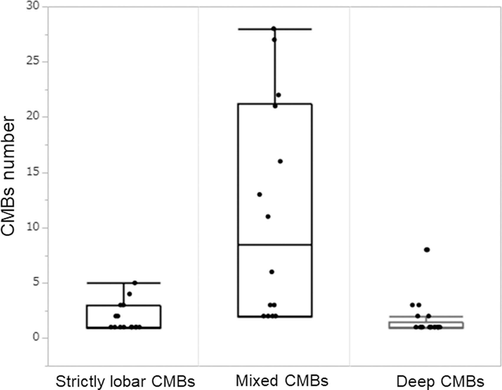
Follow-up imaging at 22 to 36 h and 7 to 14 days
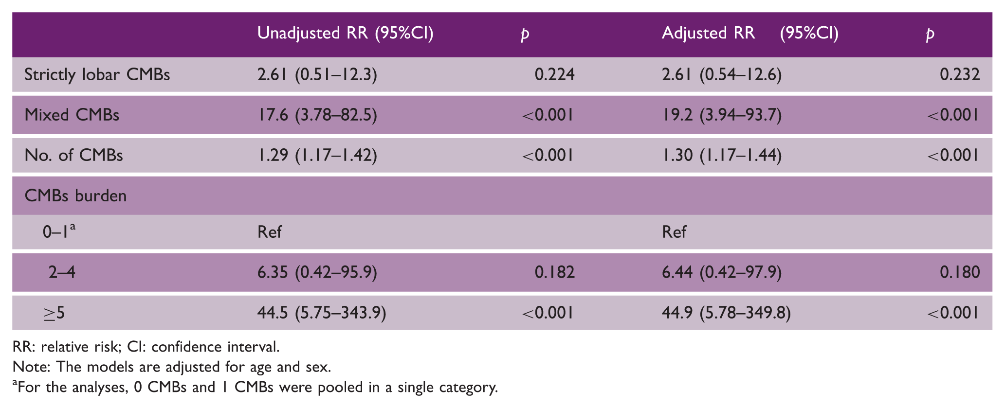
New CMBs were identified in seven patients (6%) at 22–36 h (median 28.3 h); all of these patients had received IVT (7/62, 11%) (Figure 2). Regression of baseline CMBs was not identified. New CMBs were single in all but one patient who had two new CMBs. Of a total eight new CMBs, five were located in strictly lobar regions (parietal-occipital lobe), two in strictly deep regions (basal ganglia), and one in mixed regions (parietal lobe and basal ganglia). The patients with and without new CMBs had comparable baseline characteristics, except for the allocation to the alteplase group, NIHSS score at seven days, and baseline CMBs findings; presence, numbers, burden categories, and mixed CMBs (Table 1). PVH (grade 3), DWMH (grade ≥2), and acute lacunar infarction were marginally associated with new CMBs, respectively. In age- and sex-adjusted analyses, the number of CMBs (relative risk (RR) 1.30, 95% confidence interval (CI): 1.17–1.44), mixed CMBs (RR 19.2, 95%CI: 3.94–93.7), and ≥ 5 CMBs (RR 44.9, 95%CI: 5.78–349.8) were each significantly associated with new CMBs (Table 2). Neither new additional or regression of CMBs was identified at 7–14 days (median 7.35 days). In the univariable analysis using Firth penalized maximum likelihood estimation, the association between alteplase and new CMBs did not achieve formal statistical significance (odds ratio: 13.9, 95% CI; 0.78–249.9, p = 0.074)
New CMBs after intravenous alteplase. Baseline diffusion-weighted imaging (DWI), T2*MRI, and follow-up T2*MRI at 24 h after IVT for acute ischemia in the territory of the left middle cerebral artery (yellow arrow): new CMBs, (green arrow): hemorrhagic transformation of infarcted brain tissue. Associations between baseline CMBs findings and new CMBs RR: relative risk; CI: confidence interval. Note: The models are adjusted for age and sex. For the analyses, 0 CMBs and 1 CMBs were pooled in a single category.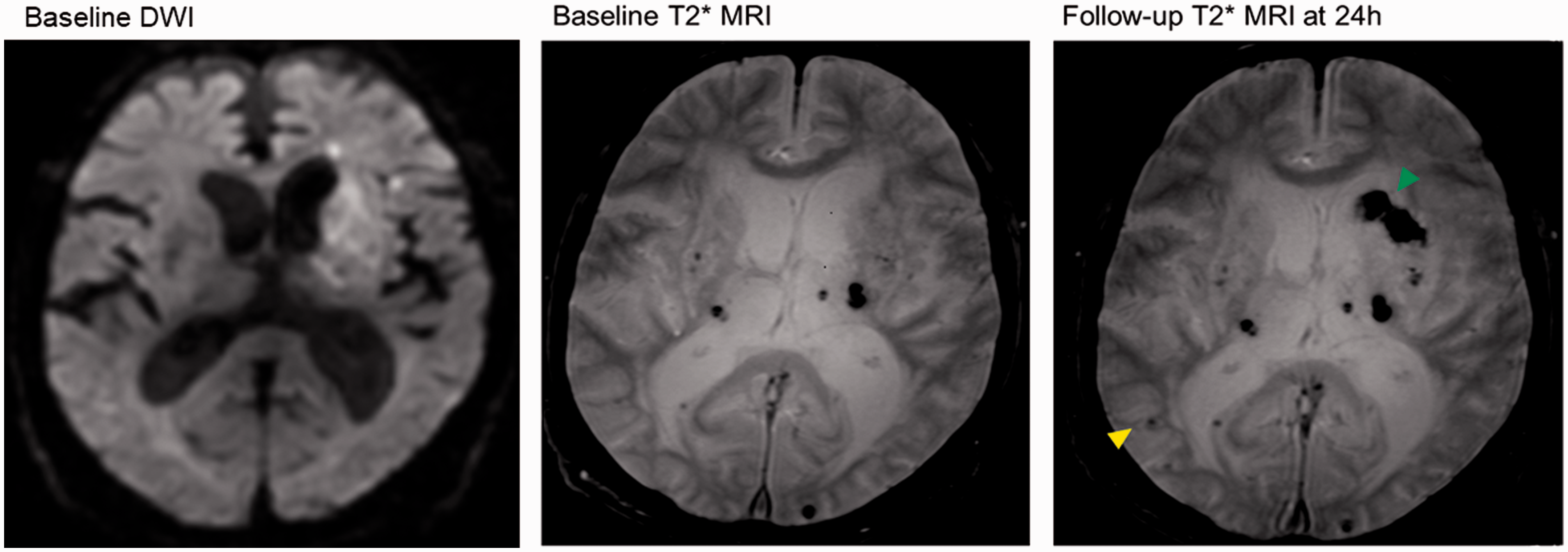
Associations between CMBs findings and clinical outcomes
Clinical outcomes by CMBs findings
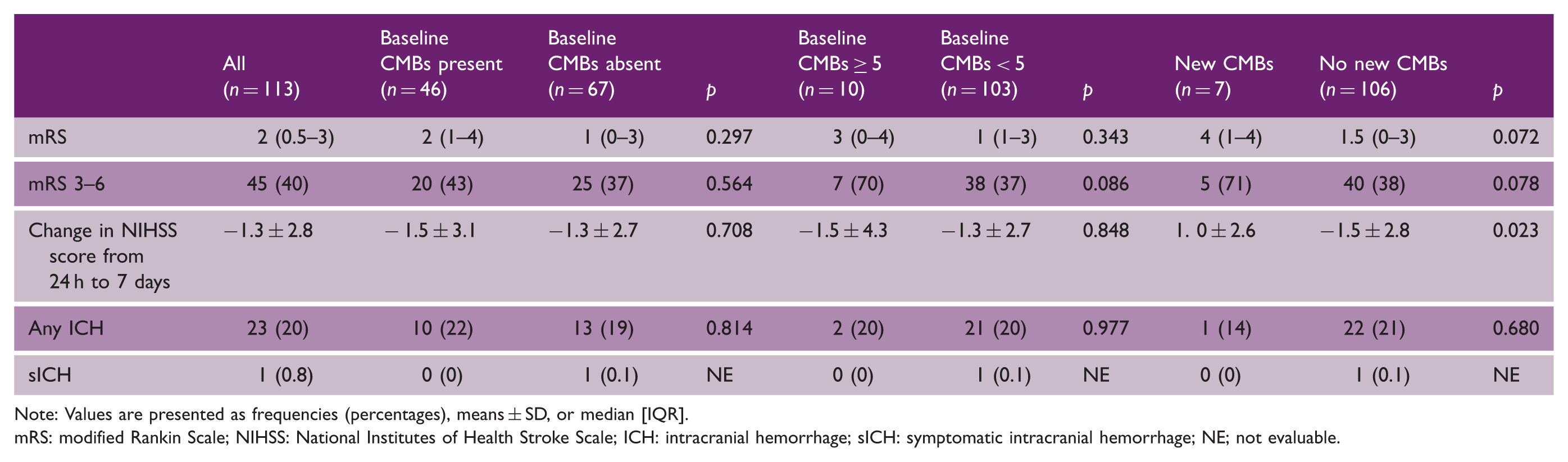
Note: Values are presented as frequencies (percentages), means ± SD, or median [IQR].
mRS: modified Rankin Scale; NIHSS: National Institutes of Health Stroke Scale; ICH: intracranial hemorrhage; sICH: symptomatic intracranial hemorrhage; NE; not evaluable.
Treatment with alteplase in patients with baseline ≥ 5 CMBs resulted in a numerically higher rate of unfavorable outcome (86% vs. 33%) and a numerical shift toward worse outcomes on ordinal mRS (median [IQR]; 4 [3–4] vs. 0 [0–3]), compared with those with < 5 CMBs (Supplementary Table 2). Interactions were significant between ≥5 CMBs and ordinal mRS score (p = 0.042) (Supplementary Table 2). Unfavorable outcomes were observed in five of seven patients (71%) with new CMBs that developed in the alteplase group and in none of the patients in the control group in which new CMBs did not emerge (Supplementary Table 2).
Any ICHs were identified in 23 patients (hemorrhage infarction type 1 (HI1): 9, HI2: 10, PH2: 4). There were no differences between patients with and without any CMBs findings regarding ICH (Table 3).
Associations between other SVD markers and clinical outcomes
There were no significant differences in clinical outcomes between each SVD groups, except that mRS score at 90 days was higher in patients with severe DWMH (grade ≥2) than those with mild DWMH (grade < 2)(median [IQR]; 2 [1–4] vs. 1 [0–3], p = 0.036) (Supplementary Table 3).
Discussion
We showed that new CMBs appeared in 11% of the patients after IVT for stroke, all CMBs emerged within 22–36 h, while that new CMBs did not develop in any of the patients who did not undergo IVT. New CMBs after IVT were significantly related to the high CMBs burden or a mixed distribution. New CMBs were significantly associated with an increase in the NIHSS score. We also demonstrated a significant interaction for unfavorable outcome between high CMBs burden and IVT.
Current knowledge regarding the temporal dynamics of CMBs is limited due to the scarcity of studies with repetitive MRI assessment. In this trial, new CMBs were only detected within 36 h after IVT by applying serial MRI. The overall frequency of new CMBs (11%) was higher in the current study than that reported rate from a meta-analysis of three observational studies (4.4%). 4 Also, a high CMBs burden was consistently associated with new CMBs; this finding concurs with the results from above-mentioned meta-analysis. 4
The predominance of non-lobar distribution of baseline CMBs was higher than those reported from previous meta-analyses,4,15 presumably due to ethnical deviation only to Asians in the present cohort, who frequently have non-lobar CMBs. 16 Previous studies have shown that preexisting strictly lobar CMBs were significantly associated with new CMBs or rPH after IVT, which indicated more specific effects of CAA-related injury.4,17,18 In contrast, most new CMBs after IVT linked a mixed distribution, which might be partly due to the greater CMBs count in the mixed distribution. Furthermore, higher number of CMBs was observed in patients with mixed CMBs compared with those with strictly lobar CMBs, reflecting the cumulative microangiopathy. Overall, the proportion of patients with ≥5 CMBs was higher in the present cohort (21%) compared with the rates reported from previous studies (4–5%).4,15 Higher CMBs burden may be a potential risk factor for new CMBs development, which might explain the number-dependent effects.
Of note, the observed new CMBs were associated with baseline CMBs burden, but which was not attributable to higher age, sex, hypertension, stroke severity, and admission blood pressure. Rather new CMBs tend to occur in preexisting vascular fragility due to microangiopathy, irrespective of the severity of the current ischemic stroke. Of eight new CMBs, five were identified in strictly lobar distributions. Pathologic studies have shown that hypertensive vasculopathy can also result in reduced vascular reactivity and structural damage, namely the disruption of the internal lamina and thin adventitia, of the cortical arterioles,19,20 rendering corresponding parenchymal tissue more vulnerable to CMBs development. However, we could not draw definitive conclusions about the origin of new CMBs due to the lack of histological verification to rule out.
As the common pathogenesis, SVD is associated with endothelial dysfunction, that contributes to blood–brain barrier dysfunction and smooth muscle cell injury, 21 which might increase susceptibility to thrombolysis. There was a borderline significant difference between acute lacunar infarction and new CMBs. These findings might support the small vessel hypothesis of new CMBs development in patients with SVD. CMBs are presumed to reflect minute intraparenchymal bleeding regions resulting from microvascular fragility or increased permeability.22,23 Moreover, the vasculature is left relatively unprotected to the acute insult of ischemic injury to vessel integrity, and ties to hemorrhagic-prone. 24 Given the mechanisms of action of alteplase, 25 the early development of CMBs might signify the cumulative synergistic effects of both factors. Furthermore, a third follow-up imaging session with an interscan interval of approximately seven days did not detect additional CMBs, which partly suggests they might not modify the parenchymal tissue response to alteplase.
Regarding the clinical impacts of CMBs, new CMBs were likely to be inversely associated with neurological improvement, which was indicated by the NIHSS score thereby reflecting vulnerability to acute ischemia due to impaired microcirculation, collateral function, and reduced plasticity because of impaired cerebral connectivity from more advanced SVD.
We demonstrated a significant interaction between CMBs burden and alteplase, indicating that ≥5 CMBs may modify the treatment response to alteplase. Our results reinforced previous observations of the heterogeneity in treatment with alteplase between patients with and without CMBs burden. 15 To our knowledge, this is the first RCT to investigate the association of CMBs with clinical outcomes following IVT. Although CMBs burden might affect the efficacy of IVT, larger trials will be needed to clarify this ill effects.
The strengths of this study include the THAWS trial relied on MRI-based for patient enrollment and thus enabled the serial MRI investigation for distinguishing CMBs development. Available data on IVT in MRI-confirmed new CMBs stem from an RCT, which could address the effect of IVT by the comparison with a control group. Stroke volume and imaging features were carefully rated by experienced specialists using standardized and validated image procedures, thereby minimizing risk of misclassification.
This study has some limitations. First, the sample size limited the numbers of comparisons and adjustment variables, and the study was probably underpowered for detecting associations. Second, the generalizability of our findings might be limited, due to the use of alteplase at 0.6 mg/kg officially approved dose in Japan.
Our study demonstrated that after IVT, new CMBs developed predominantly in patients with mixed CMBs and/or a high CMBs burden. Our findings proposed the clinical concern that the critical factor relevant to IVT appears to be preexisting diffuse arteriolar injury and a number-dependent effect may be the leading underlying pathology in early developing CMBs. Since CMBs development has been linked to increased risk of any stroke, cognitive decline, and dementia, our findings from new CMBs might help to elucidate the pathological processes of underlying CMBs features. Future larger prospective studies that include histopathological examination of CMBs are needed to elucidate the pathogenesis of new CMBs.
Supplemental Material
sj-pdf-1-wso-10.1177_17474930211035023 - Supplemental material for Cerebral microbleeds development after stroke thrombolysis: A secondary analysis of the THAWS randomized clinical trial
Supplemental material, sj-pdf-1-wso-10.1177_17474930211035023 for Cerebral microbleeds development after stroke thrombolysis: A secondary analysis of the THAWS randomized clinical trial by Kaori Miwa, Masatoshi Koga, Manabu Inoue, Sohei Yoshimura, Makoto Sasaki, Yusuke Yakushiji, Mayumi Fukuda-Doi, Yasushi Okada, Taizen Nakase, Masafumi Ihara, Yoshinari Nagakane, Shunya Takizawa, Koko Asakura, Junya Aoki, Kazumi Kimura, Haruko Yamamoto and Kazunori Toyoda in International Journal of Stroke
Footnotes
Declaration of conflicting interests
The author(s) declared no potential conflicts of interest with respect to the research, authorship, and/or publication of this article.
Funding
The author(s) disclosed receipt of the following financial support for the research, authorship, and/or publication of this article: This work was supported by the Japan Agency for Medical Research and Development (AMED) (JP16ek0210025h, JP18ek0210091h, 20lk0201094h0002, and 20lk0201109h0001)
Data availability
The data are available from the corresponding author upon reasonable request.
References
Supplementary Material
Please find the following supplemental material available below.
For Open Access articles published under a Creative Commons License, all supplemental material carries the same license as the article it is associated with.
For non-Open Access articles published, all supplemental material carries a non-exclusive license, and permission requests for re-use of supplemental material or any part of supplemental material shall be sent directly to the copyright owner as specified in the copyright notice associated with the article.