
Abstract
A retrospective study of drug abuse patients who developed arterial and venous complications in the upper extremity during 2002–2006 was performed. Twenty-two patients were admitted to hospital on 24 occasions over this period for treatment by our hand clinic. The drug most frequently causing complications was midazolam. The predominant clinical findings were increasing pain and loss of sensitivity in the hand, followed by oedema, cyanosis and marbling of the skin. Treatments included brachial block anaesthesia, low molecular weight heparin, embolectomy and fasciotomies. Despite these measures, amputations, mainly of the fingertips, were necessary in 15 patients. Complications in the upper extremity after self-injection by drug addicts are increasing; information and preventive procedures to minimize these complications are important and demanding tasks for health care bodies.
INTRODUCTION
Intravenous drug injections often occur with anaesthesia or during medical treatment. Considering the large amount of drugs in daily use, the complication rate is relatively small. However, drug addicts who make suspensions of crushed tablets for intravenous injection are a particularly vulnerable group (Charney and Stern, 1991) who may inadvertently inject a variety of drugs into arteries. These injections have been associated with numerous local and systemic problems, including cellulitis, local abscess formation, thrombophlebitis, formation of talc and microcrystalline cellulose granulomata, pseudoaneurysms and pulmonary embolism (Binswanger etal., 2000; Goldberg et al., 1984). If the drug is accidentally injected intra-arterially or into the extravascular spaces, other more severe complications may occur. The high incidence of Human Immunodeficiency (HIV) and Hepatitis B (HBV) and C (HCV) viral infections in these patients further complicates the situation in a variety of ways, not least by putting at risk the paramedical and medical staff involved in their treatment.
The number of intravenous drug users worldwide is estimated to be approximately 13.2 million (Coughlin and Mavor, 2006). In Finland, with a population of just over five million people, drug addicts using amphetamine and opiates are estimated to number 16 000–21 000, with 30–40% of these individuals residing in the region of Helsinki. In 2000, the health education stations for drug users in larger cities in Finland had 4800 customers. This number increased to 10 400 in 2004. The number of needles and syringes distributed free of charge for prevention of viral infections by these stations was 0.56 million in 2000 and 1.8 million in 2004 (Partanen et al., 2006).
In recent years, an increase has also occurred in the number of patients in need of hand surgery care. This article presents our experiences with these patients.
PATIENTS AND METHODS
This is a retrospective study of all drug abuse patients admitted to the Department of Hand Surgery of theHelsinki University Central Hospital during 2002–2006.
Patient data
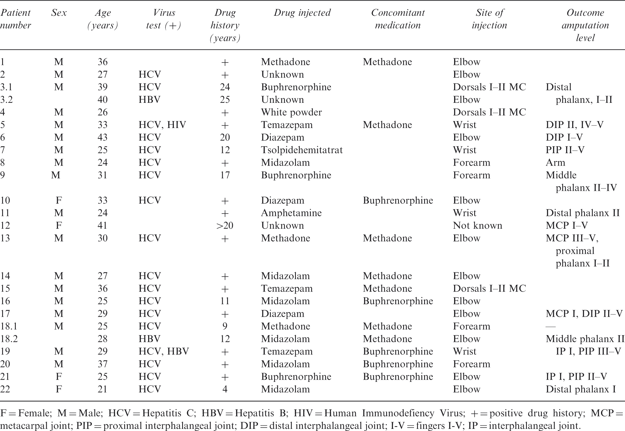
F = Female; M = Male; HCV = Hepatitis C; HBV = Hepatitis B; HIV = Human Immunodefiency Virus; + = positive drug history; MCP = metacarpal joint; PIP = proximal interphalangeal joint; DIP = distal interphalangeal joint; I-V = fingers I-V; IP = interphalangeal joint.
The drug that most frequently caused a complication was midazolam (six episodes) followed by diazepam (three episodes), temazepam (three episodes), buphrenorphine (three episodes), methadone (three episodes), amphetamine (one episode), tsolpidemhemitartrat (one episode), ‘a white powder’ (one episode) and an unknown drug (three episodes).
The time that elapsed between injection and onset of symptoms was described by most patients as very short.
Clinical findings
All patients had developed arterial and venous complications caused by intra-arterial self-injections of addictive drugs. The injection sites are illustrated in Fig 1.
(A,B) Self-injection sites of the narcotic addictive drugs.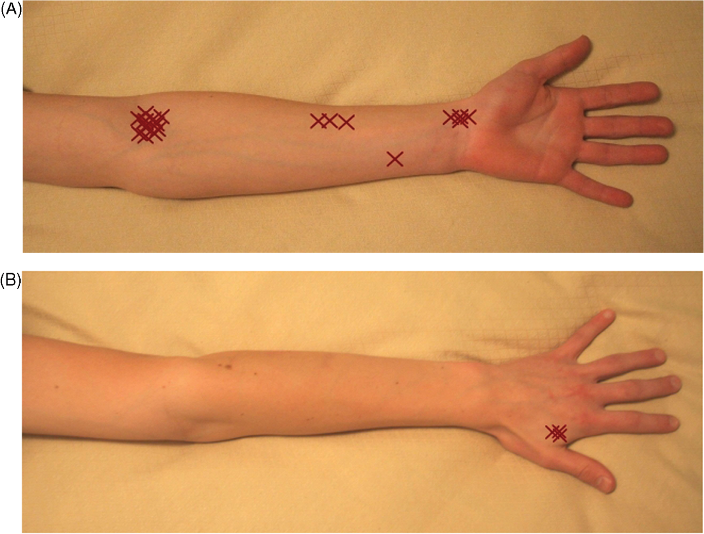
Most of the intra-arterial injections were at elbow level (12 episodes). The rest of the injections were into the radial artery of the forearm (three episodes), into the ulnar artery of the forearm (one episode), at wrist level (four episodes), and on the dorsal side of the hand between the first and the second metacarpal bones (three episodes). One patient did not know the injection level.
The predominant clinical finding was severe increasing pain in the limb distal to the injection site (19 episodes). Moreover, the patients were often unable to move the fingers (12 episodes) and reported a loss of sensitivity in the hand (14 episodes). Oedema (11 episodes) with variable amounts of cyanosis (nine episodes) with blue patches (12 episodes) and marbled skin (12 episodes) were also noted in many limbs. Signs of compartment syndrome were seen in the hand (four episodes) and the forearm (two episodes).The radial and ulnar arteries were not palpable (three episodes/one episode), and the hand felt cool (ten episodes).
Infections were present in two patients. One patient developed a 10 cm fistulous abscess on the dorsal side of the hand. Another patient developed a large necrotic area at the forearm with subsequent septic infection and was saved by an amputation near the shoulder.
Treatment
Current treatment after intra-arterial drug injections varies. The main principles include control of pain and vasospasm, prevention of thrombosis, promotion of vasodilatation and restoration of blood flow. A number of treatments have been used, including analgaesia, elevation, heparinization, vasodilators, sympathetic antagonists, anticoagulants, corticosteroids, prostaglandin inhibitors, platelet agents (Funk et al., 1999; Treiman et al., 1990) and hyperbaric oxygen therapy.
In this study, the following treatments were used in various combinations for intra-arterial injections:
Brachial plexus anaesthesia with ropivacaine was provided to 18 patients for vasodilatation and pain relief using an in-dwelling catheter and a perfusion pump. However, one of the patients removed the catheter, and another patient increased the administered dose eightfold by changing the pump settings. Low molecular heparin (Fragmin® or Klexane®) was given to 21 patients on 22 occasions for a mean of 12days. For one patient with a septic infection, the treatment lasted 90 days. On 17 occasions, the medication was started on the first day, on three occasions on the second day and on two occasions on the third and fourth day of treatment. Two patients were also treated with iloprost, a prostatacyclin analogue, infusion (Ilomedin®). Both patients had developed compartment syndrome at the forearm, and the medication was given as a part of treatment, that also included fasciotomy at the level of the forearm. An exploration of the arteria brachialis was also performed in one of the patients and a slow intra-arterial flow was found as the artery was obstructed by fibrosis. A catheter was inserted, and although no embolus was observed, the intra-arterial flow increased after the procedure. Another patient underwent embolectomy 2 days after she had self-administered an intra-arterial injection at the level of the antebrachium. A fasciotomy was performed and distally, near the wrist, an 8 cm embolus was removed from the radial artery. A fasciotomy was also performed on 12 admission occasions on 11 patients. The fasciotomies were performed on the palmar side of hand (four episodes), on the dorsal side of the hand (one episode), on both the dorsal and volar sides of the hand (one episode), and at the forearm (six episodes). A dissection of the carpal canal was also performed on all patients who underwent fasciotomy at the level of the forearm. Nineteen patients were given intravenous antibiotics according to the severity of the clinical situation. On nine occasions, a patient was given cefuroxime, on four occasions clindamycin and on four occasions the antibiotic changed between kefuroxime and clindamycine during the treatment. Ceftriaxone, sulphonamide, trimethoprim, levofloxacin, moxifloxacin, ciprofloxacin, amoxicillin, phenoxymethylpenicillin, cloxacillin, piperacillin, tazobactam and tobramycin were also prescribed on two occasions because of septic infections. An angiography was performed on three patients, and two underwent trombolytic treatment with 20 mg actilyse.
Steroids were not used in the treatment. Renal function was not measured, except for the patient with a severe septic infection.
RESULTS
The incidence of patients in need of hand surgery care increased during the study period, and the number of patients treated was one in 2002, two in 2003, two in 2004, nine in 2005 and ten in 2006. The mean delay in seeking medical help was 13.5 (range 0–180) days.
The treatment of 22 patients on 24 occasions resulted in 38 hospitalization periods, with a mean time in hospital of 9 (range 1–38) days.
Treatment was successful only for eight patients on nine occasions. The patients recovered fully, except for one who developed joint stiffness in all fingers, resulting in a finger flexion deficit of 4 cm. The patients had performed the intra-arterial injections at elbow level (five episodes), at the level of the forearm (one episode) and on the dorsal side of the hand between the first and the second metacarpal bones (two episodes). The injected drugs were midazolam (three episodes), methadone (one episodes), diazepam (one episode), temazepam (one episode), an unknown substance (two episodes). Brachial plexus anaesthesia was performed on eight occasions, low molecular weight heparin was given on nine occasions, fasciotomy was performed on four occasions, antibiotics were given on six occasions and ilomedin was given on one occasion.
Despite treatments given, amputations were performed on 15 occasions on 13 patients (mean 59 days after injection). Injections at elbow level (12 episodes) caused seven amputations at the level of the phalanges; for two patients in one finger through the distal interphalanx, for one patient to five fingers at the distal interphalangeal joint level, for one patient in one finger through the middle phalanx, for one patient at the proximal interphalangeal joint level in all fingers, for one patient at the metacarpal joint level in one finger and at the distal interphalangeal joint level in four fingers, andfor one patient at the metacarpal joint level in three fingers and through the proximal phalange in two fingers. Injections into the radial artery at the level of the forearm (three episodes) caused one amputation of the arm for one patient and amputations of three fingers for another patient. The single injection into the ulnar artery at the level of the forearm did not lead to any amputation. Injections at the wrist level (four episodes) caused amputations; for one patient at the distal phalanx level in one finger, for one patient at the distal interphalangeal joint level in three fingers, for one patient at the proximal interphalangeal joint level in three fingers and one for patient at the proximal interphalangeal joint level in four fingers. The three injections on the dorsal side of the hand between the first and second metacarpal bones caused one amputation at the distal interphalangeal joint level to two fingers. One patient did not know the injection level, but the injection caused amputations at the metacarpal joint level to all fingers.
Revisions of necrotic or infectious tissue approximately 115 days after the injection were performed on nine patients on ten occasions: on the dorsal side of the hand (two episodes), at the fingertip (five episodes), on the palmar side of the hand (one episode), and on the palmar side of the forearm (one episode). No difference in the amount of necrosis caused by different drugs was observed.
Angiography showed no blood flow on the ulnar side of the arcus palmaris in two patients, and no flow on theulnar and the radial side in one patient. Two of the patients underwent trombolytic treatment. Despite the treatment, amputations were necessary for both patients, at the level of the middle phalanges in three fingers and at the level of the proximal phalanges in five fingers.
Skin cover with free skin grafts were performed on seven patients on nine occasions (mean 17 days after injection): on the dorsal side of the hand (one episode), at the amputated arm (one episode), at the wound region of the fasciotomy on the palmar side of the forearm (five episodes), at the wound region of the fasciotomy on thedorsal side of the hand (one episode), and at the amputated finger (one episode).
Intra-arterial self-injection resulted in stiffness of the fingers and hands of six patients. All patients had performed the injection at the level of the elbow or the forearm. One patient developed an adductor deficit between the thumb and the index fingers, one patient developed an adductor deficit between the thumb and the index finger in combination with a 90° flexion deficit in the metacarpal joint, one patient’s fingers (II–V) were contracted in a rigid position, one patient developed a flexion deficit of 4 cm in the fingers (II–V), one patient developed joint stiffness in all the distal interphalangeal joints and one patient developed a 20° extension deficit in the proximal interphalangeal joint of the ring finger.
One patient gave birth during the hospitalization period.
DISCUSSION
In recent years, we have noted an increase in patients who by self-injection of narcotic addictive drugs have sustained intra-arterial complications in the upper extremity. In 2000–2003, risk-analysis research on intravascular drug addicts in Finland was conducted for the first time, supported by the A-Clinic Foundation, Stakes (National Research and Development Centre for Welfare and Health) and The Institute of Occupational Health. Four hundred and ninety-three drug addicts (70% male, 30% female) with a mean age of 28 years who were visiting health education stations to exchange their used needles and syringes were requested to undergo an interview. Most confirmed injecting drugs into their forearm, but about 15% injected into the hand or wrist. For an unknown reason, injections into the hand were more common in subjects who had abused drugs for more than five years.
Buphrenorphine was the most common drug injected intravascularly in Finland (∼70%) (Alho et al., 2006; Partanen et al., 2006). In a study of vascular complications of drug abuse in India, intravenous use of buphrenorphine was the commonest cause of vascular complications, followed by heroine. This was explained by the low cost and ready availability of buphrenorphine in India (Behera et al., 2003). Interestingly, buphrenorphine was not the drug causing most complications in our study. Although many of the addicts admitted using buphrenorphine, the complications seemed to arise from injections of other substances, indicating that the patients were ‘mixed users’ and had been using drugs for a long time. This was also confirmed by the fact that of the 12 patients receiving buphrenorphine or methadone to treat their drug abuse only four of the recorded episodes were caused by these drugs (methadone three, buphrenorphine one).
The pathogenesis of the ischaemia following intra-arterial injections is not fully understood and is still debated. Van der Post (1942) described gangrene of a finger following an intra-arterial injection of a barbiturate. Since then intra-arterial injections of many drugs, such as diazepam, amphetamine, heroin, cocaine, barbiturates, methadone and other narcotic agents, have been reported to cause acute vascular insufficiency (Shukla, 1995). Goldwell et al. (1984) reported a case in which a man who had prepared a solution of codeine phosphate performed an inadverted intra-arterial injection. The injection was followed by dry gangrene, requiring amputation at the wrist. To clarify whether the gangrene resulted from the codeine itself or the additives in the tablets, each component of the tablet was injected into the femoral arteries of dogs. Pure codeine did not affect the dogs, but injection of microcrystalline cellulose caused gangrene of the leg. Because our patients used many different drugs, determining whether the crushed tablets or the trauma caused by the injection itself led to subsequent complications is difficult. Only a few patients claimed to have used a filter. The dilution liquid was also unknown in all instances, but was assumed to be water. No difference in the time interval between injection and onset of symptoms between different drugs was observed.
The distal pathological damage may be caused by direct injury to the intima of the artery, resulting in activation of tissue thromboplastin, with subsequent vascular thrombosis, rather than an arterial spasm. Other mechanisms, including noradrenaline release, platelet aggregation and haemolysis, particulate embolism and toxic or hypersensitivity vasculitis, have also been implicated in the pathogenesis of ischaemia (Shukla, 1995).
Although an accidental injection is intra-arterial, changes may also occur in the venous circulation. Adir et al. (1991) and Funk et al. (1999) postulated that the consequent swelling depended on both arterial and venous endothelial injury, with the rapid development of venospasm as a result of the release of noradrenaline, changes in pH and acid crystal formation, possibly leading to acute compartment syndrome of the hand.
‘Puffy hand syndrome’ is a less common complication of intravenous injections of drugs. This is considered to be due to lymphatic obstruction and can appear years after drug abuse has ceased. The hand is usually swollen from the proximal segments of the fingers to the wrist. Differential diagnosis from deep palmar infections is necessary. Buphrenorphine, in particular, is thought to cause this syndrome (Simonnet et al., 2004). There were no instances of puffy hand syndrome in our study, and all our patients presented with acute swelling and severe pain of the hand.
Our treatment was successful in eight patients on nine occasions. The main treatments in these cases included brachial plexus anaesthesia (eight occasions), low molecular heparin (nine occasions), fasciotomy (four occasions) and antibiotics (nine occasions). Unsuccessful treatment leading to different levels of amputations in the upper extremity was seen in 13 patients on 15 occasions. The main treatment for these patients included brachial plexus anaesthesia (ten occasions), low molecular heparin (13 occasions), fasciotomy (eight occasions) and antibiotics (13 occasions).
In this study, it is not, therefore, possible to observe any correlation between treatment and outcome. Also the amputation level did not correlate with the level of the injection site. Seventeen patients had injected the drug in the forearm, and ten underwent amputations most often at the finger level.
Funk et al. (1999) reported two cases of compartment syndrome of the hand and the forearm as a result of intra-arterial injection of heroin. These were managed successfully by surgical fasciotomies alone. As a part of our treatment on 12 occasions, fasciotomy was performed on 11 patients. Later, eight of these patients underwent amputations of the fingers and the remaining three required excision of necrotic tissue at the level of the hand (one episode), the fingertip (one episode), or the forearm (one episode).
Angiography combined with trombolytic treatment did not save patients from amputation. This is in accordance with the results reported by Rautio et al. (2006), who found that patients treated with local thrombolysis had no better results than other patients.
Cefuroxime and clindamycin were used in 17 episodes. However, marked variation was present in the choice oftreatment and other antibiotics were administered such as ceftriaxone, sulphonamide, trimethoprim, levofloxacin,moxifloxacin, ciprofloxacin, amoxicillin, phenoxymethylpenicillin, cloxacillin, piperacillin, tazobactam and tobramycin. The variation is partly explained by two of the patients suffering from severe infections.