
Abstract
Dorsal adipofascial flaps have been used in the surgical reconstruction of complex injuries distal to the eponychial fold. Such injuries produce nail matrix devascularization/necrosis so that nail bed reconstruction can be a challenging technical problem. Irregular scarring of the nail bed and regrowth anomalies of the nail lamina can result, with both functional and cosmetic impairment of the finger. This study aimed to define the precise vascular anatomy of the dorsal adipofascial flap that has previously been used to reconstruct such complex soft tissue defects. Specifically, the purpose was to identify the key points of safe dissection for these flaps. Anatomical dissections were performed on 32 long fingers. The vascular tree was injected with suitable contrast and the distal dorsal region of the long fingers was studied.
Keywords
INTRODUCTION
Topographical vascular anatomy of dorsal digital structures has been previously studied (Braga-Silva et al., 2002; Braun et al., 1979; Endo et al., 1992; Kostopoulos et al., 2006; Strauch and De Moura, 1990), and a number of clinical applications, including several flaps (Lai et al., 1992), have been based on these descriptions. However, clinical reports on these flaps have suggested that they were not always reliable in practice, and we decided to look again, in a systematic manner, to define the key landmarks for flap dissection.
Tremolada et al. (1998) first described the subcutis of the dorsum of the finger as a source of flaps to be considered for repair of dorsal digital losses. Their anatomical and clinical studies, however, together with subsequent papers from Braga-Silva et al. (2004), focused on repair of proximal digital defects.
For repair of defects distal to the eponychial fold, Ozdemir et al. (2001) described a dorsal adipofascial flap, which should be particularly suitable for repair of oblique traumatic defects.
Since then, a larger use of this flap for traumatic fingertip defects has been reported (Laoulakos et al., 2003; Hosnuter et al., 2004; Laoulakos et al., 2004; Netscher, 2004; Rampazzo et al., 2007; Voche et al., 2008).
In this paper an anatomical study has been carried out to identify the vascularization and reliability of this flap. The aim of this study was to find precise landmarks and standardize the technique making the dissection of dorsal adipofascial flaps easy and safe.
MATERIALS AND METHODS
Anatomical dissections of 32 long fingers from six hands was performed in the Anatomy Laboratory of Bordeaux University. The vascular supply was studied by injection of both the radial and ulnar arteries at the wrist level with White Micropaque® in two hands, Blue Latex in two hands, and Red Latex in four hands. Each hand was then frozen at −4°C for 24 hours. Freezing of the injected specimens was needed to avoid leakage of hot Micropaque through smaller calibre vessels, which could produce artifacts in the vascular tree.
A cutaneous flap was elevated on each digit employing 3.5× loupe magnification. A longitudinal midaxial access incision was made on the ulnar side, along with a transversal dorsal incision following a digital crease at the base of the second phalanx and a third cutaneous access incision proximal to the eponychium.
The vascular supply of the skin flaps came from the radial side in the second, third and fourth finger, and from the ulnar side in the fifth finger, respectively.
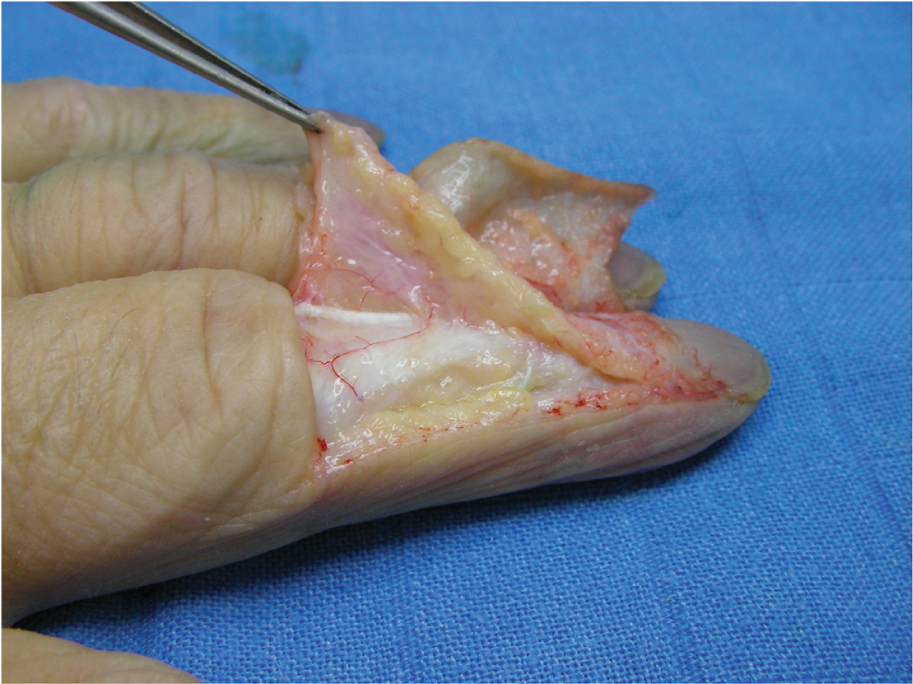
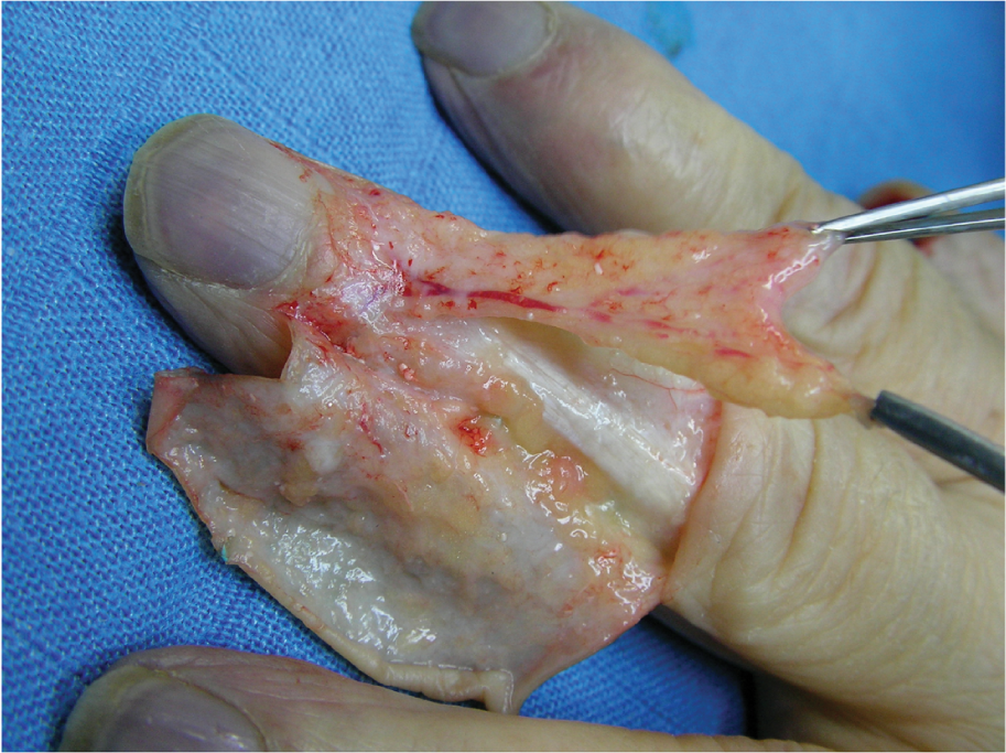
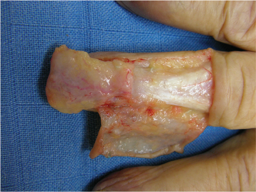
Complete exposure of the subcutaneous adipose tissue was obtained and an adipofascial flap was elevated from the dorsum of PIPJ of each finger (Fig 1), leaving intact the underlying paratenon (Fig 2) and extending from the PIP (Fig 3) to the base of the distal phalanx. Flap elevation was limited at 10 mm proximal to the eponychial fold and did not transgress this more distal zone (Fig 4).
Red latex injection showing the cutaneous window flap elevated and a recurrent dorsal vessel originating from the anastomotic dorsal arch proximal to the eponychium. Red latex injection: subcutaneous (adipofascial) flap elevation, sparing the underlying paratenon; a perforator vessel to the paratenon is shown. Red latex injection: the flap has been elevated including its main vascular supply, represented by the recurrent branch. Note its proximal bifurcation. Red latex injection: the flap has been rotated into its extreme distal final position: the rotation arch is quite wide. The recurrent main vessel and its bifurcation are evident in transparence.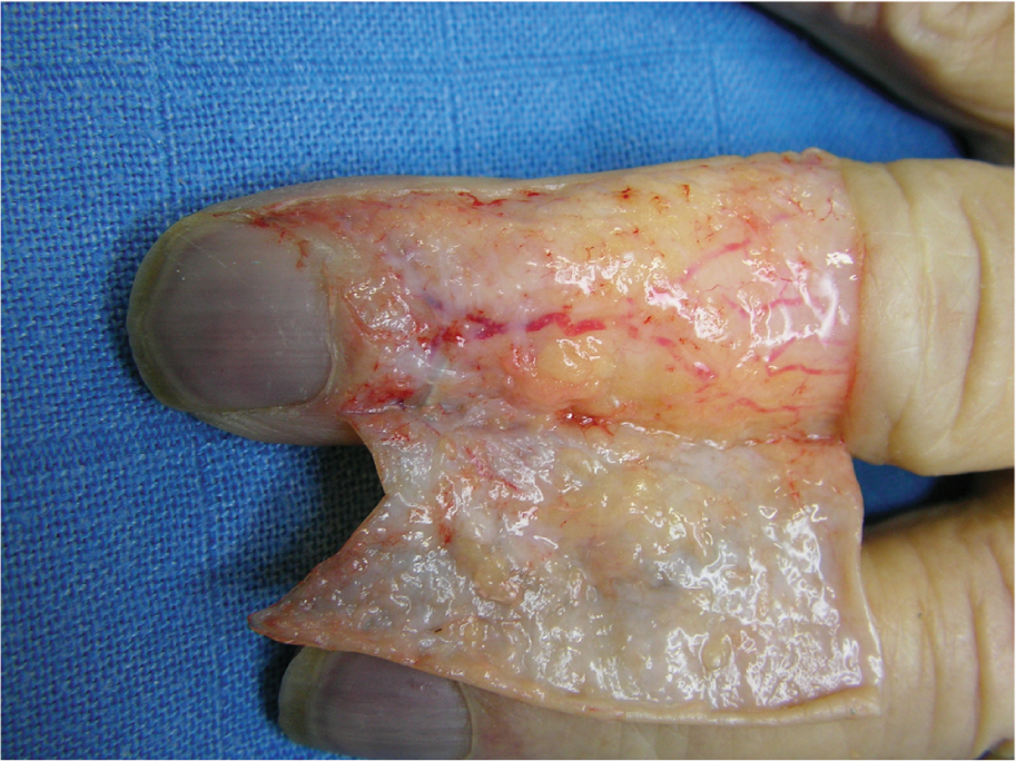
The vascular supply to the distal dorsal region of the long fingers was studied, with particular attention to the adipofascial tissue at the DIPJ level. Images of the dissections were recorded on a digital camera Sony T70.
Four groups of eight different long fingers, were examined.
RESULTS
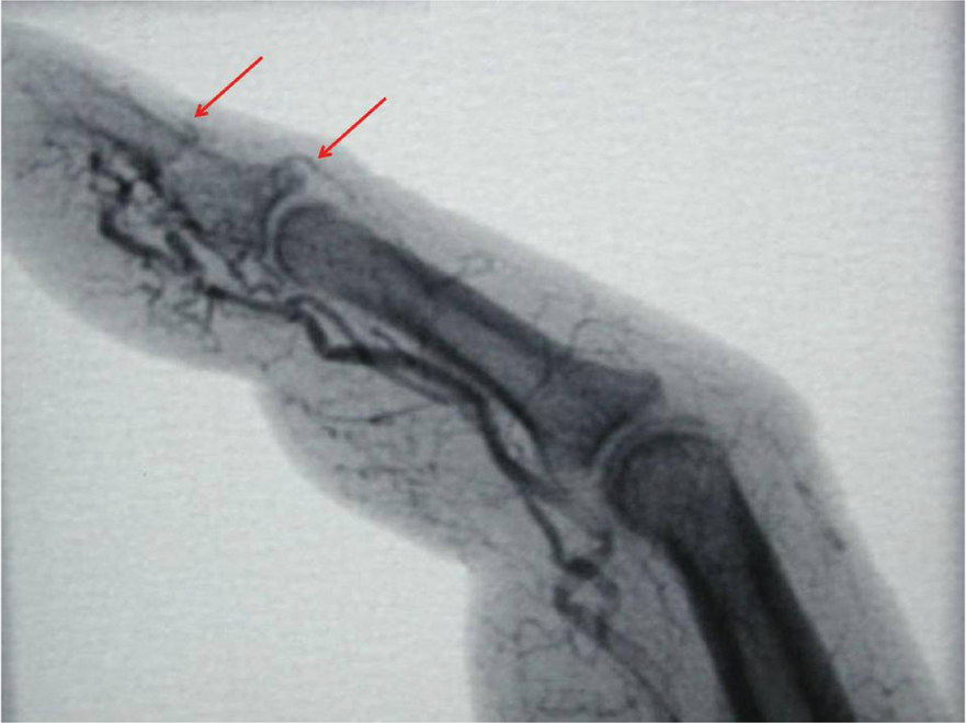
Constant distal perforators originating from palmar digital trunks, and branching to the dorsal region, were found in the area extending from 5–15 mm proximal to the eponychial fold (Figs 5 and 6).
Blue latex injection after skin removal: the inferior and oblique view clearly shows the origin of the perforators. Contrast X-ray picture showing several perforators branching from the digital trunks: they are more represented distally and even their size in the pre-eponychial region is larger.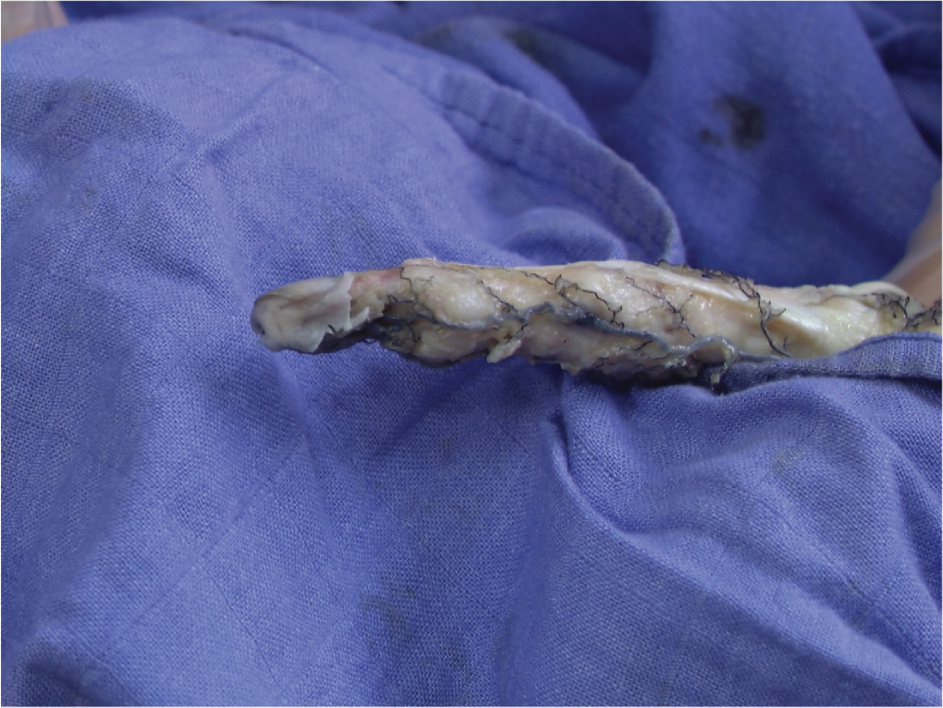
The distance of the origin of the perforator branches ranged from 0–15 mm from the eponychial fold. Analysis showed that the most constant distal perforators were found proximal to the eponychial fold, 6.62 mm (mean distance from the radial side), and 8.8 mm (mean distance from the ulnar side), respectively (mean distance for both side 7.73 mm (Fig 7).
White micropaque© injection: a ruler shows the safe distance from the eponychial fold is 10 mm; dissecting more distally both in the median and in the lateral regions could hamper the vascular supply of the flap. A safe dissection should exclude the point (situated laterally) where the perforators enter the flap and the whole anastomotic arch.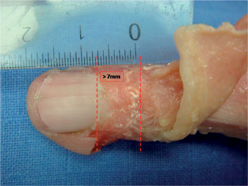
Our injection and angiographic studies confirmed that at least one perforator branched constantly from each digital artery, turned dorsally and anastomized with the contralateral branch beneath the pretendinous tissue supplying the flap by reverse flow (Fig 8). These branches, together with the eponychial network, contributed to the dorsal subcutaneous vascular supply of each finger. In particular, a constant pattern of branching was observed, as the perforator vessel originating from the radial digital artery anastomoses with the contralateral perforator coming from the ulnar digital artery in a crown-like pattern parallel to the eponychium. From this anastomotic arrangement a few (at least one for either radial and ulnar side) longitudinal recurrent trunks arise, running proximally inside the subcutaneous tissue at least as far as the PIPJ (Fig 8).
White micropaque injection: a clear view of the vascular supply of the flap is seen in this picture, showing the perforator entering the flap and branching into an anastomotic arch towards the other side; from this arch, which is organized in a crown-like way, a recurrent longitudinal branch is detected, following the main axis of the flap proximally. The injection also demonstrates how the main perforator reaches the adipofascial layer.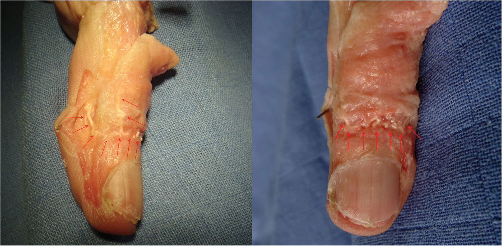
Distal perforators
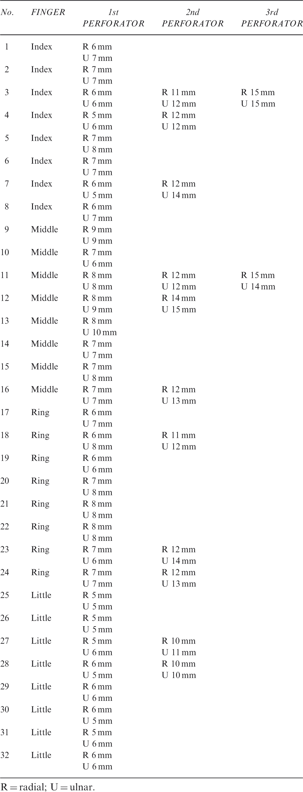
R = radial; U = ulnar.
DISCUSSION
Since the first description by Strauch and De Moura (1990), few anatomical reports of the anatomy of the dorsal branches of the digital arteries have been added. These authors demonstrated that the dorsal digital trunks are numerous and constant, and their pattern is regularly repeated in all three phalanges. The largest are found at approximately the level of the middle and distal thirds of the proximal phalanx, at the midpoint of the middle phalanx, with others at the level of the distal interphalangeal joint. In addition to these, they report numerous small bilateral branches connecting to the longitudinal dorsal arterial network.
Vascular supply to the subcutaneous tissue of the dorsum of the long digits has been first described by Tremolada et al. (1998). They were able to show that some perforating branches from the digital arteries supplied the subcutaneous tissue and skin of the dorsum. They stated that these branches were constant especially proximally to the PIP and to the DIP, and proposed that subcutaneous flaps supplied by these branches could be raised and used as reverse flow flaps for digital repair, taking care to leave a soft tissue cuff around the pedicles and maintaining a 5 mm safety distance from the eponychium.
They proposed that flap survival could be explained with the presence of venae comitantes or small veins in the subcutaneous fat cuff.
Lai et al. (1992) described a reverse digital fascial flap for proximal defects, but their report did not add any novel anatomical element to the literature. Other reviews of this flap, together with variations, tips and pitfalls have been proposed (Hosnuter et al., 2004; Laoulakos et al., 2004; Netscher, 2004; Rampazzo et al., 2007).
These applications stimulated our interest in developing a safe, easy and reliable version of the flap for repair of defects of the nail bed: thus, we made a systematic review of the anatomy in order to facilitate safe dissection.
We believe that from this study, two original messages should be retained by the hand surgeon: first, place the skin incisions on the ulnar side of the finger to raise the skin flaps and expose the subcutaneous tissue. By this means the scar is hidden on the less visible side of the finger and also spares the volar aspect used for grip. Thereby both cosmesis and function are preserved.
Secondly, plan the flap length as needed, avoid undermining the distal part of the flap and preserve at least a 10 mm cuff of tissue proximal to the eponychial fold. It has been demonstrated that 10 mm is the minimum safe length for pivoting a turnover flap. We would take issue with the 5 mm ‘safe distance’ from the eponychial fold proposed by Tremolada et al. (1998); this length is insufficient in our view.
Further, it is recommended that dissection is confined in the region of the lateral space where the distal perforator enters the flap. Avoiding the 10 mm just proximal to the perforator gives an added margin of safety (Fig 7).
This study contributes to the knowledge of dorsal digital anatomy assisting the surgeon performing such procedures to do so with better understanding of the position of the perforators supplying these flaps.
Footnotes
Acknowledgements
The authors wish to thank Epaminondas Kostopoulos, MD, for his technical help in carrying out the study.
Conflict of interests
None declared.