






Abstract
Revision carpal tunnel surgery varies from 0.3% to 19%. It involves a delayed neurolysis and prevention of perineural fibrosis. Despite numerous available procedures, the results remain mediocre. The aim of this study is to evaluate the results of the Canaletto implant in this indication. Our series includes 20 patients (1 bilateral affection) reoperated for carpal tunnel between October 2008 and December 2009. After the first operation, the symptom-free period was 112 weeks, on average. The average incision was 27 mm. After neurolysis, the Canaletto implant was placed in contact with the nerve. Immediate postoperative mobilization was commenced. Sensory (pain, DN4, and hypoesthesia), motor (Jamar, muscle wasting), and functional (disabilities of the arm, should, and hand; DASH) criteria were evaluated. Nerve conduction velocity (NCV) of the median nerve was measured. Average follow up was 12.1 months. All measurements were improved after insertion of the Canaletto implant: pain (6.45–3.68), DN4 (4.29–3.48), Quick DASH (55.30–34.96), Jamar (66.11–84.76), NCV (29.79–39.06 m/s), hypoesthesia (76.2–23.8%), wasting (42.9–23.8%). Nevertheless, four patients did not improve, and pain was the same or worse in six cases. Our results show that in recurrent carpal tunnel syndrome, Canaletto implant insertion gives results at least as good as other techniques, with the added advantage of a smaller access incision, a rapid, less invasive technique, and the eliminated morbidity of raising a flap to cover the median nerve.
Introduction
The failure of treatment of carpal tunnel syndrome is not rare. The frequency of a second intervention varies from 0.3% to 19% (Botte et al., 1996; Chang et al., 1993). Persistent syndrome, where symptoms never disappear, is differentiated from recurrent syndrome, where symptoms reappear after a 3-month period (Botte et al., 1996; Cobb et al., 1996; Plancher et al., 1996). The most common cause of persistent syndrome is incomplete release of the flexor retinaculum, less commonly an iatrogenic nerve lesion. The most common cause of true recurrence is perineural fibrosis.
In both cases, it is agreed that revision surgery must involve delayed neurolysis of the median nerve as well as a gesture of prevention of perineural adhesions. Numerous procedures have been described, such as the interposition of a biological implant (Duché et al., 2003) or flap to restore a gliding plane for the nerve (Chang et al., 1993; Plancher et al., 1996). Whatever the procedure, the results remain mediocre — even risky because 43–90% of revision cases have persistent symptoms, 20% of which are failures (Cobb et al., 1996; Craft et al., 2007; Duché et al., 2003).
Thus, the aim of our study was to evaluate the outcome of Canaletto implant interposition between the nerve and retinaculum. The concept is to avoid contraction of the retinaculum during its scarring and favour a gliding plane for the nerve by inhibiting union of the two sides of the retinaculum. We have been using this technique for 2 years for recurrent carpal tunnel syndrome.
Material and methods
Between October 2008 and December 2009, 1098 cases of carpal tunnel syndrome were operated on in our unit by four surgeons. The technique used was identical: open carpal tunnel release through a 15–20 mm incision (miniopen) under local anaesthesia and a forearm tourniquet in day cases. During this period, we included patients that showed persistence or reappearance of carpal tunnel symptoms (distal paraesthesias) after a primary release. Patients with other complications (complex regional pain syndrome 1, infection, iatrogenic nerve injury, etc.), but without distal paraesthesias, were excluded. Patients who presented with persistent distal paraesthesia related to cervical compression were excluded. Patients who presented with distal paraesthesia and carpal tunnel with major preoperative sensorimotor dysfunction and improved or even normalized postoperative electromyography were also excluded.
Our retrospective series thus included 20 patients — 21 hands (1 bilateral affection) — who had revision surgery for recurrent carpal tunnel symptoms (Table 1). Five patients ( 2, 4, 10, 14, 18) were operated on before revision in our centre and 15 in another centre. All had recurrent carpal tunnel syndrome with a symptom-free period of 112 weeks. Mean age was 54 years (range 29–82 years). There were 5 men and 15 women. All patients except one were right handed. The dominant side was affected in 15 cases. Ten patients were manual workers and the rest were unemployed or retired.
Casuistic of our 21 case series of Canaletto implant
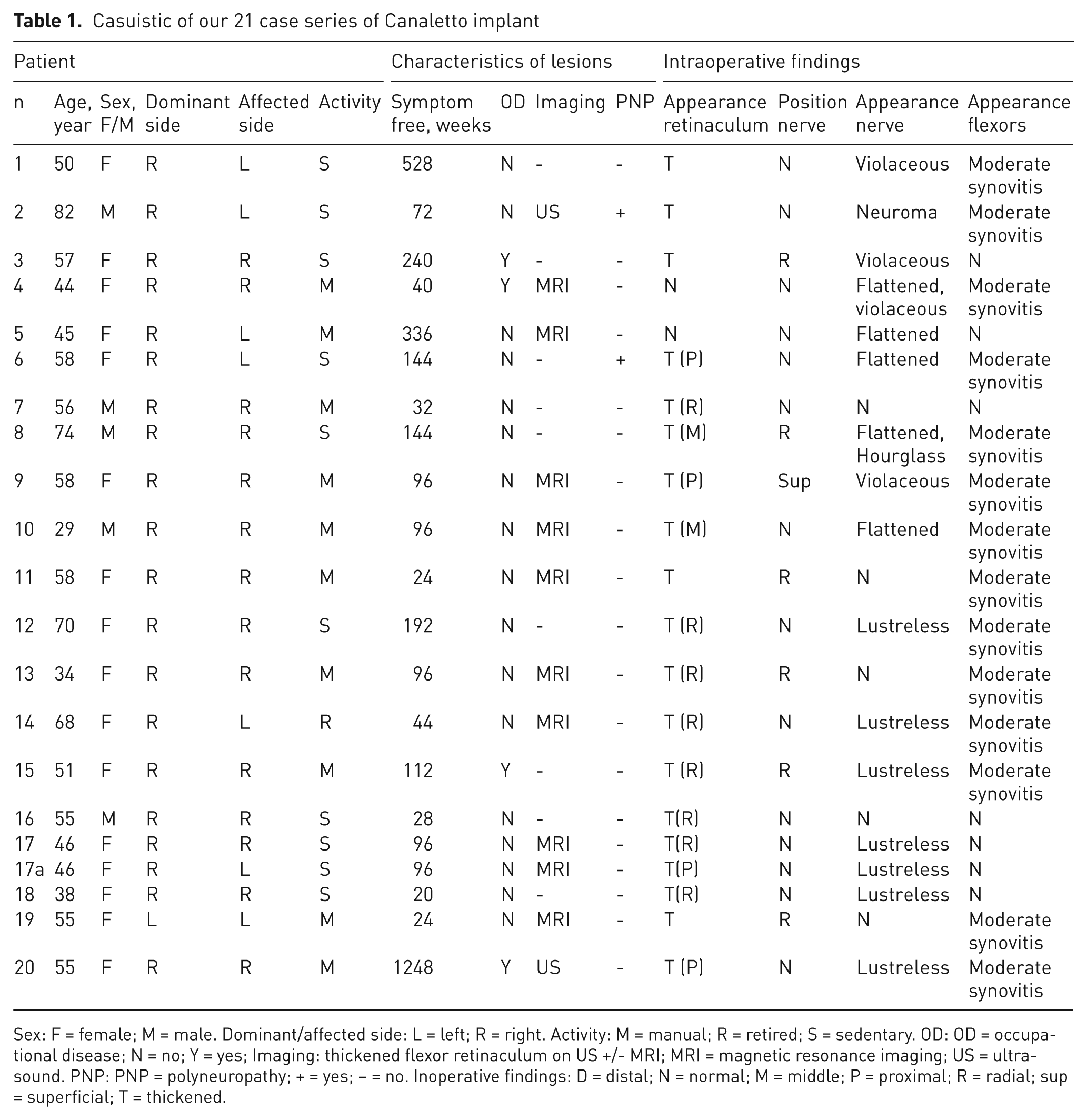
Sex: F = female; M = male. Dominant/affected side: L = left; R = right. Activity: M = manual; R = retired; S = sedentary. OD: OD = occupational disease; N = no; Y = yes; Imaging: thickened flexor retinaculum on US +/- MRI; MRI = magnetic resonance imaging; US = ultrasound. PNP: PNP = polyneuropathy; + = yes; – = no. Inoperative findings: D = distal; N = normal; M = middle; P = proximal; R = radial; sup = superficial; T = thickened.
Four patients had a polyneuropathy. Decision for revision surgery was based on EMG (electromyography) in all cases associated with MRI in 10 cases (Figure 1) and ultrasound in 2 cases.

Preoperative appearance. MRI showing thickening of the retinaculum and superficial prolapse of the median nerve.
All patients in our series were operated on under locoregional anaesthesia as day cases with upper arm tourniquet through a 27 mm incision (range 20–40 mm). The flexor ‘neoretinaculum’ appeared thickened in all except 2 cases, especially at its distal end (Table 1). After complete longitudinal incision of the retinaculum, the position of the median nerve in the carpal tunnel was normal in 14 cases, radially deviated in six cases, and superficial in one case. Macroscopically, it appeared normal in only five cases and violaceous, flattened, or lustreless in all others. Moderate flexor synovitis was noted in most cases (14 of 21 cases).
The next step was extrafascicular neurolysis of the median nerve without flexor synovectomy. The size of the Canaletto (Eurymed, Nimes, France) implant was chosen according to the defect between the two edges of the sectioned retinaculum to avoid its contraction in scarring (Figure 2). The implant was applied with its siliconized deep surface in contact with the nerve, and its edges were sutured to the edges of the retinaculum using 2 stitches of 5/0 Prolene. Skin was closed with 4/0 nylon. Full wrist and finger mobilization was encouraged immediately postoperatively. Forceful movements were allowed 6 weeks postoperatively (Figure 3).

Canaletto implant. (A) Four sizes are available. The dorsal aspect, made of polyethylene terephthalate, is flat. The palmar aspect, made of high-density silicone, is curve. (B) X-ray lateral view. The Canaletto implant is visible due to barium sulphate. (C) X-ray anteroposterior view. The Canaletto implant is visible due to barium sulphate.

Intraoperative appearance. (A) Palmar scar of 20 mm in the axis of the third interdigital space. (B) Thick ‘neoretinaculum’. (C) Appearance of the nerve after release. (D) Measuring the size of the implant. (E) Inhibition of union of the two sides of the retinaculum by suturing the Canaletto implant. (F) Canaletto in place.
Evaluation after the implant involved sensory, motor, and functional tools. Pain was evaluated using a visual analogue scale (VAS) of 0 (no pain) to 10 (maximum conceivable pain). DN4 score is based on 10 questions meant to evaluate the neuropathic character of pain (Bouhassira et al., 2009). Pain was considered neuropathic if there were more than 4 out of 10 positive answers. Quick disabilities of the arm, should, and hand (DASH) is a functional upper-limb evaluation; a score of 0 denotes no dysfunction and 100 denotes useless upper limb. Grip strength was measured in kg using the Jamar dynamometer and expressed as a percentage of the contralateral side. Nerve deficit was diagnosed with the presence of hypoesthesia or anaesthesia in median nerve territory or amyotrophy of the thenar muscles. NCV was measured in m/s. Complications and times of return to activities were noted.
The aim of the study was to detect any significant difference between pre- and postoperative measurements at final follow up for all paired quantitative (DN4, pain, quick DASH, grip strength, and NCV) and qualitative (hypoesthesia, wasting) variables. For quantitative variables, Wilcoxon for paired series was used, with an alpha risk type 1 error of 5%. For qualitative variables, we used the Chi-squared test of McNemar with an alpha risk type 1 error of 5%.
Results
Results are presented in Table 2. Mean follow up was 12.1 months. Global results showed that all variables were improved after Canaletto implant insertion. Pain significantly improved from 6.45 to 3.86 postoperatively (p=0.004, SD=1.83). DN4 changed from 4.29 to 3.48, and the difference was not significant (p=0.145; SD=1.554). Quick DASH significantly changed from 55.3% to 34.96% (p=0.002; SD=15.26). Grip strength significantly changed from 66.11% to 84.76% (p=0.014; SD=26.531). Mean NCV was 29.786 m/s preoperatively and 39.06 m/s at final follow up; the difference was statistically significant (p=0.012; SD=21.100). Hypoesthesia changed from 76.2% preoperatively to 23.8% postoperatively (p=0.001). Thenar muscle wasting changed from 42.9% preoperatively to 23.8% postoperatively; the difference was nonsignificant (p=0.219).
Results of our 21 case series of Canaletto implant
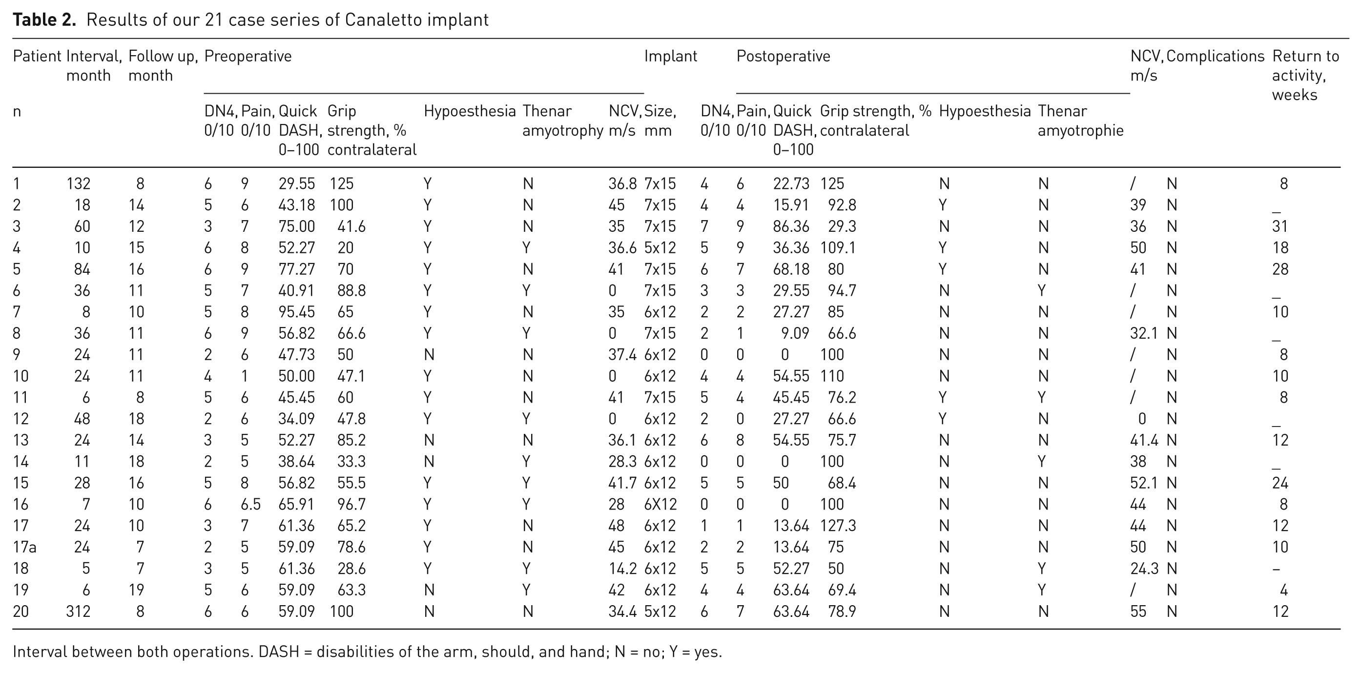
Interval between both operations. DASH = disabilities of the arm, should, and hand; N = no; Y = yes.
Case-by-case analysis of our results showed that only 4 out of 20 patients showed no improvement. Pain did not improve in one case and worsened in 5 cases. Only one patient (12) was reoperated on in another unit, but the removal of the implant did not improve her symptoms, and it is, thus, difficult to attribute the failure to the implant. The pre-existence of polyneuropathy is often incriminated in resistant cases. In fact, two cases in our series dramatically improved and in one patient (6), hypoesthesia disappeared altogether.
Discussion
Primary carpal tunnel surgery is mostly successful in the majority of cases (Steinberg et al., 2002). Many operative techniques have been described, but the principle technique remains that described by Phalen: complete longitudinal division of the whole length of the flexor retinaculum. This is often complicated by relatively decreased grip strength, which led some authors to recommend the systematic reconstruction of the retinaculum, either by local reconstruction (Hunter 1996, Kapandji, 1990; Jakab et al., 2991) or an implant (Duché et al., 2003). Some authors perform an epineurotomy of the median nerve (Chapell et al., 2003), but we consider this useless and dangerous.
Recurrence of carpal tunnel syndrome is a major complication that has been described. We can only diagnose recurrence if the NCS had improved after surgery, or even more so if it had worsened after a 3 month ‘free period’ following primary surgery, and after having eliminated double crush syndrome (Hurst et al., 1985). This period must be respected, as often the normalization of NCS is delayed after the disappearance of clinical signs. Sometimes NCS never becomes normal despite excellent clinical results. Important clues are signs of axonal affection: abductor pollicis brevis denervation with poorer quality signals, as well as decreased amplitudes of sensory response over several fingers (Botte et al., 1996).
Ultrasound and magnetic resonance imaging (MRI) are especially useful in localizing the site of recurrence of compression. MRI can show incomplete release of the retinaculum, palmar prolapse of the median nerve, intraneural nerve abnormalities, and nerve adhesion to the edges of the ‘neoretinaculum’, especially radially. It can also show flexor tenosynovitis, flexor positional abnormalities, or intra- or extraneural tumours (Murphy et al., 1993).
The incidence of recurrence is very low. In our series, recurrence was 5 out of 1098 primary cases (0.46%). The analysis of causes of failure and revision surgery was first described in the literature only in 1972 (Langloh et al., 1972). No surgical technique has proven effective for prevention or treatment.
To prevent carpal tunnel recurrence, in a recent comparative study, some authors proposed the insertion of the Canaletto implant as the primary intervention, with apparently excellent results (Duché et al., 2010). We do not share this view for three reasons: we find this unjustified due to the high success rate of classic techniques, invasive because it involves foreign body insertion, and expensive involving ‘miniopen’ surgery (Figure 4).

Clinical result
Conversely, it is agreed that radial approaches to the retinaculum are the principle cause of adhesions even endoscopically, especially if an oversized scope is inserted into a small carpal tunnel. The fibrosis sometimes forms a gangue adherent to the median nerve and surrounding it, which Hunter called neurodesis (1996). It forms a mould of the nerve reaching to the skin and local irritation syndrome is evoked. The condition is diagnosed by the presence of hypertrophic scar crossing the palmar crease, but is masked in endoscopic cases where the skin remains supple (Figure 4).
Revision surgery using the same technique does not give good results. (Amadio, 2009). Other techniques aim to restore the gliding plane of the nerve, which is an essential physiologic quality of peripheral nerves (Wilgis et al., 1986; Van Doesburg et al., 2010). The first step is neurolysis and the second is to isolate the nerve from surrounding structures often using a flap. Some techniques use the flexor apparatus and its synovium (Wulle, 1996; Jones et al., 1997), some use a muscle flap (Dellon, 1984; Spokevicius et al., 1996; Rose, 1991), and some a fat flap (Pagliei et al., 2003; Mathoulin et al., 2000). They all employ large access incisions and extensive tissue dissection, with the risk of new adhesions and unsightly scars.
Thus, the superiority of Canaletto becomes evident. The mean length of the access incision in our series was 27 mm. No dissection was necessary to insert the implant. The limited access does not limit the performance of a good neurolysis under good conditions. There is no need to extend incisions to perform neurolysis beyond the carpal tunnel, which is the site of the adhesions. When the nerve was deviated radially, this incision always allowed its centralization.
The fixation technique of the Canaletto implant to the retinaculum edges is still to be perfected. Absorbable sutures are to be avoided due to the risk of secondary implant migration. Nonabsorbable sutures bear the risk of stitch knot conflict with surrounding soft tissue structures. We have never encountered this complication in our series.
The advantage of the Canaletto implant is its siliconized deep face in contact with the nerve, which forms a gliding plane by inducing a vascularized neomembrane (Hunter et al., 1970). Other authors have used implants of similar constitution (Fissette et al., 1981) that have different forms. The aim is always to encompass the nerve forming a gliding plane, isolating it from surrounding structures. The Canaletto implant does this by interposition between the edges of the retinaculum. It fulfils both objectives of reconstruction of the retinaculum (to preserve force) and reconstitution of the gliding plane of the nerve (restoring its intracanalicular mobility) (Duché et al., 2010).
Overall, our results showed that the 7 evaluation criteria improved after implant insertion (Table 2). In recurrent carpal tunnel syndrome, insertion of the Canaletto implant after neurolysis of the median nerve gives results better or at least equal to other techniques, with the advantage of minimally invasive access and a rapid technique, without the morbidity of raising a flap to envelope the median nerve. Prospective, comparative, randomized studies of the Canaletto implant with long-term results remain necessary to determine superiority.
Footnotes
Acknowledgements
Pierre Meyer, MD, PhD, Unité d’informatique médicale, Service de Santé Publique, Hôpitaux Universitaires de Strasbourg, France.
Conflict of interests
None declared.
Funding
This research received no specific grant from any funding agency in the public, commercial, or not-for-profit sectors.