






Abstract
We report the patient-scored Health-Related Quality of Life (HRQoL) and functional outcomes of a cohort of 21 consecutive patients undergoing nerve transfer surgery for traumatic upper brachial plexus injuries. Outcomes were assessed using the British Medical Research Council power grading system, Short-Form 36, Disability of Arm, Shoulder and Hand questionnaire, and Pain Visual Analogue Scale (PVAS). The mean age of our cohort was 29.8 years (range 18–53 years), with a mean follow-up period of 42.9 months. At follow-up, elbow flexion ≥ M3 strength was achieved in 17/21 patients. Shoulder abduction ≥ M3 was achieved in 14/19 patients. External rotation ≥ M3 strength was achieved in 11/15 patients. Delayed surgical repair correlated negatively with HRQoL outcomes. Higher injury severity scores and smoking were associated with higher PVAS scores. These findings provide key prognostic information for patients and peripheral nerve surgeons embarking upon this intensive pathway to potential recovery.
Introduction
Traumatic brachial plexus injuries (TBPIs) are devastating and debilitating, with a high incidence in young male polytrauma victims (Choi et al., 1997; Estrella, 2011; Midha, 1997). In addition to motor and sensory deficits, pain and functional limitations can be equally as debilitating, rendering previously healthy individuals both physically and socioeconomically restricted (Ahmed-Labib et al., 2007). These complex injuries pose a significant management challenge for peripheral nerve surgeons. Over the past three decades, microsurgical reconstructive options for the injured brachial plexus has been revolutionized by the versatile concept of nerve transfer surgery (neurotization). Nerve transfer surgery prevails over traditional neurolysis and nerve grafting procedures in facilitating nerve repair on unscarred tissue close to the target muscle, thereby minimizing reinnervation time and permitting repair of otherwise irreparable preganglionic root avulsion injuries. From its introduction by Tuttle in the early 20th century, this innovative technique has been transformed from a laborious, technically demanding procedure, to a safe, reliable, and highly successful standard of care in the restoration of shoulder and elbow function (Addas and Midha, 2009; Bertelli and Ghizoni, 2004; Choi et al., 1997; Estrella, 2011; Ferraresi et al., 1994; Hou and Xu, 2002; Narakas and Hentz, 1988; Oberlin et al., 2009; Shin et al., 2005; Venkatramani et al., 2008).
The therapeutic goals of reconstructive surgery are to restore function and achieve an optimal quality of life for patients (Hanson et al., 2007). Surgical outcomes in functionally limited patients thus far have failed to adequately assess pain and loss of dexterity, which may be key predictors of health-related quality of life (HRQoL) in the TBPI population. The success of microsurgical reconstruction should incorporate patient self-assessment of functional outcome (Choi et al., 1997). Whilst motor and sensory recovery have been extensively examined, assessment of HRQoL and pain outcomes as endpoints following the treatment of these devastating injuries remains largely under-evaluated (Addas and Midha, 2009; Choi et al., 1997).
In the current environment of limited healthcare resources, it is imperative that patients are appropriately selected for therapeutic interventions, in order to optimize the quality of healthcare delivered and outcomes achieved (Shakespeare and Cole, 1997). The aim of this study was, firstly, to assess motor functional outcomes following nerve transfer surgery to restore shoulder and elbow function and, secondly, to use statistically validated scales to quantify HRQoL, upper extremity function, and pain following nerve transfer surgery for TBPIs. Finally, we attempted to assess the relationship between select patient, injury, and treatment factors, and functional outcomes.
Methods
Study design and sample
Twenty-one consecutive patients with upper TBPIs underwent nerve transfer procedures performed by a single surgeon (KC) between November 2003 and April 2010 at the Mater Misericordiae University Hospital (MMUH). The Department of Plastic and Reconstructive Surgery at MMUH is the national tertiary referral centre for brachial plexus injuries in the Republic of Ireland, with a population catchment area of 4.5 million.
The Hospital Inpatient Enquiry system and MMUH brachial plexus database were used to identify our patient cohort. All patients provided informed consent to participate in this study and were subsequently assessed in the out-patient setting. All patients were invited to score the SF-36, DASH, and PVAS questionnaires. The medical records of all patients were systematically reviewed to obtain demographic data, diagnostic evaluations, and surgical procedure details, and to identify patient, injury, and treatment factors.
Patient factors included age at injury, sex, employment status, and smoking status. TBPIs were defined according to mechanism of injury, pattern of root involvement, and associated injuries. Associated injuries were quantified in terms of injury severity score (ISS), as previously described (Baker et al., 1974). Treatment factors included trauma-surgery interval (time from trauma to surgical intervention), and number of donor (extraplexal/intraplexal) and recipient nerves for neurotization. Post-operative analgesic requirements and requirement for specialist chronic pain management were assessed.
Inclusion criteria for the study were patients with upper-type brachial plexus root avulsion injuries undergoing nerve transfer procedures for restoration of elbow flexion, shoulder abduction, and shoulder external rotation. A detailed preoperative assessment was performed for all patients. Elbow flexion, shoulder abduction, and external rotation range of motion were 0° in all cases. Deltoid, teres minor, supraspinatus, infraspinatus, biceps, and brachioradialis muscle were all paralyzed and scored M0 on British Medical Research Council (BMRC) scoring. Trapezius muscle scored M5, and grip and pinch strength in the hand was normal in all patients. With a minimum follow up of 12 months, all patients were evaluated for range of movements at the shoulder and elbow, and motor power functional assessments. Patients with complete C5-T1 plexus avulsion injuries presenting with a flail limb were excluded. Patients requiring secondary reconstructive procedures (tendon and muscle transfers, shoulder arthrodesis) were also excluded from the study.
Standard technique of brachial plexus exploration
All patients underwent brachial plexus exploration under general anaesthesia without the use of muscle relaxants. Supraclavicular, infraclavicular, or both surgical approaches were employed, depending on the pattern of injury suspected upon preoperative evaluation. The nerve was stimulated at 10–20 mA using a portable handheld peripheral nerve stimulator (Fischer & Paykel, Auckland, New Zealand) proximal to the neuroma, and distal contraction was assessed. If the roots were judged to be avulsed and non-graftable (the current cohort), a nerve transfer was performed. Assessment of somatosensory evoked potentials or spinal evoked potentials was not routinely performed. We judged the nerve root to be avulsed or non-graftable if (1) there was an obvious discontinuity of the root as it left the neural foramen, with no available nerve material proximal to the neuroma, and (2) on microscopic assessment, there were no normal fascicle structure following external and internal neurolysis, up to the neural foramen. Intraoperative findings were correlated with clinical, radiological, and electrophysiological data to establish a definitive diagnosis prior to surgical planning.
Nerve transfers to restore shoulder and elbow function
Selection of extraplexal and intraplexal donor nerves for neurotization was performed on a case-to-case basis, depending on the pattern of root avulsion and availability of functional nerves. Deficits in shoulder external rotation were repaired using phrenic or spinal accessory neurotisation to suprascapular nerve. Deficits in shoulder abduction were repaired using pectoral branches, intercostals nerves, and/or superior branch to triceps nerve coapted to the axillary nerve. Deficits in elbow flexion were repaired using either a single-nerve transfer using a fascicle or fascicles of the ulnar nerve (Oberlin transfer), while the remaining underwent double-nerve transfers using partial ulnar and partial median nerve neurotisation to reinnervate the biceps and brachialis, respectively. A number of patients had additional intercostal nerve transfers if ulnar or median nerves were unavailable. All nerves were coapted using 10/0 non-absorbable nylon without tension under microscopic magnification. The wounds were closed with drain placement distant to the nerve repair site. A soft collar and arm sling were worn for 4 weeks postoperatively. Gradual passive range of motion exercises were commenced at the end of week 4 until the first signs of muscle contraction were observed. At this point, a strengthening program for targeted muscle groups was commenced.
Outcomes assessment
All patients had follow-up assessments performed at the national multidisciplinary Brachial Plexus Clinic. Post-operative muscle strength was evaluated using the modified British Medical Research Council (BMRC) grade assessed by an independent observer, as previously validated (Bengtson et al., 2008). BMRC grades ≥ 3 were considered a good functional outcome. Additionally, range of movements were recorded with goniometry. The range of elbow flexion was measured as the angle formed between the long axis of the arm and forearm. The range of abduction was recorded by measuring the angle formed between the arm axis and parallel to the spinal cord axis. External rotation was measured with the patient standing with the shoulder fully internally rotated and forearm placed transversally over the abdomen. Any rotation from this position was measured and noted as the range of external rotation.
Post-operative health-related quality of life (HRQoL) outcomes were assessed using the Short Form 36 (SF-36) Health Survey Questionnaire (McHorney et al., 1993; Ware, 1999), Disability of Arm, Shoulder and Hand (DASH) Questionnaire, and pain visual analogue scale (PVAS) (Holger et al., 2004).
SF-36
HRQoL is the extent to which a patient’s day-to-day life is affected by their health, including the physical, emotional, and social impact of disease (Charlson et al., 1987). The acute version of the SF-36 assesses eight aspects or domains of HRQoL during the past week: physical functioning, role limitations due to physical problems, pain, health perception, vitality, social functioning, role limitations due to emotional problems, and mental health (McHorney et al., 1993). Physical and mental component summary scores (PCS and MCS, respectively) were created using standard norm-based scoring methods (Hays et al., 1993). The SF-36 has been extensively validated and previously used in outcome assessment of interventions in plastic and reconstructive surgery (Cocquyt et al., 2003; Loge and Kaasa, 1998;Veiga et al., 2004). The scales were scored numerically from 0 (lowest level of functioning) to 100 (highest level of functioning). Lower scores on HRQOL scales indicate limitations in the ability to perform daily activities and work due to reduced physical or emotional functioning, interference with normal social activities, feelings of nervousness and depression, body pain that is severe and limiting, poor perception of personal health, and persistent feelings of fatigue. Mean scores for all eight domains, including the physical and mental component scores, were compared in patients who experienced postoperative flap-related complications and those who did not. The Norwegian SF-36 acute version 1.0 was used in this study.
DASH questionnaire
The DASH questionnaire was designed to assess disability experienced by patients with upper extremity musculoskeletal disorders (Beaton et al., 2001, Hudak et al., 1996). It is composed of 30 general activity, symptom, and social function items, ranked on a 5-point Likert scale. The score is calculated from the responses; higher scores indicate greater disability. As recommended by the questionnaire developers, missing items are replaced with the mean value for all subjects for that item and, if more than three items are missing, the total score should not be calculated (Beaton et al., 2001). In the present study, no patient had more than two missing responses. Good validity and reliability have been reported for the use of DASH in the assessment of peripheral nerve disorders (Beaton et al., 2001; Hudak et al., 1996; Novak et al., 2011; SooHoo et al., 2002).
PVAS
Using a 100 mm VAS scale ranging from 0 (no pain) to 100 (worst possible pain), patients were invited to score post-operative pain intensity, as previously described (Estrella, 2011). Using receiver operator curve analysis, a cut-off score of 30 mm was used to dichotomize patients into higher and lower pain intensity groups. Patients who scored ≥ 30 mm were categorized as ‘higher pain-intensity scores’ and those who scored < 30 mm were deemed ‘lower pain-intensity scores.’
Statistical analysis
Descriptive statistics demonstrating patient characteristics were presented using means with standard deviation (SD) or numbers with percentages. SF-36, DASH, and PVAS scores were compared using two-tailed t-test for independent samples. Correlation coefficients (r) were used to evaluate the association between SF36, DASH, and PVAS scores and continuous variables (age, ISS). A p value < 0.05 was considered statistically significant. Statistical analyses were performed using SPSS version 15.0 (SPSS Inc., Chicago, Illinois, USA).
Results
Demographics
Twenty-one consecutive patients underwent nerve transfer procedures following TBPIs from November 2003 to April 2011 (Table 1). The mean age of our cohort (n = 21) was 29.8 years (range 18–53 years), with a male preponderance (M:F = 9.5:1). All patients were right-hand dominant. The injured limb involved the dominant hand in 18/21 patients (85.7%). Regarding employment status, 15/21 patients (71.4%) were working at the time of injury, with 6/15 patients (40%) returning to work within 1 year of surgery. Of the 9 patients who were not working, 6 associated their unemployment with their injury. The mean follow-up period from time of surgery was 42.9 months (range 12–82 months). The trauma-surgery interval (mean time from initial trauma to nerve transfer procedure) was 6.6 months (range 0.2–13 months). Trauma-transfer interval (mean time from initial trauma to transfer to MMUH brachial plexus service) was 5.4 months (range 0.2–12 months). Seven patients (33.3%) were referred from other institutions ≥ 6 months from the initial injury.
Patient demographics of the 21 patients in the reported series*
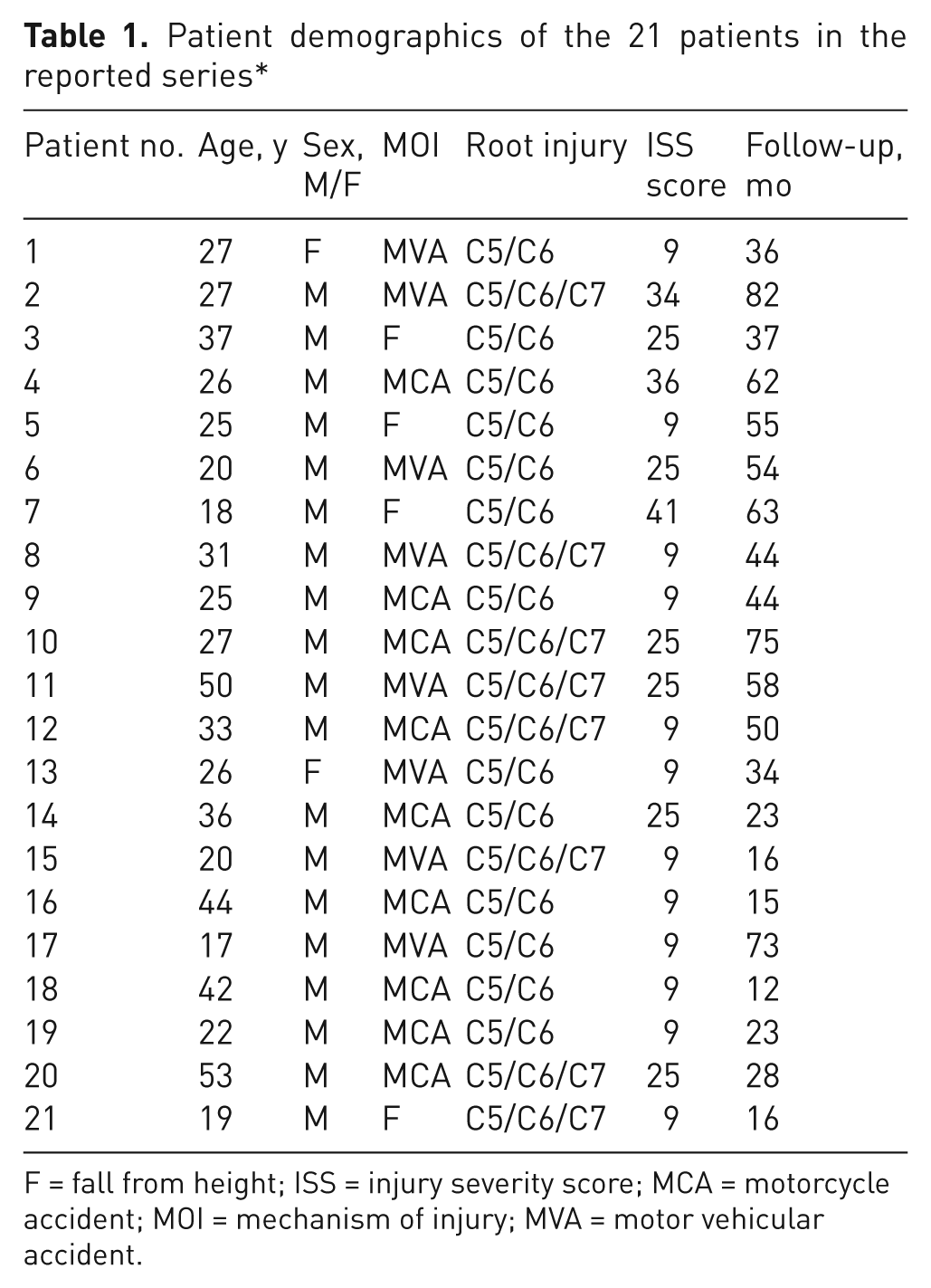
F = fall from height; ISS = injury severity score; MCA = motorcycle accident; MOI = mechanism of injury; MVA = motor vehicularaccident.
Characteristics of brachial plexus injuries
The most common mechanism of brachial plexus injury was closed traction injury as a result of motorcycle accidents in 9 patients (42.9%) and motor vehicle accidents in 8 patients (38.1%). Falls from a height resulted in plexus compression injuries in 4 patients (19%). Upper-type brachial plexus nerve root avulsion injuries were reported in all 21 patients. Root avulsion injuries were dichotomized into 2 patterns of involvement: 13 patients with C5-C6 complete nerve root avulsions (61.9%) and 8 patients with C5-C7 complete nerve root avulsions (38.1%). Concomitant major injuries with injury severity scores ≥ 20, indicative of poor prognosis, were reported in 9 patients (42.9%). These included 6 shoulder/clavicle/upper limb fractures or dislocations; 2 unstable cervical fractures; 2 major vessel injuries; 2 head injuries; 2 thoracic injuries; and 1 lower limb fracture. The mean injury severity score was 17.6 (range 9–41). Acute surgical exploration was performed in 3 patients (14.2%) for suspected vascular injuries.
Motor functional outcomes
Restoration of elbow flexion, shoulder abduction, and external rotation was performed on a case-by-case basis, depending on the injury pattern (Figure 1). The functional outcomes for each patient in terms of range of motion and BMRC power grading are shown in Table 2. Restoration of elbow flexion was indicated in all 21 patients. At latest follow-up, elbow flexion strength of ≥ M3 was achieved in 17/21 patients (80.9%), with an average of 135° (range 90–170°). Restoration of shoulder abduction was indicated in 19 patients. Shoulder abduction strength ≥ M3 was achieved in 14/19 (73.7%) of patients with an average of 93° (range 40–130°). Restoration of shoulder external rotation was indicated in 15 patients. Shoulder external rotation strength ≥ M3 was achieved in 11/15 (73.3%) of patients with an average of 86° (range 10–130°).

A 55-year-old gentleman sustained a complete C5/C6/C7 avulsion injury following an road traffic accident. He underwent spinal accessory to suprascapular, superior branch of triceps to axillary and Oberlin transfers. Results at 1 year follow-up are shown. (A) Elbow flexion against resistance (M4). (B) Reasonable external rotation (M3). (C) Good shoulder abduction (M3).
Summary of functional outcomes for shoulder and elbow reconstruction
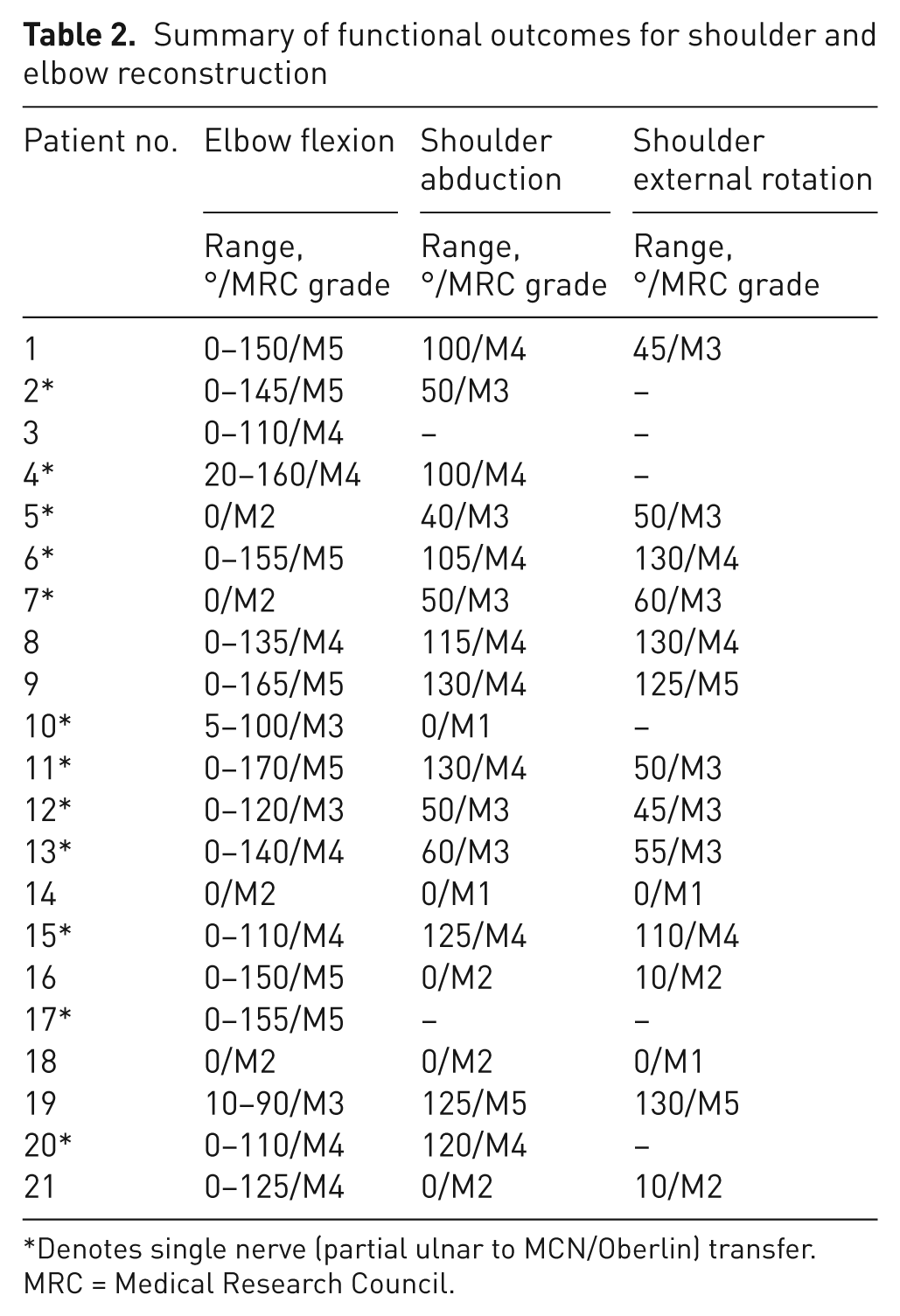
Denotes single nerve (partial ulnar to MCN/Oberlin) transfer.
MRC = Medical Research Council.
In terms of neurotization with donor nerves, 25 extraplexal and 40 intraplexal nerve transfers were performed. The mean number of nerve transfers per patient was 3 (range 1–5). Regarding extraplexal nerve transfers: 11 patients underwent spinal accessory to suprascapular nerve transfers. At latest follow-up, 8/11 patients (72.7%) obtained ≥ M3 strength. Phrenic to suprascapular nerve transfers were performed in 4 patients. Of these, 3/4 patients (75%) obtained ≥ M3 strength. Intercostal to axillary nerve transfers were performed in 5 patients. Of these, 3/5 patients (60%) obtained ≥ M3 strength. Regarding intraplexal nerve transfers: superior branch of triceps to axillary nerve transfers were performed in 10 patients. At latest follow-up, 8/10 (80%) of patients obtained ≥ M3 strength. Medial pectoral to axillary nerve transfer was performed in 1 patient, obtaining ≥ M3 strength at follow-up. Oberlin nerve transfer (partial ulnar to musculocutaneous nerve transfer for biceps neurotization) was performed in 12 patients. Of these, 9/12 patients (75%) obtained ≥ M3 strength. Partial ulnar and median nerve transfer to biceps and brachialis branches of musculocutaneous nerve, respectively, were performed in 9 patients. Of these, 7/9 (77.8%) patients obtained ≥ M3 strength. Following this double nerve transfer, paraesthesia in the ulnar and median dermatomes was detected in 2 patients, using the 2-point discrimination test. This fully resolved 3 months post-operatively. No additional complications were noted in this cohort.
HRQoL outcomes
Of the 21 patients invited to complete the SF-36, DASH, and PVAS questionnaires, the response rate was 100%. Mean level of functioning scores for each SF-36 domain was obtained. The mean DASH score for this cohort was 76.3 (SD 17.5) (range 51.1–98.2). With respect to patient-scored PVAS, 10/21 patients (47.6%) reported higher pain intensity scores. The mean PVAS score was 30.0 mm (SD 37.0) (range 0–73 mm).
Mean patient-scored SF-36, DASH, and PVAS scores were correlated with select patient, injury, and treatment variables. Trauma–surgery interval ≥ 6 months was reported in 11 patients (52.4%). This delay was attributed to lack of timely referral from other institutions, in all cases. Patients who underwent nerve transfer procedures < 6 months from the initial injury scored consistently better in the DASH and SF-36 than an injury–surgery interval ≥ 6 months (Table 3). Mean raw SF-36 scores were significantly higher in the role-physical (p < 0.01), general health (p < 0.03), and physical component score domains (p < 0.04) for an injury–surgery interval < 6 months. Mean raw DASH scores were significantly lower for an injury-severity score < 6 months (p < 0.04). There was no correlation between injury–surgery interval and patient-scored PVAS scores. There were no significant differences in patient-scored SF-36 and DASH scores in relation to patient age, ISS, or smoking status. In relation to patient-scored PVAS, mean scores were significantly higher in smokers (p < 0.031) and patients with ISS ≥ 20 (p < 0.001) (Table 4). There was a strongly positive correlation between smoking status (r = +0.466, p < 0.022) and ISS (r = +0.750, p < 0.001) when correlated with PVAS scores (Table 4).
Time delay to surgery and health-related quality of life (HRQoL) outcomes
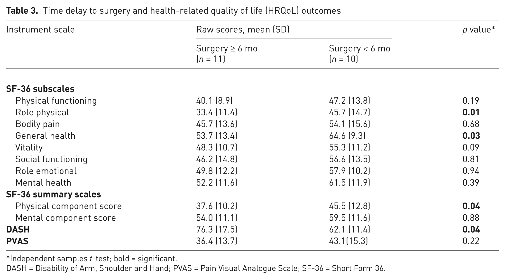
Independent samples t-test; bold = significant.
DASH = Disability of Arm, Shoulder and Hand; PVAS = Pain Visual Analogue Scale; SF-36 = Short Form 36.
Association of PVAS scores with select patient, injury, and treatment variables
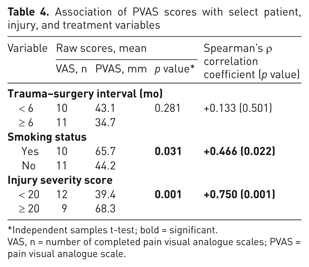
Independent samples t-test; bold = significant.
VAS, n = number of completed pain visual analogue scales; PVAS = pain visual analogue scale.
Discussion
Adult TBPIs are devastating, debilitating injuries, and result from high-speed motor vehicular accidents in the majority of cases (Choi et al., 1997; Estrella, 2011). These injuries are disproportionately prevalent in young males and pose a significant reconstructive challenge for peripheral nerve surgeons (Ahmed-Labib et al., 2007; Choi et al., 1997; Estrella, 2011). Nerve transfer surgery, since its establishment by Tuttle in 1913, has revolutionized the management of these challenging injuries, employing the creative use of applied surgical anatomy to allow the prompt restoration of physiological neuromuscular function, with superior functional outcomes (Bertelli and Ghizoni, 2004; Estrella, 2011; Hou and Xu, 2002; Oberlin et al., 1994; Oberlin et al., 2009; Rohde and Wolfe, 2007; Venkatramani et al., 2008). Patients with TBPIs seek reconstructive procedures not only to recover functional status, but also to alleviate pain and improve their quality of life (Ahmed-Labib et al., 2007). As surgical outcomes thus far have failed to adequately assess pain and loss of dexterity, we feel that the success of microsurgical reconstruction should incorporate the patient’s self-assessment of their functional recovery using statistically validated tools (Choi et al., 1997).
In this consecutive case series from a national tertiary referral centre for TBPIs, we evaluated patients presenting with upper brachial plexus root avulsion injuries treated with nerve transfers for restoration of elbow and shoulder function. In cases of otherwise irreparable avulsion injuries, where traditional nerve grafting and repair would be suboptimal, nerve transfers offer a safe and reliable reconstructive option with good functional outcomes reported internationally (Bertelli and Ghizoni, 2004; Leechavengvongs et al., 1998; Leechavengvongs et al., 2006; Oberlin et al., 1994; Sungpet et al., 2000a; Teboul et al., 2004; Venkatramani et al., 2008). In this series, return of elbow flexion was a direct result of nerve transfers to the biceps and/or brachialis branch of the musculocutaneous nerve. Seventeen of 21 patients recovered ≥ M3 elbow flexion strength, with an average range of motion of 135°. As anticipated, the partial ulnar and median nerve transfer to biceps and brachialis branches of musculocutaneous nerve was most successful, recovering ≥ M3 strength in 77.8% of cases. Whilst these outcomes are largely similar to other published series, superior outcomes have been presented in larger series reporting ≥ M3 flexion strength in 93–96.8% of cases (Leechavengvongs et al., 1998; Oberlin et al., 1994; Sungpet et al., 2000a; Teboul et al., 2004). However, three out of four patients who not achieve ≥ M3 elbow flexion strength underwent nerve transfers > 6 months from the initial injury due to delayed transfers from other institutions, and this may account for inferior outcomes in these patients. Whilst these double-nerve transfers have been reported as superior to single-nerve transfer in terms of reinnervation time and maximum power grade achieved, similar motor outcomes and DASH scores have been reported in a recent controversial study (Carlsen et al., 2011; Leechavengvongs et al., 2006; Mackinnon et al., 2005).
In this study, successful restoration of shoulder abduction was achieved in 73.7% of patients with an average range of motion of 93°. Optimal outcomes were achieved using superior branch of triceps nerve transfer to axillary nerve, with 80% of patient achieving ≥ M3 shoulder abduction strength. Intercostal to axillary nerve transfer was the least successful transfer in restoration of shoulder abduction, with 60% of patients achieving ≥ M3 shoulder abduction strength. Despite this, similar results have been reported in other series in which this transfer procedure has produced limited shoulder function (Nagano et al., 1995). Successful restoration of shoulder external rotation was achieved in 73.3% of patients, with spinal accessory to suprascapular (SSN) transfer the most commonly employed. Phrenic to SSN transfer was the most successful transfer in restoration of shoulder external rotation, with 75% of patients achieving ≥ M3 shoulder external rotation strength. There was no evidence of pulmonary compromise at most recent follow-up and numerous studies have reported similar motor outcomes (Chuang et al., 1993; Sungpet et al., 2000b).
There is a broad consensus that assessing outcomes in terms of patients’ subjective reports and psychological functioning provides a more meaningful understanding of the effects of treatment (Choi et al., 1997; Katz et al., 1994; Levine et al., 1993). In this series, HRQoL and pain outcomes were determined by means of validated assessment tools to elucidate the full scale impact of chronic pain and loss of dexterity following these devastating injuries. In the present study, the mean trauma–transfer interval was 5.4 months, with 7 patients (33.3% of the cohort) referred to our national service from other institutions ≥ 6 months from the initial injury. Because most peripheral nerve surgeons agree that the optimal time for brachial plexus reconstruction of closed injuries ranges from 3–5 months, long delays in referral compromise prompt expert management and optimization of functional outcome (Carlsen et al., 2009; Dubuisson and Kline, 2002; Kline and Judice, 1983; Narakas, 1985; Shin et al., 2005). These delays are largely unacceptable, given that a large epidemiological study noted that 78% of polytrauma patients with TBPIs were diagnosed immediately or within 3 days of the injury (Midha, 1997). In the context of a TBPI, these data highlight the need for prompt referral and transfer of the polytrauma patient to a tertiary referral centre with multidisciplinary team management where expert assessment, investigation, and surgical intervention can be instituted in a timely fashion. As a result of our findings, to address this issue in Ireland, we have developed an online tertiary referral system to expedite the transfer of patients warranting surgical intervention.
In this series, a trauma–surgery interval ≥ 6 months was reported in 12 patients (57%) and was attributed to a lack of timely referral from other institutions, in all cases. Importantly, patients undergoing nerve transfer procedures < 6 months from the initial injury scored consistently better in the DASH and specific SF-36 subscales relative to an injury–surgery interval ≥ 6 months. Delaying surgical intervention > 6 months leads to prolonged denervation with progressive muscular atrophy, fibrosis, and joint stiffness (Bentolila et al., 1999; Narakas, 1977). We postulate that these mechanisms account for higher DASH and lower HRQoL scores and, thus suboptimal outcomes, for those undergoing surgery ≥ 6 months from injury.
The debilitating nature of pain reported following TBPIs provides strong motivation to identify prognostic indicators of pain intensity (Bertelli and Ghizoni, 2004; Choi et al., 1997; Estrella, 2011). Pain following TBPIs has been attributed to deafferentation of the spinal cord subjected to root avulsion (Bertelli and Ghizoni, 2008; Loeser et al., 1968; Parry, 1980; Sindou et al., 2005). In concordance with international findings, 47.6% of patients in the present study reported significant pain following surgical intervention (Ahmed-Labib et al., 2007; Choi et al., 1997). In this context, it is postulated that nerve transfers do not relieve pain, as these are primarily motor nerve transfers, independent of sensory pain generation (Bertelli and Ghizoni, 2008). Thus, a greater onus must be placed on early pain management intervention in attempt to optimize HRQoL and pain outcomes. Nonetheless, pain intensity is only one component of pain, and the impact of pain in the context of disability should be considered in patients with chronic nerve injury (Novak et al., 2010). In this present study, we have identified ISS and smoking status as biomedical factors associated with higher pain intensity scores. The identification of these factors in association with higher pain intensity allows us to provide key prognostic information for patients in the preoperative setting.
We do recognize several limitations to this study. Firstly, we acknowledge the inherent frailties associated with a retrospective study design and unique patient sample. Additionally, the cross-sectional nature of such a study means that causality cannot be determined. Furthermore, the inclusion criteria in our series included patients who had attended clinics during the study period. This may represent a biased sample of patients who continue to seek assessment and treatment, and may not be representative of all patients following TBPIs. However, patients with physical impairments, pain, and disability often are the most difficult to treat, overwhelming the resources available, and can ultimately have diminished HRQoL. A further limitation is the obvious difficulty associated with assessing HRQoL and functional outcomes in the post- operative setting without an appropriate pre-operative comparison. This present study does, however, provide strong preliminary data for a subsequent, comprehensive, longitudinal study measuring baseline pretreatment biomedical and psychosocial characteristics; documenting the nature, duration, and cost of treatment; and following patients’ post-treatment status. In response to our findings, we have established a patient registry to track patients and their outcomes in an effort to optimize management and appropriate patient selection for these devastating injuries.
This study suggests that despite optimal motor functional outcomes following nerve transfer surgery, patients with TBPIs still report substantial disability and pain. Delayed surgical repair correlates negatively with HRQoL outcomes. Higher pain intensity is associated with a number of biomedical factors, including higher ISS scores and positive smoking status. These findings provide key prognostic information for patients and peripheral nerve surgeons embarking upon this intensive pathway to potential recovery.
Footnotes
Acknowledgements
This paper was presented in part at the XVII International Symposium on Brachial Plexus Surgery, 20 May 2011, Lisbon, Portugal.
Conflicts of interests
None declared.
Funding
This research received no specific grant from any funding agency in the public, commercial, or not-for-profit sectors.