






Abstract
We describe a method to restore active palmar abduction of the thumb and report its functional impact in tetraplegia. At 54.2 (SD 42.8) months after cervical spinal cord injury (12 traumatic, 3 nontraumatic), the extensor digiti minimi (EDM) tendon was transferred to the abductor pollicis brevis (APB) through the interosseous membrane in 15 tetraplegic patients (age range 19–70 years) in addition to a mean 3.2 procedures to restore key pinch. According to International Classification, the operated upper extremities were in the OCu4 to OCu8 (1 patient X) group. The maximum distance between thumb and index finger tips during active or passive opening of the hand, maximum angle of palmar abduction, grip and key pinch strength, and active finger range of motion were measured. All patients were re-examined after 38.4 (SD 22.7) months. The active thumb-index opening increased significantly from 2.5 (SEM 1.0) cm before to 9.0 (SEM 0.8) cm after surgery. Nine patients without previous active opening of the first web space recovered a mean thumb-index opening of 9.1 (SEM 1.7) cm, whereas this distance increased by an average of 2.9 (SEM 0.8) cm in six patients who had active thumb index distance of 6.3 (SEM 1.6) cm before surgery. All but one patient were able to direct and coordinate key pinch and perform tasks using the restored APB function, including five patients whose EDM strength was rated as grade 3 before transfer. This EDM-to-APB transfer meets the theoretical requirements of architecture matching between donor and recipient muscles, the principles of tendon transfer, and our surgical expectations. We strongly recommend that an active EDM is transferred to the APB to restore opening of the hand and help in key pinch control in patients with tetraplegia.
Keywords
Introduction
Reconstructive surgery in tetraplegia can significantly restore basic upper extremity movements, such as elbow extension, wrist extension, and hand grip in the majority of these mostly young, previously healthy, and productive individuals. This substantially improves the patients’ independence, mobility, and quality of life (Hentz, 2002; Moberg, 1978). Tendon transfer, tenodesis, and joint stabilization are strategically planned, based on the muscles remaining under voluntary control, and provide stability and strength. The goal of surgery is to restore strong lateral pinch, often referred to as key pinch, because its reconstruction requires only a limited number of donor muscles. But a stable pinch between the pad of the thumb and the radiolateral aspect of the index finger is very versatile in many activities of daily life (House, 1985; House et al., 1992).
Surgical options to enhance thumb function depend on the number of motors available for tendon transfer and include procedures such as tenodesis of the flexor pollicis longus (FPL) tendon to the radius, active transfers to the FPL, extensor tenodeses, adductor- and opponensplasty, split distal FPL tenodesis, and carpometacarpal (CMC) arthrodesis (House, 1985). These procedures seek to increase joint stability and pinch force, assuming that these are the two most critical factors to improve function.
However, the ability to increase pinch force may not necessarily improve dexterity, because many hand activities, especially to secure and manipulate objects that are of small size or with convex surfaces, need precise control, coordination, and control of direction of the force exerted by the thumb tip (Johanson et al., 2001; Smaby et al., 2004). Also many tetraplegic patients lack the ability to actively open their first web space to grasp large objects, such as a can of drink (Hentz, 2002; House, 1985).
Both these functions are normally performed by the abductor pollicis brevis (APB), which is usually paralyzed after injury to the cervical spinal cord. This muscle is uniquely able to abduct the CMC joint of the thumb perpendicular to the plane of the palm and move the thumb in the opposed position for pinch and grasp against the finger tips (Brand and Hollister, 1999). Rehabilitation of this small, weak muscle in tetraplegic patients has been mentioned only rarely, and no study has investigated the results (Hentz and Leclercq, 2002; Landi et al., 2002).
The extensor digiti minimi (EDM) is usually active in International Classification category 6 patients and above (Hentz and Leclercq, 2002), but sometimes is also active in weaker groups (Revol et al., 2002). It is a possible donor motor to restore APB function in the paralyzed thumb. We report the indication, technique, and clinical efficacy of the EDM-to-APB tendon transfer to improve reconstruction of pinch in a defined group of 15 patients with tetraplegia.
Methods
Fifteen patients with tetraplegia (7 women, 8 men) scheduled for tendon transfers were specifically screened for an EDM with muscle strength of at least Medical Research Council (MRC) grade 3 with movement against gravity (MRC, 1943; Hentz and Leclercq, 2002). Mean age at the time of surgery was 43.9 (range 19–70) years, 4 were older than 60 years, of which 1 was aged 70 years. In 12 patients the cord disorder was caused by trauma due to motor vehicle or bike accidents, falls, or by diving into shallow water, and in 3 patients the cause was nontraumatic. Time from paralysis to surgery was 54.2 (SD 42.8) months. International Classification of patients’ upper extremities ranged from OCu4 to OCu8, and one woman with a nontraumatic incomplete lesion was categorized as X (exceptional) (Table 1).
Patient information
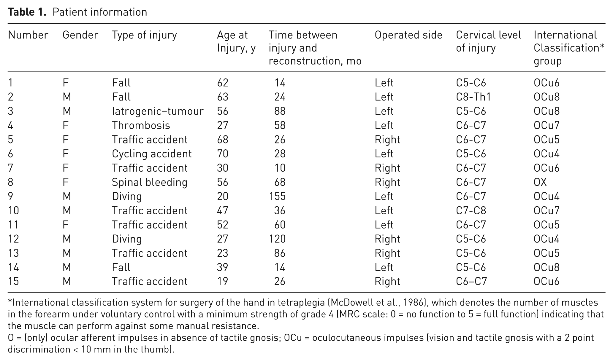
International classification system for surgery of the hand in tetraplegia (McDowell et al., 1986), which denotes the number of muscles in the forearm under voluntary control with a minimum strength of grade 4 (MRC scale: 0 = no function to 5 = full function) indicating that the muscle can perform against some manual resistance.
O = (only) ocular afferent impulses in absence of tactile gnosis; OCu = oculocutaneous impulses (vision and tactile gnosis with a 2 point discrimination < 10 mm in the thumb).
In addition to and at the same time as the EDM-to-APB transfer, patients also had between 2 and 4 additional procedures to reconstruct grip on the same hand, accounting for a total of 51 operations. Thirteen patients had restoration of thumb and finger flexion (Lamb and Chan, 1983) by transfer of the brachioradialis to the FPL and extensor carpi radialis (ECRL) to flexor digitorum profundus. We excluded the deep flexor of the little finger to prevent hyperflexion due to the small excursion needed to obtain full finger flexion in this finger. A split FPL distal thumb tenodesis was added in 12 patients to balance thumb flexion and counter the problem of a hyperflexed interphalangeal joint (Mohammed et al., 1992). Nine patients had a Zancolli lasso plasty for intrinsic substitution. The patient whose pattern of paralysis was rated as exceptional had activation of her deep finger flexors by the brachioradialis and a distal thumb tenodesis. The OCu8 patient had tendon transfer to restore thumb flexion using the pronator teres, while the brachioradialis was sutured to the Zancolli lasso plasty of the superficial finger flexors to activate the Zancolli lasso plasty (Table 2).
Pre- and postoperative data
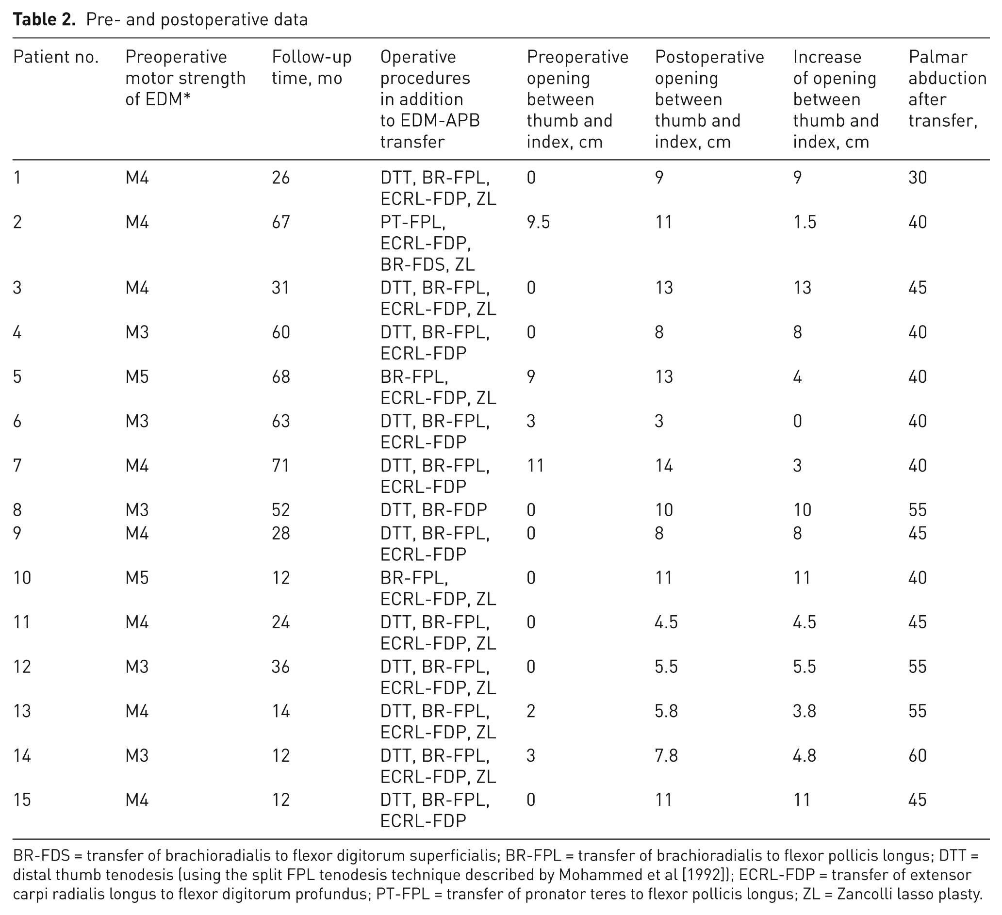
BR-FDS = transfer of brachioradialis to flexor digitorum superficialis; BR-FPL = transfer of brachioradialis to flexor pollicis longus; DTT = distal thumb tenodesis (using the split FPL tenodesis technique described by Mohammed et al [1992]); ECRL-FDP = transfer of extensor carpi radialis longus to flexor digitorum profundus; PT-FPL = transfer of pronator teres to flexor pollicis longus; ZL = Zancolli lasso plasty.
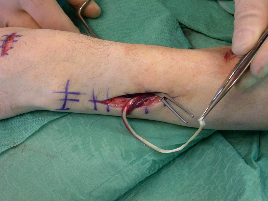
The EDM-to-APB transfer was then done. The most distal portion of the EDM tendon was exposed through a transversal incision over the fifth metacarpophalangeal joint and detached (Figures 1–3). EDM tendon usually consists of two individual tendon slips that are both harvested. Through a separate incision just distal to the dorsal aspect of the ulnar styloid, fascial bands connecting the EDM to the extensor digitorum communis (EDC) tendon of the little finger were divided. Through a 5 cm longitudinal incision on the dorso-ulnar aspect of the mid-forearm, the common extensor fascia was incised and the EDM muscle was identified and retracted. Because this procedure was usually combined with reconstruction of finger flexion and key pinch, the flexor tendons of the distal forearm were already exposed through a separate curvilinear incision proximal to the wrist. By retracting and securing the flexor tendons of the thumb and fingers as well as the median nerve ulnarly, the interosseous membrane (IOM) was exposed 2–3 cm proximal to the proximal border of the pronator quadratus muscle. An incision was made in the IOM, an area measuring 1 x 1.5 cm removed, and the hole widened using blunt dissection. At this stage, it was necessary to protect the anterior and posterior interosseous vessels and nerves. Using a tendon passer or hemostat, the EDM tendon was retrieved through the hole of the IOM and tunnelled subcutaneously over the thenar region after passing ulnar to the flexor carpi radialis (FCR) tendon. The EDM tendon was brought to the insertion of the APB. As the distal part of the EDM tendon consists of two portions, its tension can be balanced so that true palmar abduction is obtained initially, followed by a slight opposition moment, carefully avoiding any metacarpophalangeal flexion or extension movement of the arms. Tension was set so that the pulp of the thumb just touched the proximal interphalangeal joint of the index finger, while the wrist was kept in 20° of extension and maximum thumb-to-index finger distance was achieved with full passive flexion of the wrist. The reconstruction was protected in a well-moulded plaster slab.

Harvest of the extensor digiti minimi (EDM) tendon from the fifth extensor compartment. The interconnection bands to EDC V are cut and the EDM tendon is retracted to the level of the distal forearm.
Tunnelling of the extensor digiti minimi (EDM) tendon through the interosseous membrane (IOM). An oblique course is secured through the IOM.

Operative field before retrieving the extensor digiti minimi tendon to the attachment of the abductor pollicis brevis muscle. The brachioradialis and extensor carpi radialis tendons are seen after rerouting and before transfer to the flexor pollicis longus and flexor digitorum superficialis tendons, respectively.
All patients had the same postoperative regimen divided into two training periods according to the standardized protocol used by our hand therapists. The first started on the first postoperative day and focused on mobilization of the flexion apparatus to enable the patient to learn how to activate the new grip. Specifically, palmar abduction of the thumb was trained by extending the little finger against light resistance applied by the therapist just distal to the MCP joint. During this period, protective splints were used between training sessions. Task-oriented training was introduced after 4 weeks parallel to functional training and was designed to integrate the new grip function into activities of daily life. All patients were routinely evaluated 4 weeks, 3 and 6 months, and 1 year after surgery.
Preoperative evaluation included sensibility testing, measurements of joint range of motion, and grading the power of the triceps and all muscles below the elbow, allowing for categorization of each upper extremity according to the International Classification system. Preoperative strength of the EDM muscle was judged as MRC grade 3 (full range of motion against gravity) in 5 patients, grade 4 (full active range of motion against moderate resistance) in 8 cases, and normal or grade 5 (full active range of motion against full resistance) in the remaining in 2 patients. All EDM tests were performed by blocking the EDC action with MCP joints of the other fingers held flexed. EDC strength of MRC grade 3 or higher was always verified before surgery.
Using a goniometer, palmar abduction was measured between the thumb and plane of the palm. Passive movement due to tenodesis or gravity was avoided by resting the dorsum of the hand and forearm on the table.
Opening of the first web space was measured as the maximum distance between the radial corner of the nail of the index finger and distal ulnar corner of the thumb nail during wrist flexion (usually gravity-driven, unless FCR function existed) before and after the operation. Opening was considered 0 cm when ulnar aspect of thumb and radial aspect of index finger were in direct contact during active or passive wrist flexion. Key pinch was assessed using a Preston pinch gauge (European Bissel Health Care Ltd., Winchester, UK). Grip strength—the function of the finger flexors (grasp)—was measured using a standard Martin Vigorimeter (Gebrüder Martin, Tuttlingen, Germany) (middle-sized balloon) and the distance of the index, middle, ring, and little finger tips to the proximal palmar crease was recorded before and after each stage of surgical rehabilitation. Pre- and postoperative comparisons were made using paired Student’s t-test. Significance level was set at p < 0.05. Data are expressed as mean (SEM), unless stated otherwise.
Results
All patients were re-examined at a mean of 38.4 (SD 22.7) months. They reported not only improved grip, but also a substantial improvement of opening of the hand, found they were able to perform many activities of daily life, and were able to eat and dress independently. After surgery, active thumb-index opening significantly increased from 2.5 (SEM 1.0) cm preoperatively to 9.0 (SEM 0.8) cm (p < 0.001; Figures 4, 5). Nine of the 15 patients who had no active opening of the first web space (preoperative distance 0 cm) recovered a mean thumb-index opening of 9.1 (SEM 1.7) cm. Of the 6 patients who had an opening of 6.3 (SD 1.6) cm before surgery, this distance increased by 2.9 (SEM 0.8) cm. After reconstruction, the average active palmar abduction of the thumb was 45° (SEM 2°) (range 30–60°).

Maximum palmar abduction of the thumb in a patient with extensor digiti minimi (EDM) transferred to abductor pollicis brevis. Arrow indicates contour of the transferred EDM tendon.

Magnitude of the opening of first web space before and after reconstruction of active palmar abduction by extensor digiti minimi to abductor pollicis brevis tendon transfer. The opening after surgery was more than three-times greater than preoperatively (p < 0.001). Data expressed as means with whiskers indicating SEM.
All but one patient were able to direct and coordinate pinch of the thumb tip on the radial aspect of the index finger for different tasks using APB function, including five patients whose EDM strength had been rated as grade 3 before transfer.
Preoperatively, grip strength was 0.01 and 0.2 kg in 2 cases, and another patient had a key pinch of 0.3 kg, while strength was not recordable in all other patients. After surgery, all patients had active grip strength, 2.2 (SEM 1.3) kg, and key pinch, 1.3 (SEM 0.3) kg. The mean active finger flexion positions, measured from tip to the distal palmar crease were 1.0 (SD 0.5), 1.2 (SEM 0.4), 1.0 (SEM 1.1), and 0.7 (SEM 0.3) cm for index, middle, ring, and little fingers, respectively.
One EDM tendon ruptured, possibly due to excessive load bearing by the patient in the early postoperative period. However, this did not adversely affect the key pinch of 3.4 kg and grip strength of 3.0 kg.
Discussion
We found that transfer of the EDM to APB is a reliable procedure to replace lost palmar abduction of the thumb in tetraplegia. The EDM can be transferred without a significant loss of function, as the EDC retains extension of the little finger. Due to its small size and strength, EDM has not been considered a potential source for tendon transfer in the International Classification until recently, when a division of the group 6 category according to the presence of the ulnar or radial extensors of the wrist or EDM was proposed (Landi et al., 2002). Transfer of the EDM to replace thumb abduction or extension or finger extension was mentioned in this context (Landi, 2003). The purpose of this study was to prospectively investigate the effect of restoring active palmar abduction of the thumb to refine key pinch reconstruction in the tetraplegic hand by transferring the EDM to the APB and to describe its technique, indications, and clinical results.
Spinal cord injury can result in absent or weak and poorly controlled grip between the thumb and fingers, and cause great handicap. Tendon transfer and joint stabilization procedures seek to restore the ability to produce adequate key and tip pinch to accomplish tasks (Hentz and Leclercq, 2002; Moberg, 1978). The success of these procedures is usually quantified by measuring maximum static thumb pinch strength using dynamometers. This assumes that increased pinch force of the weakened or paralyzed thumb will directly lead to improved function (House and Shannon, 1985). However, we are now increasingly aware of the critical need to position the thumb and index fingers relative to each other, control the direction, and adjust the magnitude of force exerted by the thumb tip (Johanson et al., 2001; Smaby et al., 2004). Objects must be held securely to prevent them from rotating out of pinch, especially in tetraplegic individuals who frequently have weaker and misdirected thumb-tip force during key pinch compared with normal subjects.
As the most activated muscles during inherently unstable tasks were the extensor pollicis longus (EPL) and APB, these authors recommend their action is restored by tendon transfer whenever possible, and thus provide forceful pinch combined with the ability to dynamically produce grasp with different combinations of strength and dexterity.
Hentz and Leclercq (2002) agree that the APB should direct thumb force anywhere on the radial aspect of the index finger, as this greatly improves precision pinch to hold small, round, and slippery objects. This may enable tetraplegic patients to pick up pills, open buttons, use instruments, and even manage sophisticated tasks such as self-catheterization with little or no assistance. This not only preserves urinary continence and renal function, but also greatly boosts patients’ morale and self-esteem (Kiyono et al., 2000). The superficial part of the APB continues distally and dorsally to reach the EPL; the APB supports the extension mechanism of the distal phalanx, and thus acts as an antagonistic to thumb flexion (Doyle and Botte, 2003). The strategy to restore APB function may also increase lateral stability when the thumb has the tendency to slip medially or laterally, and pronate and supinate under load. For example, during grasping long objects, such as typing sticks or writing and eating implements, tend to twist (Johanson et al., 2001; Kaufman et al., 1999; Smaby et al., 2004).
We think that this dynamic stability has distinct advantages over CMC arthrodesis, which provides fixed positioning of the thumb and can be useful in OCu4 to OCu8 patients, especially in mobile or unstable joints (House and Shannon, 1985; Zancolli, 1975). Stable extension and pronation of the thumb increases the efficiency of the extrinsic thumb muscles by reducing their required excursion and allows the thumb to align and make contact with the index finger for lateral pinch using wrist extension (Ejeskär et al., 1999). Yet, fusion of the CMC joint abolishes active adduction and abduction. Painful stiffness and limited opening of the first web space may result and interfere with important load-bearing activities, such as transferring from bed to wheelchair or grasping large objects. These disadvantages are avoided with the EDM-to-APB transfer and active opening of the first web space is enhanced. Acknowledging the importance of APB function, muscle physiology and biomechanics need to be considered to decide whether this technique is superior to other methods (Brand and Hollister, 1999; Fridén and Lieber, 2002; Lieber and Friden, 2000). Studies of the cross-sectional area, muscle length, muscle fibre length, and muscle mass show that the there is a very close match between the muscle architecture of the EDM and APB, suggesting a similar ability of generate force, velocity, and excursion of the donor and recipient muscle (Jacobson et al., 1992; Lieber et al., 1992).
It is generally agreed that only donors with at least full range of motion against moderate resistance (MRC grade 4) will reliably produce active motion, as the muscle chosen for transfer must be strong enough to perform its new function in its altered position. In addition, loss of one strength grade is generally noticed after transfer. However, in our five patients who had an EDM muscle strength of only MRC grade 3 (motion against gravity), all recovered valuable APB function, probably because the thumb is not abducted against resistance. The APB positions the thumb for action, rather than performing the action itself, and requires only a tension fraction of 1.1%, which is weaker than any of the interossei and about the same as the extensor proprius of the index and little fingers (Brand and Hollister, 1999).
In the most effective transfer, the muscle passes in a direct line from its origin to the insertion of the tendon being substituted. This configuration of straight line of pull is almost ideally met by the EDM-to-APB transfer through the interosseous membrane, probably better than through alternative pathways of transferring the EDM, such as around the ulnar border of the forearm or using other donors, such as the palmaris longus (Hentz and Leclercq, 2002). In comparison with a two-step procedure to create thumb abduction involving the EPL, EDC, abductor pollicis longus (APL), and extensor pollicis brevis (EPB) tendons, and with the sacrifice of the brachioradialis muscle as a power source, the EDM-to-APB tendon transfer is straightforward and achieves wider opening of the first web space (Goloborod’ko, 1999). We have also, in the past, transferred the ring finger superficialis tendon to the APB insertion and powered it using either the brachioradialis or flexor carpi ulnaris muscle, but none of these alternatives produced as good active palmar thumb abduction as the EDM transfer (Ejeskär, 2005). This procedure can improve force direction and coordination of the restored thumb flexion, and we strongly recommend this additional step to refine key pinch reconstruction in tetraplegic patients with an active EDM.
Footnotes
Conflict of interests
None declared.
Funding
Funding for this project was provided by Swedish Research Council Grant 11200 and the University of Gothenburg. This investigation complies with the University of Gothenburg Human Research Protection Program guidelines.