






Abstract
There has been a considerable evolution of screws used for internal fixation of scaphoid fractures. We discuss here, early results of a recently introduced implant Synthes 3.0 mm headless compression screw used for scaphoid fracture fixation. Twenty eight patients with scaphoid fractures (five acute and 23 nonunions) were treated with internal fixation by this non-variable pitch screw over a period of 18 months. All nonunions had pedicle vascularized bone grafting. All five patients with acute scaphoid fracture fixation had radiological healing at a mean of 8 weeks. Fifteen of 23 scaphoid fracture nonunions showed definite signs and a further seven showed probable signs of radiological healing at a mean of 8 months. One nonunion has failed to unite after surgery.
Introduction
There are several techniques and implants to fix scaphoid fractures. Various studies have described the clinical results and biomechanical comparison of screws used in scaphoid fracture fixation (Adla et al., 2005; Beadel et al., 2004; Inaparthy and Nicholl, 2008; Panchal et al., 2007; Pensy et al., 2009). We discuss here the non-variable pitch 3.0 mm headless compression screw (3.0 HCS) (® Synthes GmbH, Oberdorf, Switzerland).
Patients and methods
Twenty eight patients with scaphoid fractures were treated with 3.0 HCS fixation over a period of 18 months (January 2009–June 2010). We treated five acute fractures and 23 established nonunions of the scaphoid. There were four B2 (acute complete waist) and one B3 (acute proximal pole) fractures in the acute group. Fifteen waist and eight proximal pole nonunions were among the 23 D2 (displaced nonunion – unstable) type fractures. All operations were performed directly by, or under supervision of, the senior author. All patients received a single dose of intravenous antibiotic before the start of the procedure. An arm tourniquet at 250 mm Hg was used and the screw was placed using an image intensifier in all cases.
The 3.0 HCS is a cannulated counter-sinkable compression screw with an identical pitch of head and shaft (1.25 mm). It is available in two materials (titanium alloy and stainless steel) and two shaft thread lengths (short and long). The head of the screw has a 3.5 mm thread with self tapping flutes. It has a self drilling tip facilitating screw placement across the fracture over a 1.1 mm guide wire. The screw achieves compression across the fracture in three steps. First, a compression sleeve is used for screw insertion over a guide wire. Second, the fracture gap is closed and compressed by turning the sleeve. Third, counter-sinking of the screw into bone is done by a screwdriver, while the compression sleeve is held stationary. The correct length of thread is selected to avoid the thread crossing the fracture gap.
There were four male and one female patients with acute fractures. Their mean age was 27 years (range, 20–32). The female patient had associated injuries and needed internal fixation of a distal radius fracture. There was one proximal pole fracture and four displaced waist fractures that were fixed between 2–14 days after injury. The screw was placed antegrade for the proximal pole fracture and retrograde for the waist fracture.
Of the 23 patients with a nonunion, one was female and 22 were male with a mean age of 25.3 years (range, 16–55 years). There were eight proximal pole nonunions and 15 waist fracture nonunions. All had vascularized pedicle bone grafting. The proximal pole nonunions were approached through a dorsal approach using a pedicled distal radial bone graft based on the 1,2 intercompartmental supraretinacular artery (ICSRA). The nonunions of waist fractures were approached through a palmar incision extending proximally to enable harvesting of a distal radial bone graft based on the pedicle from palmar carpal anastomosis.
All patients were immobilized in a palmar plaster slab, including the thumb, until review for suture removal at 10 days. A thumb spica cast was used for 6 weeks. Patients were reviewed clinically and radiologically at 6 weeks, 3 months, 6 months and 1 year. Patients had standardized supervised rehabilitation. Patients were given a removable wrist splint for use after six weeks, if no signs of union on the radiograph were seen. A standard scaphoid radiograph series was obtained and reviewed by two observers at each visit. Radiological signs of fracture healing was categorized as definite, probable or no signs of union (Dias et al., 1989). Definite sign of union was recorded when no fracture gap was seen and bridging trabeculae were seen on both posteroanterior and lateral projections. Probable union was recorded when continuation of trabeculae was seen in at least one view and when no other signs of failure, such as lucency, around the screw was noted.
Results
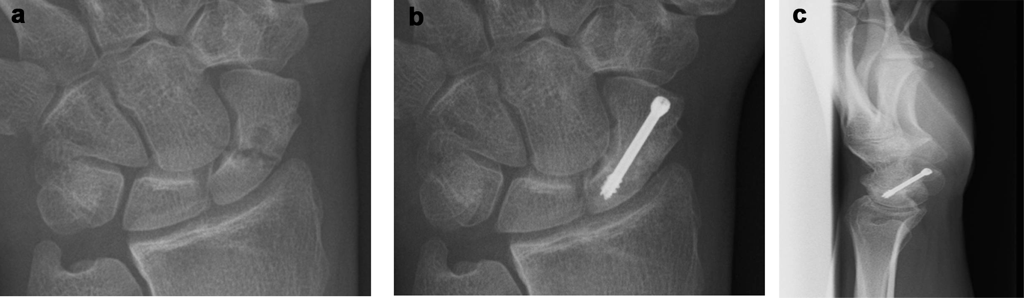
All five patients with acute scaphoid fracture fixation had radiological healing at a mean of 8 weeks. One patient had partial backing out of the screw after fracture union (Figure 1). There was a restriction of 20° of pronation in the patient with the distal radius fracture, but she had a satisfactory outcome for the scaphoid fracture.

(a) Acute scaphoid fracture – preoperative. (b) Acute scaphoid fracture – intraoperative. (c) Acute scaphoid fracture – postoperative (screw backed out but union achieved).
Of the 23 nonunions, 15 united (Figure 2) and seven showed probable signs (Figure 3) of radiological healing at a mean of 8 months (range 6–18 months). One nonunion failed to unite with the vascular pedicle graft (signs of loosening of the screw and no signs of radiological healing at 12 month) (Figure 4).
(a) Nonunion scaphoid – preoperative. (b) Nonunion scaphoid – postoperative (definite union) at 6 months. (c) Nonunion scaphoid – postoperative (definite union) at 6 months.

(a) Nonunion scaphoid – preoperative. (b) Nonunion scaphoid – postoperative (probable union) at 6 months. (c) Nonunion scaphoid – postoperative (probable union) at 6 months.

(a) Nonunion scaphoid – preoperative. (b) Nonunion scaphoid – intraoperative. (c) Nonunion scaphoid – postoperative (failure of union) at 12 months.
Three patients had problems with a tender scar, but this gradually resolved over a period of time with desensitization measures. No other complications, such as infection, nerve damage and complex regional pain syndrome, were seen.
Discussion
We used scaphoid radiographs to categorize fracture healing as definite, probable or no signs of union (Dias et al., 1989). Similar categorization was also used in other recent published studies (Inaparthy and Nicholl, 2008; Trezies et al., 2000; Tu et al., 2008). Union rate after scaphoid nonunion in our series was 95.6 % (if both definite and probable signs of healing are included) or 65.2% (if only definite healing are included). We are unable to attribute the state of union to the use of this screw alone as we also performed a vascularized graft in all cases. Our numbers are too small to arrive at any conclusion but feel that the technique of getting compression is unique and our early results do not suggest that the implant may be inferior to other implants.
Several different implants have been used for scaphoid fracture fixation. Various screws for scaphoid fixation have evolved and they are described as first and second generation screws (Fowler and Ilyas, 2010). The Herbert screw is a first generation screw and is a non-cannulated, variable pitch HCS. The compression jig and differential pitch of the Herbert screw helps achieve compression across the fracture site. The Whipple–Herbert screw (® Zimmer) is a cannulated modification with self-tapping leading threads.
The second generation of scaphoid screws include the Acutrak screw (® Acumed), which is a headless, self tapping, cannulated screw; the Kompressor screw (® Integra), which is a two piece headless, self tapping, cannulated screw; the Stryker Twinfix (® Stryker), which is a headless, cannulated, self tapping screw that allows for independent rotation of distal threads. The 3.0 HCS (® Synthes) used in our cases is a mono-block headless, self drilling, self tapping, cannulated screw with a uniform pitch. It uses the sleeve to achieve compression. It is suitable for a percutaneous fixation.
The 3.0 mm HCS with a vascularized bone graft has adequate early results for scaphoid fracture fixation.
Footnotes
Funding
This work received no specific grant from any funding agency in the public, commercial, or not-for-profit sectors.
Conflict of interests
None declared.