






Abstract
Normally the metacarpals have an epiphysis at one end — distally for the second to fifth and proximally for the first. Pseudoepiphyses are notches or clefts that occur at the non-epiphyseal ends of bones where an epiphyseal plate would be expected and are common incidental findings in the metacarpals of the growing hand. We aimed to identify the prevalence of pseudoepiphyses on serial radiographs of 610 healthy asymptomatic children. Pseudoepiphyses in the form of notches or clefts were common, identified most often in the second metacarpal (15.25%), fifth metacarpal (7.21%), and third metacarpal (0.49%). Complete pseudoepiphyses, in which the cleft extended across the full width of the metacarpal, were seen in the first metacarpal (1.97%) and the second metacarpal (1.31%). Pseudoepiphyses are a normal variant of metacarpal ossification and should not be confused with fractures in skeletally immature patients.
Introduction
There is a considerable amount of variation in the development of ossification centres in the growing hand. Secondary ossification centres occur in the second to fifth metacarpal heads at age 5–6 and at the base of the first metacarpal at age 7–8, although they are often not recognizable on plain radiography until a year after they begin to form (Scheur and Black, 2000). Fusion of these centres is also subject to a large amount of variation, but occurs at age 14–15 in girls and around age 16 in boys (Broom, 1906).
Pseudoepiphyses are notches or clefts that occur at the non-epiphyseal ends of bones, in a position that corresponds to the expected position of an epiphyseal plate in a typical long bone (Scheur and Black, 2000). These are most common in the head of the first metacarpal, the lateral aspect of the base of the second metacarpal, and the medial aspect of the base of the fifth metacarpal (Levine, 1972). They were first described in the 18th century (Nesbitt, 1736) and have been linked to a number of clinical conditions, including Down’s syndrome, hypothyroidism, and achondroplasia (Lee and Garn, 1967; Moses et al., 1998). However, they do occur during normal development and represent a normal variant of physeal ossification (Ogden et al., 1994).
The incidence of pseudoepiphyses is not clearly reported, although they are documented in a number of reported series (Broom, 1906; Haines, 1974; Lee and Garn, 1967; Poznanski, 1984). They may be mistaken for fracture lines and, therefore, become clinically important when evaluating patients after trauma, with congenital hand anomalies or metabolic bone disease (Keats and Harrison, 1980). In this study we examined the incidence of pseudoepiphyses in a population of healthy children between the ages of 5 and 18 and observed the development of the pseudoepiphysis over 5 consecutive years of imaging in each child.
Methods
Serial radiographs of 610 asymptomatic children were reviewed to determine the frequency and location of metacarpal pseudoepiphyses. The radiographs were performed as part of a school screening programme for scoliosis previously completed at our unit (Dickson, 1983). Ethical approval was granted as part of the grant application process for the screening programme.
During this programme, more than 900 fit and healthy children (aged 5–13 when screening began) had serial radiographs of the left hand over a 5-year period, to provide a marker of skeletal maturity in a study of spinal development. Our study group was comprised of the 610 children (360 boys plus 250 girls) who attended every annual review radiograph over a 5-year study period. As a result, there were five consecutive images for each child and a total of 3050 images were reviewed.
Results
A complete proximal pseudoepiphysis occurred in the second metacarpal in 8 out of the 610 cases (1.31%). These appeared as a lucent cleft across the base of the metacarpal with the appearance of a second epiphyseal plate (Figure 1). All of these were present at the first of five serial radiographs at mean age of 10 years and 4 months (range 6 years and 6 months to 13 years and 8 months). At the last of the five serial images, four had fused completely and four remained apparent. Combining all eight cases, the mean age at which the complete pseudoepiphysis was last visible was 14 years and 4 months (range 11 years 4 months to 16 years 6 months).

Complete pseudoepiphysis of the second metacarpal.
Typical cleft-like pseudoepiphyses were observed in the second metacarpal in 93 cases (15.25%) (Figure 2). All of these were present on the first radiograph, taken at a mean age of 9 years and 3 months (range 6 years and 6 months to 12 years and 10 months). At the last serial radiograph, the pseudoepiphysis remained visible in 20 out of 93 cases (22%). Combining all 93 cases, the age at which the partial pseudoepiphysis was last apparent was 12 years and 2 months (range 8 years and 1 month to 16 years).
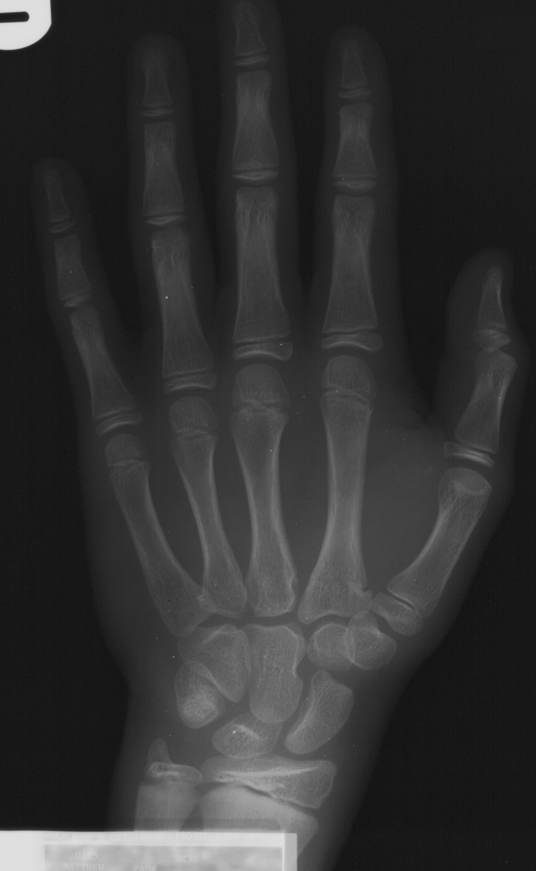
Partial pseudoepiphysis of the second metacarpal.
Pseudoepiphyses were also noted at the head of the first metacarpal in 12 out of the 610 children (1.97%). These were observed to occur as a lucent cleft across the head of the metacarpal (Figure 3). All of these were present at the first serial radiograph (mean age 10 years and 8 months; range 7 years and 8 months to 14 years and 2 months); however, none were apparent on the final radiograph. The mean age at which a pseudoepiphysis of the first metacarpal was last visible was 12 years and 11 months (range 9 years and 9 months to 16 years and 4 months).
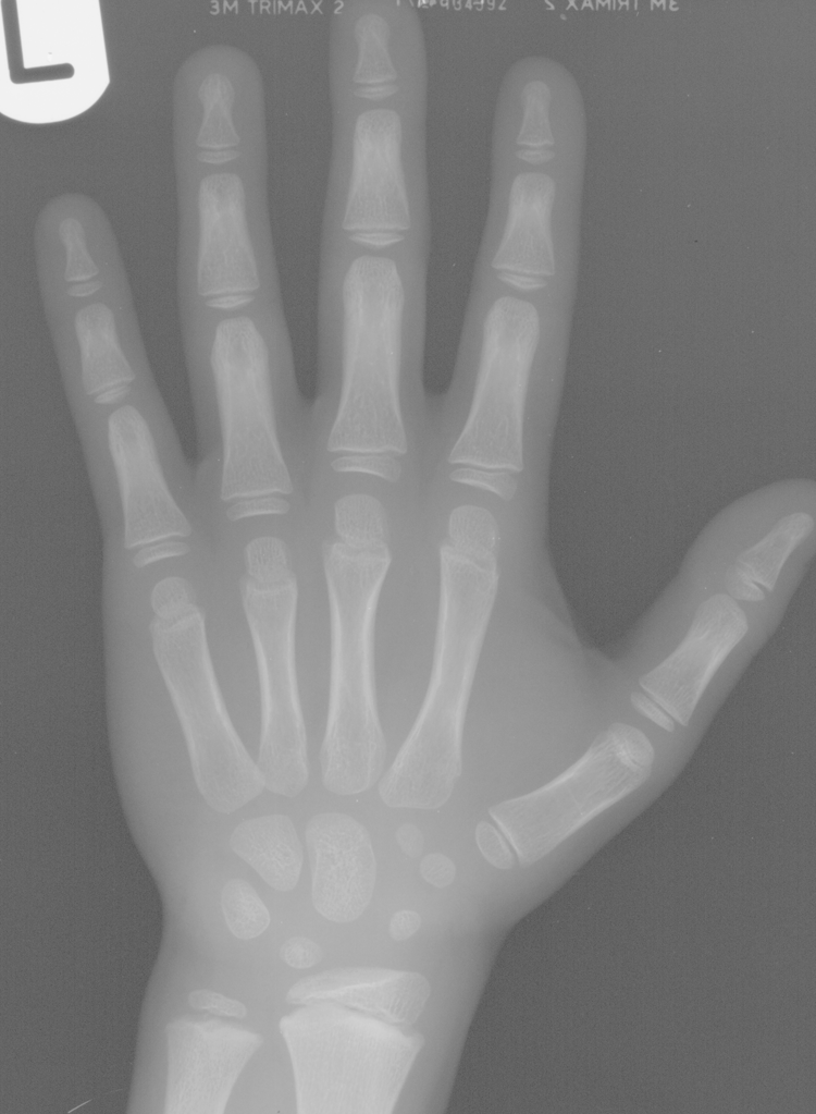
Complete pseudoepiphysis of the first metacarpal.
Cleft-like pseudoepiphyses were also noted in the fifth metacarpal (44; 7.21%), and the third metacarpal (3; 0.49%). In the fifth metacarpal, these were apparent on the first serial radiograph (mean age 9 years, range 6 years and 8 months to 13 years and 6 months), and the mean age at which they were last apparent was 12 years and 3 months (range 9 years and 4 months to 16 years and 6 months). These were only present on the final radiograph in 12 out of 44 cases. In the third metacarpal, these partial pseudoepiphyses were apparent on the first serial radiograph (mean age 9 years and 8 months, range 7 years to 11 years and 8 months) and were not present on the final image. The mean age at which they were last visible was 12 years and 11 months (range 11 years and 8 months to 14 years and 6 months).
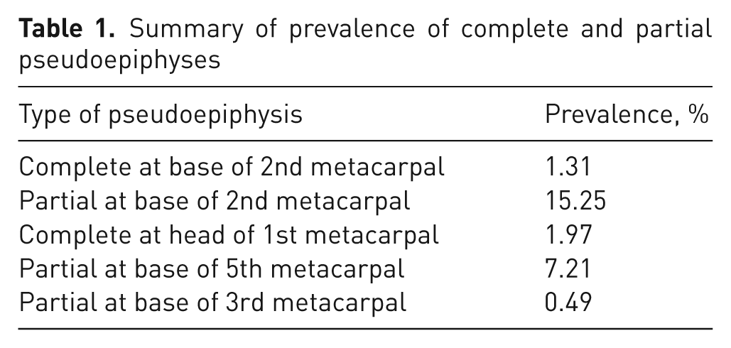
All of these lesions were asymptomatic. A summary of the prevalence of complete and partial pseudoepiphyses is shown in Table 1.
Summary of prevalence of complete and partial pseudoepiphyses
Discussion
There are a total of 48 separate ossification centres recognized in the growing hand, of which 29 are primary centres (including the thumb and sesamoids) and 19 are secondary (Scheuer and Black, 2000). Primary centres arise both in the early foetal period and after birth. The appearance of secondary centres is interspersed between the sequential primary centres; thus, both primary and secondary ossification centres may occur together on plain radiographs. Primary ossification of the metacarpals occurs between weeks 8 and 10, and commences sequentially in metacarpals 2–5 and then in the first metacarpal (Scheur and Black, 2000). Secondary ossification centres occur in the second to fifth metacarpal heads at age 5–6 years and at the base of the first metacarpal at age 7–8 years, although they are often not recognizable on plain radiography until a year after they begin to form (Scheur and Black, 2000). Fusion of these centres is also subject to a large amount of variation, but occurs in the metacarpal heads between 14 years and 5 months and 15 years in girls, and around 16 years and 6 months in boys (Broom, 1906). The base of the first metacarpal completes fusion at approximately 14 years and 1 month in girls and 16 years 4 months in boys (Broom, 1906).
Pseudoepiphyses occur at the non-epiphyseal ends of bones and are most common in the head of the first metacarpal and the base of the second and fifth metacarpals (Levine, 1972). Our study confirms that these are the most common sites, and we are able to add that in a population of children the prevalence of a complete pseudoepiphysis is low: 1.97% in the first metacarpal and 1.31% in the second metacarpal. Partial or cleft-like pseudoepiphyses were more common and occurred in the second, third, and fifth metacarpals (Table 1). Previous reports of pseudoepiphyses in healthy children are based on both plain radiography and histological examination. Lee and Garn (1967) investigated a longitudinal series of hand radiographs of healthy white children (116 boys and 118 girls) and found notching at the non-epiphyseal ends of the first, second, or fifth metacarpals in all children. They attempted to grade the intensity of notching and found a moderate relationship in intensity between siblings. The prevalence of transverse notches of this kind has been reported as around 30% at the base of the second metacarpal and 80% at the base of the fifth metacarpal (Lee and Garn, 1967; Dreizen et al., 1965; Levine, 1972). This is slightly higher than reported in the current study, and this is likely to be because we identified clefts bearing the appearance of a partial epiphysis rather than a simple indentation in the cortical surface. In contrast, the prevalence of a complete pseudoepiphysis reported in the current study is in accordance with previous descriptions (Poznanski, 1984).
The timing of pseudoepiphysis formation is not apparent from our series, as almost all were visible in the first serial X-ray. However, it has previously been suggested that they can be identified on plain radiographs from between the ages of 1–5 and remain potentially identifiable until completion of fusion within the hand (Ogden et al., 1994; Scheuer and Black, 2000). We found a wide variation in the age at which both complete and partial pseudoepiphyses appeared to fuse and noted that they were often fused before the normal epiphyseal plates within the hand.
The exact mechanism of formation of pseudoepiphyses is incompletely understood and histological descriptions are varied. It is clear that they represent a normal variant of physeal development and ultimately form a completely normal adult bone. Posener et al. (1939) initially described them as supernumerary epiphyses that appear as a separate node of ossification in an island of hyaline cartilage. Ogden et al. (1994) reported a different pattern of ossification and recognized three basic patterns of formation. In the first, a central osseous bridge extends outwards from the metaphysis and then expands into a ‘mushroom-like’ osseous structure. The circular notch proximal to this structure gives the bone the appearance of an epiphysis. If this notch is displaced to one side, it can give the appearance of a cleft or partial pseudo-epiphysis. The other two patterns recognized were of an abnormal peripheral osseous bridge, creating an eccentric notch in the bone, and of multiple abnormal osseous bridging points. In each situation, the area that appeared to be a ‘pseudo-physis’ lacked typical cell columns and these were incapable of significantly contributing to the postnatal growth of the involved bone. It seems likely that the partial pseudoepiphyses observed in our series followed this pattern of formation. However, the complete pseudoepiphyses observed are likely to have formed as de novo ossification centres within a cartilaginous epiphysis.
The prevalence of partial pseudoepiphyses in this and other studies supports the idea that these are transient structures that do not alter normal metacarpal development. Lee and Garn (1967) found that they were common in healthy children and often had no specific diagnostic significance. However, although they do not appear to alter the growth of bone, they are more common when there is a coexisting congenital abnormality or generalized growth retardation (Wood et al., 1994). It could be that the increased incidence of pseudoepiphyses in these cases is a result of the increased chance of identifying them where growth is slow. As a result, their appearance has been linked to a number of pathological conditions, including Down’s syndrome (Lee and Garn, 1967), hypothyroidism, achondroplasia (Wagner, 1956), cleidocranial dysostosis, and Laurence–Moon–Biedl–Bardet syndrome (Moses et al., 1998).
All of the variants seen in our study were noted in asymptomatic growing children. The finding of a metacarpal pseudoepiphysis on imaging of a growing child is therefore likely to be a normal variant of metacarpal ossification. Care should be taken that they are not confused with fractures in skeletally immature patients. Where a pseudoepiphysis is seen on the radiographs of a healthy asymptomatic child, the patient and their family should simply be reassured.
Footnotes
Funding
This research received no specific grant from any funding agency in the public, commercial, or not-for-profit sectors.
Conflict of interests
None declared.